- Content
Congenital malformation of the external ear. Axial CT scan shows significant narrowing of the ear canal with its obliteration due to impaired formation of soft tissues and bones
If hearing loss or hearing loss occurs, a CT scan of the ear is warranted. This is a modern, highly informative visualization method that uses X-rays and computer data processing to construct a three-dimensional image.
Contrast enhancement when performing a CT scan of the ear makes it possible to evaluate not only the bone structures, but also the soft tissues, nerves and vessels of the area, which is important for determining further tactics of action - conservative therapy or surgical treatment.
Normal ear anatomy
Computed tomography provides comprehensive information about the pathological processes of the outer and middle ear.
CT scanning is used for atresia of the external auditory canal (due to the lack of an opening, it is impossible to see the condition of the eardrum and middle ear structures during otoscopy).
The need for visualization, as an additional part of the examination, arises when therapy is ineffective or when complications are suspected. What does a CT scan of the middle ear show:
- inflammation and its consequences;
- injuries;
- pathological changes in the temporal bone;
- tumors (osteoma, schwannoma, paraganglioma, etc.) and their invasion into surrounding structures;
- congenital malformations;
- vascular pathologies;
- condition of the bone labyrinth;
- destructive and degenerative processes (osteodystrophy, fibrous osteodysplasia, post-inflammatory ossification, sclerotic changes, etc.);
- foreign bodies in the ear.
A computer scan of the temporal bone allows for differential diagnosis of tumors with inflammation, clarification of anatomical features, and determination of the need for surgery and its volume. The study is useful in assessing complications after surgical interventions.
A CT scan of the inner ear is indicated if a pathological process is suspected in the area limited by the temporal bone. The sooner a computer scan is done, the greater the chances of a successful operation, including to restore or preserve hearing.
The structures of the middle and inner ear are hidden behind the eardrum, limiting direct noninvasive assessment.
Otoscopy does not provide a full-scale picture, so CT and/or MRI are performed to determine the extent of the disease, the exact location of the pathological process, the structures involved and possible destructive changes in the bone.
The most common diseases in this area are chronic and acute inflammatory diseases (infectious and secretory otitis media), less often - tumors (middle ear schwannoma and paraganglioma). A CT scan of the inner ear with contrast shows the Eustachian tube, cochlea, auditory and vestibular nerves, blood vessels and bones, which makes it possible to identify the cause of problems with the corresponding complaints.
How to do a CT scan of the ear
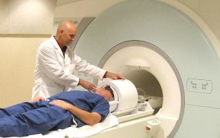
Patient position during examination
You can get a CT scan of the middle and inner ear in St. Petersburg at the Magnit diagnostic center, by appointment, using the form on the website or by calling +7 (812) 407-32-31. At the appointed time, the patient is taken to the X-ray room.
First, the doctor will clarify whether there is a concomitant pathology, allergic reactions to iodine and its derivatives in the anamnesis.
Pregnancy (even probable) should be reported in advance - in this case it is possible to resort to diagnostics not related to radiation exposure (ultrasound, otoscopy, MRI).
The patient is placed on the table (horizontal position). For convenience and fixation, elastic rollers and belts are used; this measure is necessary so that the resulting tomograms do not have artifacts (blurring, as in pictures taken with a regular camera of a moving object).
In the middle of the study, a contrast agent is injected intravenously to better visualize the smallest details. This is done by the nurse at the same time, or the radiopharmaceutical is delivered automatically using an injector during certain phases of the study (bolus enhancement).
The table slides deep into the tomograph; sensors begin to rotate around the head in the ring of the device with a characteristic clicking sound, capturing the response of organs and structures to the flow of X-ray particles emitted by the emitter.
The information is transferred to the computer, and a special program restores the resulting sections into a single whole.
There is a “panic button” at the patient’s fingertips: the staff should be immediately notified of any deterioration in health (sticky sweat, faintness, severe lack of air). When using modern contrast agents, generalized hypersensitivity reactions are extremely rare, occurring in less than 1-5% of cases.
Which is better: CT or MRI of the ear?
CT (A) and MRI (B) - picture of cholesterol granuloma
This dilemma worries many patients, but the type of examination is chosen by the attending physician, based on the results of previous diagnostics and specific complaints.
There is no point in refusing an ear CT scan due to false concerns about the harm of the procedure; modern multi-detector tomographs make it possible to minimize the effect of radiation exposure for a larger category of people (with the exception of pregnant women and children under 14 years of age).
In some observations, especially if a tumor process is visualized, both MRI and CT of the ear may be needed - these methods complement each other. Performing a magnetic resonance scan is justified if, after a CT scan, there are still doubts about the diagnosis.
MRI better demonstrates meningocele, cholesteatoma, facial neuritis, intracranial pathology caused by processes in the middle ear, which is not always shown by high-resolution computed tomography. CT of the middle ear is often unable to visualize the degree of possible obliteration of the cochlea after inflammation or the long-term consequences of a temporal bone fracture. Accurate assessment of the condition is critical for planning cochlear implantation.
A computed tomography scan of a child or pregnant woman can be performed for health reasons. Preferable during the gestation period is MRI, which does not use X-ray radiation, and the magnetic field does not pose a danger to the body.
Preparing for a CT examination of the ear
A light snack 40 minutes before CT neutralizes autonomic symptoms
A CT scan of the ear does not require any lengthy preparation; it is enough to choose comfortable clothes without metal parts and leave all jewelry at home. You will also be asked to remove removable dentures and piercings. If you plan to administer a contrast agent, you need to pay attention to the following aspects:
- Blood test for creatinine. You can take it at the clinic and bring the result with you (no later than the 10-day deadline), or do it immediately before computer diagnostics. The Magnit center provides such a service, but you must arrive for the examination 15 minutes earlier.
- A break from taking Metformin and its analogues. Patients with diabetes mellitus who control their glucose levels with medication should stop treatment with glucose-lowering drugs 48 hours before the diagnostic procedure. Before this, consultation and permission from an endocrinologist is required.
- Lactation. During this period, the study will not lead to negative consequences if you stock up on milk for the next two feedings. After a CT scan, pumping is recommended to avoid mastitis.
- A loose snack. The administration of a contrast agent may be accompanied by a number of autonomic reactions (metallic taste, nausea, dizziness). To prevent this from happening, you need to eat a light breakfast 40 minutes before the diagnostic procedure.
CT ear with contrast
The Magnit diagnostic center uses a modern contrast agent with a maximum safety profile
A native CT scan of the ear shows predominantly bony structures. To assess vessels, soft tissues, the extent of the pathological process and determine the degree of involvement of neighboring areas, contrast contrast is necessary.
Enhanced computed tomography provides excellent visualization of the differences between bony structures, air and soft tissues, coupled with high spatial resolution, and is the modality of choice for examining middle ear structures.
Contrast-enhanced CT scan of the temporal bone allows differentiation of inflammatory changes, cholesteatoma and tumor. The price for an enhanced ear CT scan is slightly higher, as it includes the cost of the radiopharmaceutical.
Indications and contraindications
An ENT doctor will refer you for a CT scan of the ear if the primary diagnostic methods show ambiguous results.
For all unexplained symptoms, diagnostic testing is warranted. Indications for CT ear are as follows:
- hearing impairment/loss with an assessment of its possible causes;
- earache;
- discharge from the ear canal;
- long-term inflammation in the middle or inner ear that cannot be treated;
- dizziness, unsteadiness of gait, nystagmus;
- injury to the ear, temporal bone;
- suspicion of a tumor process;
- identification of developmental anomalies;
- preoperative diagnosis;
- dynamic observation of changes after conservative therapy or surgery;
- diagnosis of complications: petrositis, labyrinthitis, labyrinthine fistula, intracranial abscess or brain abscess, meningitis.
A general contraindication to native CT of the ear is pregnancy; with contrast, the following are added to this:
- burdened allergic history: generalized allergic reaction to a radiopharmaceutical such as Quincke's edema or anaphylatic shock;
- urological and nephrological pathology, accompanied by chronic renal failure;
- hyperfunction of the thyroid gland.
Patients with severe obesity (body weight above 150 kg) are not tested for technical reasons.
The radiologist deciphers the received data, and the patient is given a conclusion and images in electronic form. We present to your attention several photos of a CT scan of the ear, with typical pathological processes:
Facial nerve schwannoma: Axial CT image (bone window) demonstrates ganglion expansion and a homogeneous soft tissue mass is present (arrow)
Chronic middle ear inflammation with cholesteatoma: Coronal CT scan shows a lateral semicircular canal fistula (arrow) caused by bone erosion due to neoplastic growth
Chronic inflammation of the middle ear: Axial CT demonstrates focal calcification (arrow) in the tympanic cavity - tympanosclerosis.
Source: https://spb24mrt.ru/kt-info/chto-pokazyvaet-kt-uha
What does an MRI of the ear show - indications for diagnostics MRI and CT scan of the inner ear
It is quite difficult to study all the organs and systems that are localized in the head.
Not all diagnostic methods are suitable for studying the condition of these organs, because the brain contains a lot of nervous tissue. Accordingly, radiation diagnostics is excluded immediately.
It is almost impossible to carry out invasive diagnostic methods, because the organs of the visual, auditory, and vestibular apparatus are located too compactly.
To diagnose hearing organs, experts recommend performing an MRI of the ear or a CT scan. Experts have recognized magnetic resonance imaging as the main method of visualizing the head and brain areas. Its advantage is the absence of radiation exposure and invasion. After an MRI of the middle and inner ear, the patient does not experience any consequences.
A radiologist can conduct magnetic resonance imaging in the area of the inner ear as an independent study, as well as in a comprehensive study of the eye orbits, brain, arteries, and temporal bones.
As for computed tomography, it is usually prescribed if the patient has complaints of hearing problems or pain in the ears. This diagnostic method is usually prescribed first.
It will help identify defects, anomalies, and diseases of organs, and MRI is often prescribed if it is necessary to clarify the source of the lesion and the localization of the pathology.
The specialist decides which of these two diagnostic methods to prescribe to the patient, taking into account the specifics of the situation.
In what case is it necessary to perform diagnostics?
MRI of the inner ear is usually prescribed if the patient has specific symptoms that indicate the presence of diseases of this organ. Symptoms for which a diagnosis must be prescribed include:
- frequent dizziness;
- severe hearing loss, complete loss;
- pain, itching inside the ear;
- swelling, redness of the ears;
- ringing, noise inside the ears;
- heat;
- the presence of specific discharge from the ears.
These same symptoms require a CT scan of the ear. The choice of procedure is up to the doctor. In addition, computed tomography is performed if there are the following indications:
- unsteadiness of gait;
- tingling, aching pain;
- planned observation, which allows a specialist to monitor the progress of therapy;
- otosclerosis;
- upcoming elective surgery;
- suspicion of the presence of a neoplasm inside the organ.
Suspicion of the presence of diseases of the inner ear in a patient may cause symptoms such as anemia of the facial nerve, impaired tone of the facial muscles, and impaired sense of balance. These signs are characteristic of many pathologies, but most often they are caused by pathological processes in the cerebellopontine angles.
You need to be especially careful if you have tinnitus that recurs periodically. It can be triggered by various reasons, not only pathology in the hearing organs. Therefore, MRI of the inner ear is performed to differentiate hearing impairment from disorders in the spine, disorders of the vascular system, and pathological processes inside the brain.
What can tomography detect?
A study in the middle ear area shows even the smallest changes in such parameters of the head organs as structure and functioning. Diagnostic indications give the doctor the opportunity to examine microscopic abnormalities in the area of the auditory organ. The scan provides a series of images that contain sections of the smallest areas of the middle and inner ear in various projections.
A special advantage of diagnostic methods such as magnetic resonance imaging and computed tomography is the ability to rotate a three-dimensional model of the hearing organ, enlarge the desired area, and change the clarity of the picture.
MRI can visualize:
- many inflammatory processes that occur in the area of the auditory nerve;
- tumors (benign/malignant) in this area;
- acoustic neuromas;
- metastases penetrating into the area of the auditory organ from neighboring tissues;
- deformation/destruction of bone structures;
- consequences of otitis media;
- tissue proliferation (uncontrolled).
To diagnose many of the above pathologies, MRI with contrast is required. In this case, the contrast agent is administered intravenously. It penetrates even the smallest capillaries, into all affected areas, making them more visible. Contrast also gives the specialist the opportunity to identify areas with impaired blood circulation.
Computed tomography helps detect the following pathologies:
- hearing loss. Thanks to CT, the doctor will clarify the form of the disease (congenital, acquired);
- inflammatory, infectious processes;
- fractures. Diagnostics allows you to determine the plane of the fracture and the presence of displacement;
- neoplasms (angiomas, tumors, neuromas, cholesteatomas, abscesses).
No special preparation is required for diagnosis. It is enough to remove all jewelry and clothing with metal elements. Only when using a contrast agent, the patient should refrain from drinking and eating 4 hours before the procedure.
The price of the procedure depends on the equipment used and the presence of a contrast agent. If a CT scan is performed, its cost will be between 3.5 and 8 thousand rubles. If an MRI is prescribed, it will cost more, about 5 – 9.5 thousand rubles. Contrasting will cost approximately 700 – 1000 rubles.
The entire diagnosis takes about 15 minutes. If it is necessary to use a contrast agent to diagnose tumor formations, the procedure may take 45 minutes.
Contraindications for performing computed tomography and magnetic resonance imaging
MRI cannot be performed if the patient has:
- insulin pump;
- pacemaker;
- built-in electronic devices;
- renal failure (diagnosis with contrast is prohibited in case of this);
- allergy to contrast agent;
- metal implants.
Computed tomography is not recommended:
- children under 14 years of age;
- early stages of pregnancy;
- unconscious, serious condition;
- breastfeeding (cannot be diagnosed with contrast);
- liver, kidney failure.
The specialist decides which procedure to undergo (MRI or CT) when examining the ears. You should always follow his recommendations for a speedy cure of any pathology.
Source: https://DiagnostLab.ru/mrt/golova-i-sheya/chto-pokazyvaet-mrt-uha.html
Computed tomography of the ear
Various pathologies of the inner ear lead to a decrease or complete loss of auditory activity.
The use of modern diagnostic methods allows us to determine the condition of soft and hard tissues, as well as the vascular system of the area being studied.
Ear CT is one of the most informative diagnostic methods, which is necessary for making a diagnosis and prescribing appropriate treatment.
Computed tomography belongs to the category of advanced radiographic methods. Using a special apparatus, ionized radiation is captured, after which a graphic image is displayed on the monitor screen.
Modern cone beam scanners significantly reduce examination time. It only takes a few seconds to get an accurate picture.
Content
Indications for the procedure
A computed tomography scan of the ear is prescribed if the following symptoms are present:
- The patient's hearing acuity sharply decreased. Symptoms can occur for no apparent reason, or after inflammation of the nasopharynx or otitis media.
- Discharge of purulent or serous fluid from the ear.
- Painful sensations of varying degrees of intensity.
- The patient complains of frequent dizziness, and there is a lack of coordination when walking. All this indicates a dysfunction of the vestibular apparatus.
- Suspicion of the presence of tumors in the ear area and nearby tissues.
- Suspicion of a temporal bone fracture.
In addition, computed tomography is prescribed as a monitoring measure. We are talking about planned procedures to evaluate previous treatment, as well as preparation before surgery in connection with neoplasms.
What are the contraindications?
Contraindications to CT scanning of the inner ear are explained by the specifics of the procedure itself:
- Impact of gamma radiation on humans.
- The need to use a contrast agent.
- During the procedure, the patient must remain motionless.
Based on this, a CT scan is not performed if a person is in an alcoholic, drugged or unconscious state. During the examination, the patient may make involuntary movements, which will inevitably distort the information obtained. Various implants and pacemakers are sensitive to gamma rays, so they can also be an obstacle to the procedure.
If we are talking about a contrast agent, then there are certain limitations. The fact is that the contrast contains iodine and barium, which are eliminated from the body naturally within a few days after the CT scan. If the patient has acute or chronic pathologies in the kidneys, then the contrast agent is not used.
Preparing for CT
Computed tomography is performed on an outpatient basis. It is advisable to arrive 30 minutes before the start of the examination. The main requirement is the absence of metal objects on the body and clothing:
- Bracelets, chains and other jewelry.
- Bracket systems.
- Dental jaws (if they contain metal elements).
- Hearing Aids.
Can it be done for children and during pregnancy?
Most experts agree that a child can only undergo a CT scan of the ear from the age of 14. Before this age, it is also possible to use computed tomography, but only if there is an urgent medical need.
For pregnant women, CT scanning is not advisable, especially in the first and last trimester. During the examination, the patient receives a certain dose of radiation, which can negatively affect the formation of the fetus.
Ear CT scan: what does this study show?
Every patient who has been prescribed a CT scan of the inner ear probably wants to know what this procedure shows? It allows:
- Assess the condition of the facial nerve.
- Show a clear picture of the structure of the middle ear.
- Determine the type of hearing loss in the patient (congenital or acquired).
- Visualize various neoplasms (neurinomas, angiomas, abscesses, etc.).
- Identify infectious and inflammatory processes.
- If an injury occurs, a CT scan determines the displacement of the bones and the general condition of the hard tissues.
How is the examination carried out?
Regardless of what exactly is being examined, computed tomography is performed as follows:
- The patient is positioned standing in a special scanning area (tomograph).
- The jaw, chin and head are installed statically using clamps. This is necessary to prevent involuntary movements of the patient.
- After this, the procedure itself begins. The tomograph ring rotates around the patient’s head (the operation of the device may be accompanied by characteristic sounds).
- The image is immediately transferred to the monitor. The specialist can immediately assess the presence or absence of pathologies from the images.
- The entire procedure takes 1-2 minutes, and the scanning itself takes about 15-20 seconds.
Features of CT with contrast
Certain clinical cases require the use of a contrast agent consisting of barium or iodine salts. The solution itself is administered intravenously immediately before the procedure.
Before computed tomography using a contrast agent, the following requirements must be observed:
- Restriction of food and water 4 hours before the examination.
- Diet for three days before the procedure. It is necessary to exclude any flour, fatty, spicy and smoked foods.
- It is advisable to refrain from smoking on the day of the examination.
- If you have an allergy to iodine, you need to warn about it in advance.
When ingested, the contrast gives the vessel a brighter hue, so they are better visible in photographs. This method is most often prescribed for studying blood circulation in veins and arteries, as well as for complex diagnosis of neoplasms.
How often can a CT scan be done?
A diagnostic test that would not only be accurate, fast and informative, but also absolutely safe for the human body, has not yet been invented. A CT scan of the middle ear allows you to quickly determine the pathology, but frequent use of this procedure can negatively affect the functioning of the internal organs.
The maximum permissible radiation dose is about 150 mSv per year. If this level is exceeded, the consequences for the body can be extremely negative. But if the clinical picture of the disease requires re-examination, then repeated images are possible, especially since cone-beam computed tomography at maximum load produces an exposure of 0.1500 mSv per procedure.
Decoding the results obtained
During the examination, X-ray pulses are converted into electrical signals, which undergo computer processing, resulting in an image appearing on the monitor screen.
The resulting images are interpreted by a specialist. When making a diagnosis, the radiologist pays attention to the following parameters:
- Dimensions of the internal auditory canal.
- Condition of the bone labyrinth.
- The presence of inflammation and neoplasms.
Depending on the area being examined, foci of pathology and neoplasms may appear on the image as light or dark spots.
Which is better - CT or MRI?
One of the possible alternative diagnostic methods to CT is magnetic resonance imaging. It is based on magnetic resonance, which is not accompanied by gamma radiation. In this regard, MRI has fewer contraindications, which is a definite advantage.
Additionally, middle ear cholesteatoma is not as visible on CT as it is on MRI. Magnetic resonance therapy also better demonstrates intracranial pathologies, meningocele and facial neuritis.
On the other hand, MRI is good at visualizing soft tissues, but may have problems identifying changes in hard tissues. This method is not used for emergency diagnosis because it takes about 1 hour.
Thus, answering the question of which is better - MRI or CT of the ear, it is impossible to give a definite answer. The appropriateness of a particular examination method is determined by the attending physician, but in most cases, MRI is used when confirmation of the diagnosis is required after a computed tomography scan.
Where can I get a computed tomography scan of the inner ear in St. Petersburg?
X-ray centers "LUCH" has been conducting X-ray examinations since 2015. We understand the importance of timely diagnosis, so with us you can undergo a computed tomography scan of the inner ear, as well as a number of other studies, as soon as possible.
At your request, the examination results can be recorded on digital media or sent by email. Leave your online application or call the contact phone number to make an appointment.
Sign up for a study by phone or online +7 (812) 332-52-54
Source: https://Center-Luch.ru/stati/kompyuternaya-tomografiya-uha/
MRI ear
Problems with the normal functioning of the inner ear can manifest themselves as anemia of the facial nerve, pathology of facial muscle tone, and even a violation of the normal sense of balance.
The symptoms may not seem the most terrible, but they can hide serious problems, including deformation of bone structures and tumor formations of varying degrees of severity.
An MRI of the inner ear is performed in order to identify all these disturbances in the normal functioning of this organ.
MRI image of the inner ear
There is only one way to prevent the development of complex and dangerous diseases - timely diagnosis. You should contact your doctor for a referral for a hearing examination if you experience the following symptoms:
- hearing loss or noticeable decrease in sensitivity;
- noise and ringing in the ears;
- systematic dizziness;
- itching and pain in the ears;
- swelling of the ears;
- elevated temperature;
- pain localized in the throat or nasopharynx;
- discharge from the ear.
Identifiable diseases
The advantages of MRI as a method for studying the inner ear are obvious: tomography is safe from the point of view of its impact on health, it is carried out quickly and allows you to fully examine even the smallest changes in the well-functioning system of this part of the hearing organ. Tomography images show all the details in different projections and even in a three-dimensional model, which allows you to examine the inner ear even more carefully. This diagnostic method reveals:
- cochlear neuritis (inflammatory processes in the tissues of the auditory nerve);
- tumors and metastases growing from other organs;
- uncontrolled abnormal tissue growths;
- deformations with potential destruction of bone structures;
- consequences of inflammatory processes;
- deformations of the ear labyrinth;
- inflammation of the labyrinth, which developed due to the penetration of pathogenic organisms or trauma.
You should contact your doctor for a referral for a hearing examination if you experience symptoms such as hearing loss or a noticeable decrease in sensitivity, noise and ringing in the ears, systematic dizziness, etc.
Disorders of auditory and vestibular activity, inflammatory and non-inflammatory processes, injuries and the consequences of previous diseases - all this can be detected by tomography of the inner ear in the early stages, preventing the disease from progressing to a more serious level, when damage can affect an already vital organ: the brain .
A diagnostic examination of the inner ear can be carried out either as an independent study or as part of a diagnosis of the condition of the temporal bones of the skull, the brain and the arteries that supply it, as well as the eye orbits.
All these areas are so close, and the influence they have on each other is so great that it is very convenient to conduct a comprehensive survey of them. However, such a diagnosis is carried out only in cases with an unclear diagnosis.
If the symptoms of the disease are defined to one degree or another, directed tomography is performed.
Collaboration of CT and MRI
The need for a more serious examination than a simple examination can only be determined by an otolaryngologist, who is most often consulted due to symptoms such as ringing and tinnitus.
The cause of this unpleasant phenomenon can be determined using an MRI, which will help determine whether it is a consequence of a hearing disorder or whether it is caused by more serious problems.
These include:
- pathologies developing in the brain;
- disorders of the vascular system;
- problems with the spine.
It is the MRI that will reveal the true source of noise and ringing, determine the main problems of the inner ear and all potential diseases associated with them.
MRI image of the head and inner ear complex
However, there are cases when an MRI of the inner ear only reveals pathologies, but cannot show them in sufficient detail. Such cases include diseases associated with the bone structure of the internal part of the hearing organ or the temporal bones of the skull located in close proximity to it. If ailments of this nature are identified, a CT scan will be a more effective study.
Unlike MRI, computed tomography accurately determines the condition and pathologies of bone tissue, visualizes even the most massive bones, and determines density disorders in all sections with a high level of accuracy. The images that such diagnostics provide allow us to examine the area of the inner ear in several planes, in no way inferior to MRI images.
Magnetic resonance scanning, in contrast to computed tomography, is indicated when it is necessary to examine the anatomy of the inner ear in detail, identify pathologies of its structure, and study adjacent tissues and vessels. The view of this area can be further improved by introducing a contrast agent, which accumulates in the tumor tissues, causing highlighted spots to appear on the images.
Easy preparation and short implementation
You need to prepare for an inner ear tomography in the same way as for any other type of MRI. There is no special preparation, but it is important to undergo a medical examination in advance, which will identify contraindications to the procedure in advance (pregnancy, tendency to nervous disorders, claustrophobia, renal failure, allergic reactions). Also before MRI it is necessary:
- get rid of metal parts on the body and clothing;
- warn the specialist conducting the diagnostics about the presence of metal prostheses, metal implants in the teeth or joints, which during the formation of a magnetic field, these parts can heat up and shift from their original positions;
- Warn the radiologist about electronic devices, such as electronic implants, insulin pumps, hearing aids, pacemakers, since the operation of these devices may be disrupted by the same magnetic field.
The patient is placed in a tomograph, which uses magnets to scan a specified area of the body (only the inner ear or the “brain, arteries, eye orbits” complex).
The patient, who has been examined and received a referral for the procedure, is placed in a tomograph, which uses magnets to scan the specified area of the body (only the inner ear or the “brain, arteries, eye orbits” complex). In the photographs, which depict everything scanned by a tomograph, the specialist examines the structure and condition of the internal auditory canal, the cerebellopontine angle, and the bone and soft tissues surrounding this system. The procedure lasts no more than 30 minutes, of which most of the time is spent on the body’s absorption of the injected contrast agent.
Advantages are not only in price
The advantages of magnetic resonance imaging lie in the ability of this particular diagnostic method to successfully confirm an already made diagnosis, specify the location and stage of development of the anomaly, and also identify inflammation and painful conditions of the inner ear in the early stages. The area of affected tissues and elements can be examined from all sides, which is very important when planning surgical interventions and assessing postoperative consequences.
Source: http://mrt-diagnostics.ru/mrt/golovy/mrt-uha/
MRI as a “diagnostician” of the causes of deafness
You notice that external sounds have become muffled: noise from neighbors is no longer so annoying, and the howling of car alarms at the entrance does not interfere with a restful sleep - it’s time to pay a visit to an otolaryngologist.
Hearing impairment affects almost every person of retirement age. People of this age are more likely than others to be found in line at the doctor's office.
But deafness is not only an age-related symptom; it sometimes manifests itself as a consequence of abnormal development of the child in the prenatal period, trauma during childbirth, bruise or fracture of the skull, as well as manifestations of an infectious disease.
As a result, problems with the perception of sounds may be:
- innate nature;
- acquired during life;
- permanent plan;
- periodic (spontaneously appearing and then disappearing again).
Experts gradate pathological causes into two types:
- conductive deafness (look at the integrity or damage of the eardrum, in frequent cases there is perforation), the presence of diseases of the outer or middle ear;
- sensorineural (sensorineural) hearing loss (the causes of hearing impairment are inflammation of the ear cells responsible for sound perception, nerve fibers in the pathways, and the brain's auditory center.
What impairs hearing
In addition to factors determined genetically and congenitally, hearing acuity is influenced by:
- infectious processes of a bacterial and viral nature: acute respiratory diseases, influenza strains, mumps (“mumps”), rubella, infectious mononucleosis, herpes, herpes zoster, types of sexually transmitted diseases, human immunodeficiency virus, AIDS.
- Constant sound exposure over time: listening to music with headphones at high volume, high sound decibels (exceeding 85 dB), in particular industrial noise in enterprises.
- autoimmune pathologies: systemic lupus erythematosus, Wegener's granulomatosis, ulcerative colitis, Meniere's disease;
- neurology: diseases such as acoustic neuroma, multiple sclerosis, migraine;
- oncological pathologies: blood, brain (meningioma), nerve tumor, cholesterol granulomas, cystic formations of the temporal lobe pyramid;
- vascular pathologies: vertebrobasilar insufficiency, defects in the functioning of mitochondria (mitochondriopathy).
Toxic poisoning (chemicals, drugs, food), contusion, head injuries, changes in atmospheric pressure, problems with blood vessels and the heart can contribute to deafness.
Examination methods
When ear problems appear, you should not be under the illusion that they will disappear on their own after some time. In no case should you postpone a visit to the otolaryngologist if hearing loss is accompanied by headaches, ringing in the ears, dizziness, or numbness in the ears.
Self-medication, of course, can be considered as an option for help, but usually it only temporarily eliminates unpleasant pain. But when you need to make an accurate diagnosis and carry out the correct therapy, it is better to go to an ENT specialist. Only a medical examination can determine what caused the hearing loss.
If the disease is not complicated by factors requiring emergency hospitalization, the otolaryngologist prescribes a number of diagnostic procedures. Among them:
- Anamnesis is collected, medical history, and a list of medications taken are studied;
- donating blood to establish an infection or autoimmune process;
- auditory analysis through speech audiometry;
- looks to see if the eardrum is mobile (to determine the sound conductivity of the middle part of the ear);
- otoacoustic emissions (OAE): assessment of the condition of the hair cells of the inner ear;
- purpose of an audiogram: a graph is made of a person’s perception of different levels of sounds (from quiet to loud);
- MRI of the hearing organs, skull, posterior cranial fossa, temporal bone.
Nowadays, using magnetic resonance imaging, it is possible to see with high accuracy what led to hearing loss. MRI images obtained will show the presence of:
- tumor-like formation of the cerebellopontine angle;
- pathological bone growth due to metabolic disorders (otosclerosis);
- paraganglioma of the skull base, glomus tumor of the middle ear;
- arteriovenous malformation of the brain;
- dysplasia of the bulb of the jugular blood branch.
There are two types of MRI diagnostics of hearing organs. This is due to the fact that it is easier to conduct research: prelabyrinthine (conductive) and postlabyrinthine (sensorineural).
The first type involves checking the outer and middle ear.
In this regard, the thinnest (up to 1 mm) sections are viewed through the pyramidal part of the temple bone in different coronal planes (axial and straight).
During a post-labyrinthine scan, the cochlea, nerve branches, brain stem, and cortical connections are examined. All parts of the hearing organ, the cerebellopontine angles in the axial coronal planes are examined.
With this detailed MRI examination, a detailed examination of parts of the concha of the ear is performed. This is necessary in order not to miss the smallest pathological processes that have become a source of hearing loss. This will help establish the correct diagnosis and carry out the necessary therapy.
Source: https://mrt-v-spb.ru/poterja-sluha/
MRI of the inner ear and CT diagnostics - what do images of the ear canals show?
Magnetic resonance imaging shows in detail the slightest changes in the tissues of the human body. This is a safe and informative method for determining ENT diseases.
MRI of the ears is used as an independent study, as well as in combination with a brain examination.
Let's consider what pathologies the procedure can reveal and what alternative methods exist for diagnosing ear diseases.
MRI examination of the ear - the essence of the procedure
Tomography of the inner and middle ear is a simple and painless procedure for the patient. The patient is placed on the couch, the head is usually fixed.
This is necessary to ensure that the images do not contain defects that form during movement and complicate diagnosis. The medical staff then retires to an adjacent room and observes the patient through glass.
The image generated by the device is displayed on the monitor screen.
Indications for diagnosis
An MRI examination procedure is prescribed if the patient complains of the following symptoms:
- hearing loss or complete loss;
- dizziness and loss of orientation;
- external signs of inflammation of the ears;
- pain, noise, ringing in the ears, itching;
- bloody, purulent, or watery discharge from the ears.
Middle ear diseases can manifest themselves in loss of balance and decreased tone of the facial muscles.
Applying Contrast
Contrast is used to enhance the visible differences between tissues and various organs during an MRI procedure. The contrast agent is administered intravenously using an injection syringe. The research protocol involves taking a series of images, first before the administration of contrast, and then during the process of filling the vessels with the injected drug.
An MRI with contrast usually takes longer than without it. On average, the procedure can last from 15 to 45 minutes.
What pathologies are revealed by the images?
An MRI of the inner ear can show various pathologies and also help the doctor evaluate the condition of the internal auditory canals and cerebellopontine angles. In addition, the following can be defined:
- Acoustic neuroma. A benign neoplasm that compresses the structures of the internal passages. Leads to hearing loss, dizziness, loss of orientation in space and other disorders.
- Tumors. MRI can show the development of benign and malignant diseases even at an early stage. In addition, the study will help detect the penetration of metastases into other organs and tissues.
- MRI is effective for identifying abnormalities of the hearing system and deformed structures of the inner ear.
- Inflammatory diseases. Typically, if a patient has otitis media, a doctor can make a diagnosis based on an examination. MRI is required only in case of atypical symptoms of the disease or lack of results of therapy. Diagnostics is also necessary for complications of otitis media that lead to inflammation of the cranial nerves. Such phenomena are difficult to diagnose and difficult to treat.
MRI is prescribed to assess the condition of the middle ear after complex otitis media, pathologies or deformations of the ear labyrinth. If hearing loss is suspected, a tomogram can be done even on an infant.
Difference between CT and MRI examination
Magnetic resonance imaging of the ears is one of the most popular and very informative studies, but not the only one possible. Sometimes a CT scan (computed tomography) is prescribed to identify pathologies and disorders of the structures of the inner ear. What is the difference between these examinations, what do they show and when should each of them be used?
| Study title | What is it based on? | Indications | Contraindications | Peculiarities |
| CT (computed tomography) | X-ray radiation |
|
Not for use during pregnancy or in young children. Computed tomography sometimes requires the administration of contrast - it is important that the patient is not allergic to iodine-containing drugs | The procedure is performed within 5-10 minutes. Most effective in identifying pathologies of bone structures |
| MRI (magnetic resonance imaging) | Electromagnetic field |
|
It is not recommended for people with pacemakers, metal clips on blood vessels, or those who have endoprostheses installed. MRI with contrast is contraindicated in patients with renal and hepatic impairment | The examination time can reach 40 minutes. The CT procedure is indispensable for identifying tumors and fluid in the cavities of the middle ear. |
Alternative Methods
If an MRI or CT scan of the ear is not suitable for the patient for some reason, there are alternative diagnostic methods. The doctor may recommend taking an x-ray of the inner ear or undergoing an ultrasound procedure. Let's consider when X-rays and ultrasounds are prescribed, as well as contraindications and features of these examinations.
Ultrasound
Ultrasound is used in cases of hearing impairment, patient complaints of ear pain, and inflammation of the postauricular lymph nodes. The study will help identify abscess, choleasthenoma, angioma, neuroma. At the same time, ultrasound is not able to penetrate the thickness of the temporal bone, so the procedure will not show the presence of pathologies in the middle ear.
Radiography
A targeted photograph of the head in the area of the temporal bone is quite indicative in identifying pathologies of the middle ear.
The finished image will allow the doctor to see the external and internal auditory canal, the tympanic cavity, the auditory ossicles, the semicircular canals of parts of the pyramid, the temporal bone and its cellular structure.
An x-ray will show clouding of the tympanic cavity, cave, and other cells, characteristic of acute otitis. Mastoiditis is diagnosed by a decrease in the airiness of the cavities of the mastoid process, destruction of the bony partitions separating them.
However, a targeted X-ray may be uninformative if elements of the skull overlap the area being diagnosed. In this case, it is better to do a CT scan, where this effect is excluded.
Source: https://vedmed-expert.ru/mrt/golova-i-sheya/mrt-uha.html