Diagnostics
21.08.2016
6.3 thousand
4.2 thousand
4 min.
In recent years, studies such as cardiac MRI have become increasingly common. Along with computed tomography, this is the most modern method for diagnosing various somatic diseases. Using MRI, you can evaluate not only the condition of the heart itself, but also the blood vessels. In the latter case, magnetic resonance angiography is performed.
Magnetic resonance imaging is a diagnostic method based on the phenomenon of magnetic resonance. Under the influence of the magnetic field created by the apparatus, the nuclei of hydrogen atoms are excited and energy is released. This allows you to get a picture on the monitor screen. This research method became widely used only from the end of the 20th century.
The MRI machine makes layer-by-layer sections of the heart in different planes. The person is surrounded by a strong magnetic field.
Unlike X-ray examination, there is no radiation exposure. This diagnostic method is absolutely safe.
To assess blood flow in the heart in various acute conditions (myocarditis, heart attack), an additional dye can be injected.
Not everyone knows what a cardiac MRI shows. Using this study, the following diseases and pathological conditions in humans can be identified:
- acquired and congenital defects;
- tumors;
- cardiac aneurysm;
- coarctation of the aorta;
- cardiomyopathy;
- pulmonary form of hypertension;
- affected area during myocardial infarction;
- myocarditis;
- aortitis;
- atherosclerosis of the coronary arteries and aorta;
- mediastinal tumors.
MRI allows you to determine various changes in the aorta, assess the condition of all chambers of the heart (ventricles and atria) and the contractility of the heart muscle, its structure, as well as determine the state of hemodynamics, which is often disturbed with heart defects (aortic and mitral stenosis). Using MRI, you can measure the volume of the atria and ventricles and determine the thickness of their walls. The valves are represented as a three-dimensional image.
MRI of the heart and coronary vessels is prescribed only for certain indications. MRI is performed if the following diseases and pathological conditions are suspected:
- defect of the septum between the atria and ventricles;
- damage to large arteries;
- primary and secondary neoplasms (tumors);
- thrombosis;
- heart failure;
- cardiomyopathy;
- inflammation of the aorta;
- atherosclerotic vascular lesions;
- tumors of the mediastinal organs;
- congenital anomalies of valves and aorta;
- acquired developmental defects;
- IHD with impaired coronary blood flow (if there are contraindications to radiography);
- developmental anomalies of the coronary arteries;
- aneurysm of the aorta and coronary arteries;
- pericarditis;
- right ventricular dysplasia;
- ventricular hypertrophy;
- myocardial infarction;
- aneurysm rupture;
- myocardial rupture;
- pulmonary embolism.
MRI is often performed to evaluate the effectiveness of treatment. An important feature of MRI is the ability to differentiate simple myocardial ischemia from infarction. In the latter case, areas of tissue necrosis are identified. Most heart diseases present with chest pain.
If this symptom is present, MRI can determine the underlying disease (aneurysm, heart attack, acute coronary syndrome, thromboembolism). This test is often performed on people who have had an acute heart attack. The goal is to evaluate the functioning of the ventricles of the heart.
Despite the safety of this study, there are absolute and relative contraindications to its conduct.
An MRI cannot be performed on a person with metal or electronic implants, a pacemaker, or built-in hearing aids.
Metal implants include prostheses, fixed cochlear devices, middle ear implants, metal fragments in the human body, occluders, stents, pacemakers, defibrillators, vena cava filters, intracranial clips, catheters, electrodes.
Metal has magnetic properties. When examining such individuals, unreliable data may be obtained. A similar study is not carried out for people with an Ilizarov apparatus. The following contraindications are relative:
- first trimester of pregnancy;
- fear of closed spaces (claustrophobia);
- severe renal and liver dysfunction (for contrast studies);
- serious condition of the person (being on a ventilator);
- alcohol intoxication;
- drug intoxication;
- mental illness (panic attack);
- heart failure in the stage of decompensation;
- inappropriate behavior of the patient;
- the presence of tattoos on the body made using metallic inks.
If prostheses or implants are made of titanium, then MRI can be performed, since this substance is not ferromagnetic. MRI with contrast injection is contraindicated in cases of hematopoietic anemia, contrast intolerance, renal failure, and during pregnancy. Tomography is not performed when wearing a piercing.
No special preparation is required for this study. This is a great advantage over other diagnostic procedures. Before performing magnetic resonance imaging, you must sign an informed consent and remove all metal objects (glasses, bracelets, chains, pendants, rings, watches, hairpins, earrings and other jewelry).
This is very important, since their presence will affect the operation of the MRI machine. If there are implants, the study is not carried out, since their displacement and tissue damage cannot be ruled out.
This is especially dangerous for people with pacemakers. You can drink and eat food before the tomography. The person lies down on the tomograph table, which slides in and closes.
An open tomography is performed.
Excited persons are advised to drink a sedative. It is very important to lie still during the procedure. When performing a tomography, the doctor must periodically enter into a conversation with the patient. This is necessary to assess his psychological status and condition. ECG monitoring is mandatory, blood pressure is measured, and breathing is assessed.
There is a button inside the cabin that the patient can press if the condition worsens. During contrast MRI, a dye is injected intravenously. The study is carried out within the walls of a medical institution. The operator periodically asks the patient to hold his breath and not move. The entire research procedure lasts up to an hour. During it, a person hears various loud sounds.
The generated signals are converted into a snapshot that appears on the screen. After the examination, the patient is given a medical report. After administration of contrast and MRI, some people experience nausea, vomiting, and a rash. Swelling is occasionally observed. The results obtained are transmitted to the attending physician. Based on these and the results of other studies, a final diagnosis is made.
This instrumental research method has the following advantages:
- allows you to obtain data on the state of various structures of the heart, its function and blood flow;
- non-invasive;
- painless;
- informative;
- does not require lengthy patient preparation;
- has few contraindications;
- suitable for children;
- not accompanied by body irradiation;
- allows you to detect the smallest tumors and determine their nature (to distinguish benign tumors from malignant ones).
There are few disadvantages to MRI. These include the inability to examine individuals with metal implants and the high cost. This diagnostic method is not free in every medical institution. Such equipment is expensive.
If it breaks, problems with repair may arise. Thus, magnetic resonance imaging is widely used to diagnose diseases of the heart and other organs. This is the most reliable and informative method of examining cardiac patients.
Source: https://vashflebolog.com/diagnostics/mrt-serdca.html
What does MRI of the heart vessels and coronary vessels show?
Cardiac MRI has been used in clinical practice since the early 90s of the 20th century. In 2003, P. Mansfield and P. Lauterbur received the Nobel Prize in Physiology or Medicine for the development of this diagnostic method based on the phenomenon of nuclear magnetic resonance. Contents
Frequently asked questions that arise from patients who are prescribed cardiac MRI: what does this study show, is it possible to get by with more accessible and inexpensive methods of instrumental diagnostics?
After all, for an MRI of the heart and coronary vessels the price reaches tens of thousands of rubles, while an ECG or ultrasound will cost several times less. Cardiac MRI is usually used in the final stages of diagnosis if other methods have not provided accurate, comprehensive information about structural abnormalities and functional deficits. What cardiac pathologies does MRI detect?
Heart pathologies
MRI of the heart and coronary vessels has a wide range of indications: verification of diagnosis and monitoring of the dynamics of heart defects; preoperative monitoring and analysis of changes after surgery; cardiomyopathy; pathological processes in the pericardium and mediastinum; detection of neoplasms; cardiac ischemia; detection of vascular thrombosis.
MRI of the heart with contrast allows you to get the clearest picture of the area of the heart being examined.
MRI of cardiac vessels in most cases allows you to do without direct invasive angiography, which is fraught with complications. However, if “jewelry” cardiac surgery is indicated, it is preferable for the doctor to have the clearest possible picture of the area of the operation: such as that provided by MRI of the heart with contrast.
To do this, the patient is injected with a contrast agent (gadolinium) before the procedure. Cardiac MRI with gadolinium is also prescribed for patients for whom computed tomography (CT), accompanied by radiation exposure, is contraindicated.
Unlike CT, MRI provides a clearer picture of soft tissues, the ability to examine in detail the narrowing of the coronary vessels, and identify the localization of dying areas of the heart muscle suffering from ischemia.
Recommendations for MRI examination
The main cardiovascular pathologies for which MRI is preferable in terms of information content:
- Cardiac ischemia. Contrasting with gadolinium preparations makes it possible to monitor in real time where the myocardial blood supply is reduced and to assess the ability to contract the myocardium;
- Cardiomyopathies. MRI is prescribed for suspected pathological stretching of the chambers of the heart, their thickening and deformation, replacement of muscle tissue with fatty and fibrous tissue. The method allows direct visualization of anatomical and even histological details (for example, recording fatty infiltration);
- Myocarditis. During inflammatory processes in the myocardium, the symptoms are nonspecific, which often complicates diagnosis. MRI of the heart with contrast can give an objective picture of cardiac dysfunction during inflammation of various etiologies (infectious, toxic, autoimmune) and identify the exact location of the source of inflammation;
- Pathologies of the valve apparatus. For cardiac MRI, the indication is not only the need to detect the affected valve, but also to determine the anatomical details of the valve apparatus, evaluate the functioning (stenosis, regurgitation), and identify how the valve damage affected the surrounding tissues and the functioning of the organ as a whole. A particularly vivid picture of the functioning of valves and turbulent flows in their zone is provided by the use of the MRI cinema mode;
- Neoplasms. Suspicions of cardiac tumors are intended to be definitively confirmed or refuted by MRI. The device's readings allow you to identify the boundaries of the tumor. If the tumor is in the process of growing (including beyond the myocardium), it is necessary to use a contrast-enhanced technique to accurately determine the affected areas;
- Detection of blood clots. MTR of the heart and blood vessels can be prescribed if the patient has suffered a heart attack; with symptoms of cardiomyopathy with dilatation, detection of atrial fibrillation: in these cases, the likelihood of blood clots is especially high, which must be neutralized in a timely manner. Blood clots of a cardiogenic nature, when they break off and enter the bloodstream, often cause death.
The advantages of the method traditionally include the ability to scan the heart in many planes, assess blood flow velocity, and obtain contrast images of soft tissues. Important advantages are non-invasiveness and lack of radiation exposure. Does the MRI method have any disadvantages or limitations?
Limitations for MRI
The widespread use of MRI is limited by the high cost of the method. To conduct research, expensive equipment, highly qualified medical personnel, and specially equipped premises are required.
Therefore, when asked whether cardiac MRI is performed in regular clinics to which the patient is assigned, the answer will most often be negative. Specialists from large cardiological institutions, such as the Scientific Center for Cardiovascular Surgery named after. them. A.N. Bakuleva.
When examining cardiac MRI in Bakuleva, the price ranges from 2.5 to 35 thousand rubles.
A preliminary verifiable diagnosis and the complexity of the analysis determine the cost of cardiac MRI: the price in most clinics starts at 8 thousand rubles; additional costs will be required by contrast when prescribing the most informative version of the study with gadolinium. Prices for a contrast agent range from 5 thousand rubles.
Other limitations include the impossibility of examining patients who have metal implants or artificial heart pacemakers. In these cases, CT must be prescribed instead of MRI.
How to sign up for the study?
When choosing a clinic, patients are faced with a number of questions, which makes the search a difficult task:
- Where can I get the examination, what type of tomograph do I need?
- Consultation with a practitioner is required
- You need to undergo an MRI examination with a magnetic field strength of 3 Tesla. Where can I find such devices?
- Do you need a medical center open 24 hours a day?
- It is recommended to undergo a study with contrast, what are the features, are there any contraindications?
- Where can I get an MRI for my child?
- How is pricing structured? It is necessary to search for the optimal option.
Finding a clinic for your unique case is really difficult. Some patients do not need to undergo research using expensive equipment at all, while others urgently need advice from the best specialists in a particular field.
The MRI and CT Appointment Service can help you choose a clinic. They work with a large number of clinics, and often have exclusive discounts. Thus, making an appointment through them is even cheaper than calling clinics yourself.
Phone numbers of the City Service for Appointment for MRI and CT in St. Petersburg - (812) 313-26-79
in Moscow - (499) 519-32-95; (495) 230-78-55
Source: http://venoz.ru/diagnostika/mrt/chto-pokazyvaet-mrt-sosudov-serdca-koronarnyx-sosudov.html
MRI of the heart and coronary vessels
MRI diagnostics of the heart is a very important method for studying this organ. It allows you to very accurately estimate the speed of blood flow and the size of the heart chambers. Using MRI, clear images of the heart and adjacent soft tissues and blood vessels are obtained.
A distinctive feature of MRI of the heart and blood vessels is that they cannot be stationary. In addition, it affects the quality of visualization of changes in the chest when a person breathes.
High-resolution MRI makes it possible to clearly distinguish the boundaries between soft tissues containing water. As a result, tomography is performed on devices with high field strength.
What does MRI of the heart and blood vessels show? MRI of the heart and blood vessels makes it possible to study the features of the anatomical structure of the heart and coronary system, the functioning of the heart chambers, valves, and changes in the myocardium.
Thus, MRI of the heart and blood vessels makes it possible to identify various tumors, defects, signs of atherosclerosis, heart attack, obesity of the heart, and evaluate the results of their treatment. Sometimes a special contrast agent is injected into the blood vessels to provide clearer visualization and enhance the “visibility” of the vessels. A contrast agent, gadolinium, is usually administered. The method is called MR angiography.
Indications for MRI of the heart and blood vessels:
- diagnosis of neoplasms
- cardiomyopathy study
- assessment of anatomical changes in the structure of the heart and blood vessels resulting from a stroke or heart attack
- research and assessment of congenital defects, pericardium
- monitoring recovery in the postoperative period
- diagnosis of other pathologies accompanied by chest pain.
This method is especially effective for recognizing tumors of the heart and surrounding tissues and analyzing the extent of atherosclerosis.
MRI of cardiac vessels makes it possible to diagnose vascular diseases (thoracic aortic aneurysm, etc.). The difference between an MRI and a CT scan is that there are no x-rays.
The examination is carried out inside a cylindrical chamber in which a person is placed on a movable table. The patient's chest is exposed to the magnetic field of the tomograph.
During this time, be sure to be completely still, otherwise the pictures will turn out blurry.
This especially applies to the study of the patency of the coronary vessels, in which blood circulates under high pressure.
When performing MRI of cardiac vessels, as a rule, a whole series of images is taken in different projections of the heart, surrounding tissues and vessels that are in motion, through the bone frame of the chest.
Preparing for cardiac MRI
Individual preparation for MRI of the heart and blood vessels is not required. There are no particular differences from studying other organs with this method.
Before the procedure, remove clothing with metal elements. Jewelry, piercings, costume jewelry, watches, phones must be removed or removed before examination outside the treatment room. If you perform an MRI with contrast, be sure to come to the procedure on an empty stomach.
Contraindications to examination are divided into absolute and relative. The examination is prohibited if implants are present, as well as the following devices: defibrillators; hemostatic clips; hearing aids and inner ear prostheses; ferromagnetic Ilizarov devices, metal joint prostheses.
Relative contraindications are contraindications for health reasons:
- presence of prosthetic valves
- presence of nerve stimulants
- presence of metal elements in the body
- insulin pumps
- heart and kidney failure
- pregnancy in 1st trimester
- claustrophobia
- extremely serious condition of the patient.
With intravenous administration of contrast, allergic reactions may develop. Therefore, you should definitely consult your doctor first.
MRI with contrast
Contrast is used to improve the quality of visualization of the organs, tissues and vessels being studied. Be sure to prescribe the use of contrast if the presence of cancerous tumors is suspected. The drug is administered to the patient intravenously.
After administration, the substance quickly spreads through the bloodstream and improves the visibility of organs in photographs. Contrast drugs are removed from the body within two days. Contrast agents containing iodine components are contraindicated in people with allergies to iodine and kidney disease.
The most informative option is the contrast agent gadolinium.
Cardiac MRI with gadolinium is performed in patients for whom radiation exposure to computed tomography (CT) is contraindicated.
Interpretation of MRI results
After the scan is completed, medical specialists begin to decipher the data obtained and draw up a conclusion, so the patient needs to wait in the clinic. The conclusion and photographs are handed over to the patient. They can also be recorded on digital media.
With the received conclusion and photographs, the patient must immediately contact the attending physician.
If the patient comes to the clinic on his own initiative, he will be advised which specialist he should contact after the examination: a therapist, a cardiologist, an oncologist.
Magnetic resonance imaging of the heart has been used in medicine since the 90s of the 20th century, but the widespread use of the method is hampered by the high cost compared to ultrasound or CT.
MRI uses more expensive equipment and requires highly qualified medical personnel, and contrast will also require additional costs. The initial cost of cardiac MRI ranges from 5 to 8 thousand rubles.
The cost, of course, depends on the region, the level of the clinic and specialists, the type of MRI scanners and the contrast used. Prices for contrast agents range from 5 thousand rubles.
But if it is impossible to make a correct diagnosis using other methods or to clarify it, MRI of the heart and coronary vessels will help.
This method is absolutely harmless, which allows it to be prescribed repeatedly within a short period of time, without side effects for the patient. MRI eliminates the need for invasive angiography, which can cause complications for the patient.
If cardiac surgery is planned, then the role of the MRI procedure will also play a significant role in order for doctors to compile a complete picture for the operation.
MRI, unlike CT, shows soft tissue more clearly, allows you to examine in detail the narrowing of the coronary vessels, and determine the localization of dying areas of the heart muscle suffering from ischemia.
SHARE WITH YOUR FRIENDS AND VOTE
(5
Source: https://tomografpro.ru/mrt-grudnoj-kletki/tomografyia-serdca.html
MRI of the heart
Cardiac MRI is a non-invasive, modern and highly informative method for studying the heart and pericardial structures, which is an important link in the diagnosis of diseases, defects and pathologies of the most important human organ, and also allows you to correctly select and evaluate the amount of treatment, both surgical and conservative (medicinal). .
- detection of congenital anomalies of the development of the heart muscle, arterial and venous systems and their condition;
- visualization of the right and left ventricles of the heart, assessment of their anatomical structure and determination of functional ability;
- diagnosis of heart valve diseases;
- diagnosis of diseases of large vascular structures (aorta, pulmonary trunk, superior and inferior vena cava and others), as well as pathologies of the pericardium;
- determining the presence of an inflammatory or infectious process in the heart;
- identification of malignant and benign neoplasms;
- diagnosis of post-infarction complications, visualization of scar changes and sclerotic changes;
- due to its safe and repeated use, cardiac MRI allows you to monitor the dynamics of heart and vascular diseases, change treatment tactics, and in the case of surgical treatment, assess the volume of necessary surgical intervention;
- detection of congenital heart defects, such as tetralogy of Fallot, ventricular and atrial septal defect, mitral valve prolapse and others.
Magnetic resonance imaging allows you to:
- determine the site of heart damage, assess the degree of damage and the extent of the pathological process;
- assess the size of the atria and ventricles, diagnose hypertrophic changes in the wall of the left and right ventricles;
- detect calcified and atherosclerotic deposits in large vessels of the heart (atherosclerosis of the aorta, atherosclerosis of the coronary arteries);
- see post-traumatic complications, such as hemopericardium, developed aneurysm, rupture of tissues surrounding the heart;
- determine the effectiveness of conservative and surgical treatment and approximate terms of rehabilitation.
Order of conduct
The procedure requires calm, measured work of the heart, because The faster it beats and the more movements the chest makes, the more noise there is in the image. To even out the functioning of the heart, the doctor may prescribe appropriate medications before the procedure.
During the examination, electrodes (3 pieces) are glued to the patient’s chest to take ECG readings. To do this, men often have to shave the areas of skin to which the electrodes are attached.
The examination is performed in the supine position. The examination time depends on the diagnosis and takes from 30 minutes to an hour. When using contrast, the examination time increases.
During the examination, the patient receives commands to take in air and not breathe for 10 - 30 seconds, for a better scan.
Contraindications for cardiac MRI:
At the moment, there is not a single confirmed case where MRI would have a pathological effect on the body. Although MRI is considered an absolutely harmless research method, there are still contraindications. These include:
- the presence of any non-removable metal or magnet-dependent products on the body (this may include: piercings, rings, subcutaneous implants, metal earrings, etc.);
- presence of implanted heart valves;
- conditions after joint replacement (in cases where the joint endoprosthesis is represented by a metal structure);
- installed pacemaker or nerve impulse stimulator;
- the presence of stented vessels in the body;
- all kinds of metal devices for osteosynthesis (screws, Kirschner wires, plates for intramedullary and extraosseous osteosynthesis, massive rods, combined bone fixators, etc.);
- bullets remaining in the body or not removed, metal fragments after gunshot wounds, falls on sharp metal debris and inadequate surgical treatment;
- when leaving special medical structures (clips) when clipping a cerebral aneurysm, carotid arteries and other vessels;
- if the patient has mental disorders (claustrophobia, autism) associated with the patient’s inability to remain motionless during the procedure;
- in the first trimester of pregnancy, women are not recommended to undergo an MRI examination due to the possible negative impact of a strong magnetic field on the child’s body;
- patients with a very large weight exceeding the permissible figures of the tomograph (as a rule, MRI is not performed on patients weighing over 150 kg).
This article contains general information only, is not scientific material and should not be construed as a substitute for medical advice.
Source: https://mrt-kt.info/vidy-mrt/grudnoy-kletki/serdca/
MRI of the heart and coronary vessels with contrast – what shows | second opinion
MRI of the heart and coronary vessels with contrast - which shows when used to diagnose blood supply disorders in the myocardium, visualize structural disorders of the heart valves, ventricles, atria
MRI of the heart and coronary vessels with contrast: what shows when it is performed
MRI of cardiac vessels is used only by leading cardiology clinics. The non-invasiveness and high quality of the test provides valuable information for doctors.
Non-invasiveness – absence of surgical intervention during the procedure. The absence of radiation exposure is a significant advantage of the procedure.
MRI of cardiac vessels - what is it used for?
Cardiac MRI shows images of large blood vessels, the heart. Angiography requires the administration of a contrast agent. Magnetic resonance imaging is performed to assess heart rate and myocardial dysfunction.
Indications for cardiac MRI:
- Suspicion of a heart attack;
- Cardiac ischemia;
- Heart failure;
- Heart valve pathology;
- Congenital heart defects;
- Myocardial tumors.
Magnetic resonance imaging of the heart, large vessels using X-rays and computed tomography is not of high quality, since the techniques do not allow visualization of changes in the myocardium.
Cardiac magnetic resonance imaging is a more sensitive method compared to computed tomography and radiography. To conduct the study, MR contrast agents (gadolinium) are administered intravenously, but have fewer side effects when compared with analogues used to perform computed tomography and radiography.
A contrast agent such as gadolinium has a high degree of visualization quality, which allows it to be used for diagnosing myocardial diseases. May be used for people allergic to dyes used in CT scans.
If there are serious problems with the kidneys or liver, the use of gadolinium is safer than the use of other analogues.
MRI of the heart - what shows how to prepare
Preparation for an MRI of the heart includes a preliminary study of the patient’s medical history, exclusion of the presence of metal objects in the human body, and the use of sedatives in the presence of claustrophobia.
If you are afraid of closed spaces, you need to warn the doctor in advance about the pathology, so that the specialist prescribes a sedative in time.
Avoid eating 6 hours before the procedure. Only clear liquids (water, apple juice) are allowed to drink. If it is necessary to take regular medications, the decision to take is determined by consultation of the attending physician with a radiation diagnostics physician. If necessary, take your regular medications for testing.
What you need to tell your doctor before a cardiac MRI:
- Having an implanted defibrillator or pacemaker;
- Metal clip in a brain vessel;
- Pregnancy;
- Metallic inclusions in the inner ear;
- Cochlear implant.
During the magnetic resonance examination, you wear clothing that can be easily removed. Women wear a bra, which is easily removed before the procedure.
Clothes containing metal objects - buckles, zippers, buttons - are excluded.
What does cardiac magnetic resonance imaging show?
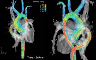
German researchers conducted a study of the effectiveness of MRI in patients with hemodynamic disorders and breathing difficulties.
A group of 17 people was taken for the study. Contrast magnetic resonance imaging allows you to evaluate the function of the left ventricle, the value of end-diastolic ejection, systolic function, and the condition of the common carotid arteries.
With the help of magnetic resonance scanning, specialists are able to monitor the functionality of the heart in real time.
Research results:
- The average duration of breath holding in the control group was 280-299 seconds;
- Duration of apnea – 3 seconds – 8 minutes – for different people;
- During the initial period of apnea, the heart begins to pump blood more strongly than in the physiological state of the heart muscle. The myocardium works more actively to ensure optimal gas supply to the blood.
MRI of the heart – what does it show?
The above examination shows that after the divers’ breathing is restored, the functioning of the heart and coronary vessels returns to normal. The study was carried out on healthy divers.
Does the functioning of the heart and coronary vessels change in pathological respiratory failure when assessed using magnetic resonance imaging.
MRI in standard horizontal projection shows left ventricular function. During the rest period, diastole is clearly visualized. When the heart contracts, tension in the heart muscle is observed.
Cardiac MRI is used not only in healthy people. Computer and magnetic resonance imaging shows the condition of internal organs in animals and people during archaeological excavations.
The possibilities of using the research for these purposes have been studied by researchers at the Chicago Institute. The results were presented at the next meeting of the Radiological Society of North America.
The destruction of tissue from the crypts compromises the safety of the find. When examining the internal state using magnetic resonance scanning, it is possible to clearly trace the condition of the heart, coronary vessels, liver, kidneys, and other organs. Molecular examination shows the age of the find and its state of preservation.
For better display, removal of balsamic materials is recommended. After cleansing the heart, the quality of visualization of the following anatomical formations increases:
- Valves;
- Ventricles;
- Coronary vessels;
- Myocardial condition.
The heart muscle is best visualized after fluid has been removed. Rehydration allows for better diagnosis of structures, since the chambers of the heart must be filled with a physiological volume of blood.
- The research team carefully studied the internal chambers, as it was necessary to determine the age of the find.
- Such results determined the possibility of using magnetic resonance imaging not only to assess dynamic changes in blood supply, but also morphological cardiac changes.
- How to do a stress MRI of the heart
Pharmacological tests of the use of MRI with pharmacological loads on the myocardium showed high intensity. The test is used to monitor blood flow to the heart muscle.
Physical activity is a way to increase the functional activity of the myocardium. The procedure is performed to determine the intensity of blood flow to the heart during rest. A pharmacological test allows you to provoke a functional load on the heart with the help of medications.
The following drugs should be used for analysis:
- Regadenoson;
- Dipyridamole;
- Adenosine.
Medicines are used to widen the coronary arteries to improve blood flow to the heart. A similar situation occurs when performing physical exercises. The drugs only cause an increase in heart rate.
The use of low concentrations of gadolinium contrast makes it possible to study the passage of blood through the myocardium. After completing the study, it is possible to produce graphic images to record changes and register pathology.
Dynamic monitoring of blood movement through the coronary artery using magnetic resonance imaging allows one to assess the speed of blood flow, visualize vessel narrowing, and neurological spasms of the artery during respiratory failure.
The procedure is slightly invasive. To better visualize the movement of blood through the coronary artery, intravenous contrast is required. Gadolinium passes through the myocardium and illuminates small and large vessels.
In case of myocardial infarction, the procedure allows you to assess the size of the affected muscle.
PET/CT can cope well with this task, but the study is difficult to perform due to the need for rapid delivery and inactivation of short-lived radioactive isotopes.
MRI of the heart with contrast – what is it used for?
MRI of the heart with contrast is used to visualize the condition of the coronary artery and other vessels if angioplasty is necessary in the treatment of coronary heart disease.
Instrumental diagnostic technologies have quickly moved from radiation techniques for detecting cardiac pathology to advanced radiation-safe methods. In addition to the absence of radiation exposure, magnetic resonance scanning makes it possible to qualitatively study not only the anatomical structure of the organ, but also to trace the nature of the blood supply.
Contrast has been used in radiography for a long time. The use of iodine helps to better trace the structure of the organ, the condition of the wall, and emphasizes malignant neoplasms.
In 1950, fluoroscopy began to be used to detect calcification of the coronary arteries. In 1958, ultrasound began to be used to determine cardiac pathology, and the principles of echocardiography were developed.
Until now, X-ray technologies with contrast of the esophagus are used, but cardiac MRI with contrast has become a more popular research technique among cardiac surgeons. Analysis solves the necessary problems more efficiently.
Source: https://secondopinions.ru/mrt-serdcza-i-koronarnyx-sosudov-s-kontrastirovaniem-chto-pokazyvaet
MRI of the heart and blood vessels in Moscow and St. Petersburg: where to do it, what it shows
Magnetic resonance imaging of the heart and blood vessels is a highly informative method that allows identifying pathological changes in the myocardium, coronary arteries, and pericardium.
MRI is based on the use of a magnetic field that creates a resonance of hydrogen atoms, which distort the radio frequency signal. Special sensors capture the converted pulses. The program processes images and creates a spatial model.
Visual display of the model allows you to study the smallest details, the subtle anatomical elements of the heart valves.
Determining changes in the coronary arteries and aorta requires a procedure with contrast. Intravenous injection of the drug helps monitor the internal and external condition of the vessel. The method reveals obstructions in patency, areas of narrowing, and locations of atherosclerotic plaques and blood clots.
Magnetic resonance scanning better shows pericarditis, cardiomyopathy, ventricular and atrial septal defects, and other defects.
Modern innovative technologies make it possible to visualize small inflammatory foci of the myocardium and impaired microcirculation in the coronary artery.
Is it possible to do MRI of the heart and blood vessels?
During magnetic resonance imaging of the heart and blood vessels, several hundred tomograms are made with a step of several millimeters. It is difficult for a doctor to analyze each image, so a software application processes the images. Innovative devices are equipped with a three-dimensional reconstruction mode.
Thus, MRI of blood vessels and the heart is not only performed, but is also recommended for suspected pathologies of the myocardium, pericardium, and heart valves. The examination allows you to verify the dissection of the arterial wall (aneurysm), thromboembolism of the vessel. After intravenous administration of contrast, it is possible to identify areas of microcirculation disturbance and areas of arterial narrowing.
Magnetic resonance imaging is not performed if there are contraindications:
- First trimester of pregnancy;
- Artificial metal heart valves;
- Mental disorders;
- State of drug or alcohol intoxication;
- Renal failure (when examined with contrast);
- Fear of enclosed spaces.
High-quality images are obtained only if you remain stationary during scanning. The duration of tomography of the cardiovascular system is 20-30 minutes. During this interval, you must remain motionless. Due to increased excitability, examination in children is difficult. Infants and preschoolers undergo MRI of the chest cavity under anesthesia.
Example of MRI tomograms of the heart and blood vessels
MRI of the heart – what does it show?
MRI scanning is the most effective method for examining the cardiovascular system. The possibilities of nuclear magnetic resonance of the mediastinum are quite extensive. Only visualization of the lungs is limited due to increased airiness of the alveoli.
What does a cardiac MRI show:
- The condition of the heart cavities, aorta, large vessels is examined by devices with a spin echo;
- Valves, ventricles, coronary artery are devices with a “gradient echo”;
- Analysis of the phases of cardiac activity is carried out with the presence of “cine-MRI”;
- Contrast magnetic resonance imaging is done after heart attacks to assess changes in cardiac blood supply;
- The speed of microcirculation is assessed by phase-contrast tomography;
- Three-dimensional reconstruction matches the existing model of the heart and blood vessels.
The procedure is performed according to indications. The harmlessness of the study allows scanning to be done at the request of the patient. The high cost of tomography limits the widespread use of the method.
Main indications for cardiac magnetic resonance imaging
A cardiologist gives a referral for an MRI of the heart. Common indications:
- Diagnosis of inflammatory changes in the myocardium and pericardium;
- Study of the condition of the heart valves after a number of diseases - endocarditis, rheumatism, congenital heart defects;
- Verification of the degree of changes in coronary heart disease;
- Study of the condition of the heart arteries;
- Study of the condition after stenting, coronary artery bypass grafting (CABG);
- Determination of the severity of cardiomyopathy.
The safety and harmlessness of nuclear magnetic resonance allows children from the age of five to perform the procedure.
What are the contraindications to MRI of the heart and blood vessels?
Metal objects inside the human body limit the scope of examination. A magnetic field causes objects to heat up or move. Pacemakers and insulin pumps are a limitation to magnetic resonance scanning. In such a situation, a CT scan of the lungs is done with contrast, which also qualitatively shows the pathology of the cardiovascular system.
Classification of contraindications to MRI of the cardiovascular system:
- Absolute;
- Relative.
The first category of properties does not allow the patient to scan. Taking medications will not help return your health to a state that allows you to conduct nuclear magnetic resonance.
Absolute contraindications to cardiac MRI:
- Presence of a cardioverter (defibrillator);
- Pacemaker;
- Rhythm imitators;
- In-ear prostheses;
- Ilizarov apparatus.
Before tomography, the location and composition of the implant should be determined. Some metals do not react to a magnetic field, so scanning can be carried out.
Relative contraindications to MRI of the heart and blood vessels:
- Heart failure in the stage of decompensation;
- Heart valve replacement;
- Decreased renal reabsorption;
- Installation of insulin pumps;
- Nerve stimulants;
- Presence of vascular clips;
- Patient's panic disorder;
- Claustrophobia;
- Dynamic scanning to assess vital signs;
- Serious human condition.
Contraindications are determined not only by the attending physician who issues the referral, but also by the radiologist who will conduct the examination.
MRI of heart valves
Heart defects lead to mixing of arterial and venous blood through additional defects of the interventricular or interatrial septum and valve apparatus. The disease leads to a lack of oxygen supply to the internal organs along with blood. To eliminate hypoxia and normalize the blood supply, artificial valves (aortic, mitral) are installed.
Three-dimensional modeling mode (3D reconstruction) helps create a spatial representation of the object with visualization of the cardiac cavities, aorta, coronary and pulmonary arteries. Intravenous administration of a contrast agent improves the visibility of the inner wall of blood vessels.
Valve replacement requires dynamic diagnostics. The position of the products may change, the formation of additional passages will lead to mixing of arterial and venous blood.
Computed tomography (CT) with contrast is capable of visualizing the condition of blood vessels, identifying atherosclerotic plaques, blood clots, and air emboli.
X-ray examinations lead to radiation exposure of tissues, so they cannot be performed several times in a row.
MRI is a safe procedure (in the absence of contraindications). Nuclear magnetic resonance does not cause any harm to health, so the examination is done an unlimited number of times.
Innovative tomographs with a power of three Tesla show even the smallest changes in the heart muscle and valves. The examination has a high price. Most types of heart disease can be detected by high-field tomography with a tube power of 1.5-3 Tesla.
There is no rationality for using more powerful devices in medicine.
What diseases can be detected by cardiac MRI?
The main diagnostic purpose of performing magnetic resonance and computed tomography of the cardiovascular region is to determine blood supply defects (aortic, mitral valve defects, septal defects).
What diseases can be detected by cardiac MRI:
- Stenosis of the coronary arteries;
- Narrowing of the aorta;
- Aortic, mitral insufficiency;
- Atherosclerosis, vascular thromboembolism;
- Pericarditis, myocarditis;
- Pericardial tamponade.
A number of cardiovascular diseases are accompanied by regurgitation - the backflow of blood. Magnetic resonance imaging is a priority study for identifying the symptom of vortex, verifying valvular dysfunction, and narrowing of the arteries.
- After coronary artery bypass grafting (CABG), MRI can be used to measure systolic and diastolic blood flow and diagnose antegrade and retrograde volumes.
- The procedure also reveals associated complications - aortic and mitral insufficiency, rheumatic changes, atherosclerosis plaques.
MRI of the heart after bypass surgery
Coronary artery bypass grafting (CABG) surgery is performed to restore the state of blood flow through the coronary artery. Artificial creation of shunts helps to eliminate the obstacle to myocardial microcirculation, prevent coronary heart disease and myocardial infarction.
Dynamic observation after surgery is performed:
- To assess the quality of manipulation;
- Determination of blood movement through the stenting site;
- Identification of complications.
Repeated scanning after a few months allows you to study the thickness of the myocardium, identify areas of hypoxia, and features of the circulation of arterial and venous blood.
MRI after CABG is contraindicated if the patient has a pacemaker or metal valve installed. The first tomography is performed 1.5 months after bypass surgery.
Where to get a cardiac MRI in Moscow and St. Petersburg
Several dozen medical centers offer magnetic resonance imaging in St. Petersburg and MSK. Private clinics are equipped with more modern tomographs compared to public institutions, but the rationality of examination with innovative installations does not always exist.
We offer you to choose where to have a cardiac MRI in Moscow and St. Petersburg in the “medical centers” sections. An optimized search using dozens of parameters allows you to find the desired medical institution by price, location, and opening hours.
Source: https://mrt-kt-legkih.ru/article/mrt-serdca-i-sosudov