Colposcopy is a diagnostic method necessary to identify benign, precancerous and malignant pathologies of the cervical canal. The study is carried out using a device - a colposcope; during the procedure, it is also possible to take biomaterial (scraping) from the cervix for subsequent cytological analysis.
Colposcopy procedure
Why is a colposcopy prescribed?
Colposcopy in gynecology is a frequently prescribed test. Its essence lies in examining the cervix using a colposcope - a special binocular microscope equipped with illumination. Colposcopic examination involves studying the structure of the mucous lining of the vulva, vagina and cervix under magnification.
Women undergo colposcopy of the cervix in order to identify oncopathologies, as well as precancerous diseases or benign conditions.
Also, during colposcopy, smears are taken from the cervix and a biopsy is taken for subsequent cytological examination. In addition, the procedure allows you to obtain photographs of the foci of the pathological process under the necessary magnification.
Using a colposcope, you can achieve an image magnification of 6-40 times. At low magnification, the specialist determines the presence of foci of pathology, evaluates their color, surface, location, and shape.
Then, with high magnification, the doctor examines suspicious areas of the mucosa. If it is necessary to better visualize the vessels of the microvasculature, the specialist uses a green filter.
This approach is necessary to detect invasive cervical cancer.
Doctor adjusting colposcope
The colposcope consists of optical and lighting systems. The device is adapted for carrying out the procedure using a non-contact method. The design of the colposcope includes an optical head, a tripod and a base.
This is necessary for ease of installation and use of the device. The optical head includes prismatic binoculars equipped with eyepieces that allow viewing of the tissues being examined.
The head also contains an illuminator that creates the lighting necessary for work.
Colposcopic examination of the cervix can be simple or extensive.
- During a simple procedure, the mucous lining of the cervix is directly studied without the use of any additional reagents.
- Extended cervical colposcopy involves examining the cervix after treatment with a 3% acetic acid solution. This manipulation is necessary to more clearly identify pathological changes in tissue structure. This is explained by the fact that acetic acid causes short-term swelling of the mucous membrane and vascular contraction.
Flora smear in women: interpretation of results
Also, during extended microscopy, treatment with Lugol's solution can be performed to determine glycogen in cells. This manipulation is called the Schiller test.
In precancerous conditions, mucosal epithelial cells contain little glycogen, and therefore are not stained with Lugol's solution. When examined, they look like whitish spots against the background of healthy tissue, uniformly colored brown.
Treatment with Lugol's solution facilitates the selection of a site for subsequent cervical biopsy.
Indications for colposcopic examination of the cervix
Discomfort may be a reason to prescribe a procedure
A referral for colposcopy is necessary if there are the following clinical indications:
- Detection of pathologically altered cells in a smear from the cervical canal.
- Suspicion of cancer and a number of other pathologies (for example, genital warts).
- Positive test result for the presence of human papillomavirus in the body.
- Extramenstrual bleeding.
- Chronic inflammatory processes of the cervix.
- Pathological discharge.
- Itching and discomfort.
- Prolonged aching pain in the lower abdomen.
The listed conditions are the answer to the question of when to do colposcopy.
Purposes of colposcopy
Why is colposcopy needed and why is it done? The goals of this procedure are:
- determination of precancerous pathologies and oncological diseases in patients with a positive Papanicolaou smear;
- detailed examination of the cervical canal and vagina;
- monitoring the effectiveness of treatment of neoplastic processes.
The doctor must explain to the patient why colposcopy is being done and how to properly prepare for it.
Rules for preparing for the procedure
When preparing for a colposcopic examination, there is no need to adjust your diet. There are some restrictions, namely:
- Two days before colposcopy you should not douche.
- Two days before the study, you should not use intimate hygiene products.
- Use a condom during sexual intercourse.
- For several days, do not use vaginal tablets, suppositories and other local medications.
Following simple recommendations allows you to get the most adequate results.
How is colposcopy performed?
Colposcopy is performed outside of menstruation. More often, the procedure is prescribed on days 7-10 of the cycle. This is explained by the fact that during this period the mucus in the cervical canal is transparent and does not cause any difficulties during the examination.
From a clinical point of view, what is cervical colposcopy and how is it performed? The procedure is performed both on an outpatient and inpatient basis.
When prescribing this manipulation, the doctor must explain to the patient why colposcopy is needed in this clinical situation, what information it allows to obtain. After this, the woman is told about the rules of preparation for the procedure.
The patient should also be notified that during the study, a biopsy of the biomaterial may be required for subsequent cytological examination.
Examination of smears for cervical cytology: interpretation
A woman may be concerned about whether it is painful or not to undergo a colposcopy. Usually the procedure brings mild discomfort and does not cause pain.
However, with cervicitis, with thrush and some other pathological conditions, painful sensations are still possible. Pain accompanies obtaining a biopsy.
There may also be some discomfort for a short time after a colposcopy, but this should go away quickly.
The question is also often asked whether colposcopy is performed on virgins. The answer can be affirmative only if serious gynecological pathologies are suspected. If there are no risks for the patient, then the procedure is not performed on girls.
Conducting research
What kind of procedure is this, if we consider directly the sequence of actions during its implementation? You should pay attention to the following points:
- during the procedure, the woman is positioned on a gynecological chair;
- first, the mirrors are inserted, then the colposcope;
- then the cervical canal and vagina are examined using low magnification, if necessary, drying the discharge with a cotton swab;
- during examination, the specialist assesses the presence, size, quantity, nature, color of pathological foci;
- using a green filter, the doctor assesses the condition of the vascular network;
- if there are formations, the specialist indicates their nature: polyps, condylomas, cysts, etc.;
- if an extended examination is necessary, the cervix is treated with a solution of acetic acid, after ten seconds the mucus is removed;
- then the foci of pathology on the surface of the mucosa are determined;
- then a Schiller test is performed (treatment with Lugol's solution containing 2% potassium iodide, 1% iodine) and identification of iodine-negative areas, which are examined in more detail using high magnification;
- if the need arises, a biopsy is taken, which is subsequently sent to the laboratory for histological examination.
What does colposcopy show?
Colposcopic picture in normal conditions and in pathologies
Why is a colposcopic examination prescribed and what results does it provide? This technique is a correct way to identify foci of ectopia, which can be either a physiological or pathological condition. With large ectopic lesions, bloody discharge after sexual intercourse is possible, as well as excess vaginal mucous discharge. In the absence of such symptoms, but in the presence of foci of ectopia, it does not require any correction.
What is a test for hidden infections and how to take it
Colposcopy is important for identifying precancerous pathological conditions - dysplasia. Thanks to precise diagnostic criteria, the doctor can assess the degree of dysplastic changes and prescribe adequate corrective measures.
Colposcopic examination makes it possible to identify atrophic foci, the inflammatory process, tissue proliferation with the formation of papillomas, condylomas. Colposcopy of the cervix during erosion is mandatory and is necessary to determine the boundaries of the pathological focus and its structural features.
Is the procedure necessary during pregnancy?
- Is colposcopy performed during pregnancy? In the early stages, it is carried out in case of questionable or poor results of a smear for oncocytology from the cervix.
- What benefits does the manipulation give in this case? It is necessary for timely diagnosis of oncological pathologies and determination of tactics for managing the patient during pregnancy until the process of delivery.
- It is worth noting that simple, rather than extended, colposcopy is usually used.
In the later stages, colposcopic examination is not prescribed due to the risk of provoking infection and bleeding.
What to do after the procedure
After the manipulation there are no restrictions, the patient leads her usual lifestyle. You should use panty liners for one or two days - discharge is possible.
- If a biopsy was performed during the procedure, for 10 days it is forbidden to take baths, visit the sauna and bathhouse, use tampons and douches, have sex, or take medications containing aspirin.
- The next day, it is necessary to remove the tampon that was inserted to stop the bleeding.
- In rare cases, complications such as cervicitis and vaginitis, bleeding, allergic reactions to iodine and other reagents are observed after the procedure.
If, after the manipulation, heavy bleeding of a bloody nature does not stop during the day, or purulent discharge appears, the body temperature has increased, the lower abdomen hurts and weakness and dizziness are observed, you should immediately seek medical help.
Colposcopy is a modern method for diagnosing cervical erosion.
You can also see what colposcopy shows in the video:
Source: https://shejka-matki.ru/diagnostika/kolposkopiya-shejki-matki.html
5 pathologies detected by colposcopy of the cervix
One of the best methods for diagnosing gynecological diseases is colposcopy. For what reasons, and for what purpose, how is it performed, and how to prepare for the colposcopy procedure? These questions concern all women who have been prescribed this manipulation.
What is cervical colposcopy
Colposcopy is a diagnostic examination of the vagina and cervix using a special colposcope device. The main indications for this examination are deviations from the norm in the results of a cytological smear. Using a colposcope under various optical magnifications, you can examine the entire area of the cervix and vaginal walls, and differentiate malignant tumors from benign neoplasms. Assess the appearance of organs (vascular pattern, epithelial disorders, boundaries of identified neoplasms, tissue color). During the procedure, if necessary, a biopsy is performed for further diagnosis.
This procedure can also be performed during pregnancy, but not for prophylactic purposes, but only if there are direct indications for this. This manipulation is safe for the fetus, a woman does not need to be afraid of it.
Those who are about to undergo an examination are concerned about the pain of colposcopy; this method is practically painless and can cause slight discomfort, as during a regular gynecological examination.
The subject is positioned on a gynecological chair, the doctor uses a mirror to expose the cervix, removes vaginal discharge with a cotton swab and then conducts an examination.
A simple examination lasts no more than 10 minutes, but there are times when the gynecologist needs detailed information about the condition of the mucous membrane of the cervix, then additional tests are carried out in which the cervix is treated with a 3% solution of acetic acid and an aqueous solution of iodine.
- When treating the cervical mucosa with a vinegar solution, an immediate reaction occurs, healthy tissues sharply contract, but if there is a pathology, the reaction will not occur. During acid treatment, the patient does not experience pain, and the mucous membrane returns to normal after 2 minutes;
- After treating the mucous membrane with an iodine solution, the doctor waits 2 minutes, during which time healthy tissue should turn the color of the solution, but tissue with pathology will not change its color;
- Fluorescent colposcopy is performed only in cases of suspected malignancy. During the examination, the mucous membrane is treated with fluorochrome, then the tissues are illuminated with ultraviolet rays. In the light of the rays, oncological inclusions glow pink.
If the examination reveals areas with pathological changes, the specialist performs a targeted biopsy for the purpose of histological examination, specifically from the area with pathology.
Indications for the study
Colposcopy is a routine examination that a woman should undergo annually, but there are cases when it may be additionally prescribed by a gynecologist if the patient develops symptoms such as:
- Constant mild pain in the lower abdomen or wave-like attacks of pain, from strong pain to weak and again to strong;
- Bleeding outside the menstrual cycle;
- Rash or single rashes, of unknown etiology, on the genitals;
- Pain during sexual intercourse and bleeding during or after intercourse;
- Discomfortable sensations in the vagina, accompanied by burning and itching.
Although colposcopy is a safe examination method, it also has a number of limitations and contraindications. Manipulation is not prescribed in the first 2 months after childbirth, if less than a month has passed since the abortion or if surgery has recently been performed on the cervix.
Contraindications to extended colposcopy are the patient’s intolerance to acetic acid and preparations containing iodine.
The procedure is prescribed with caution to women with severe inflammation, cervical bleeding, and severe atrophy of the external os of the cervical canal.
How to prepare for colposcopy
The examination does not require special preparation, everything is almost the same as during a regular gynecological examination:
- A day before the procedure, refrain from sexual intercourse;
- Do not douche or perform hygiene procedures using gels and other products other than baby soap;
- Stop treatment with any vaginal medications three days in advance.
The study is scheduled 2 days after the end of menstruation or immediately before its start; in the middle of the menstrual cycle, colposcopy is not performed due to the woman’s heavy discharge. Abundant mucus secretions make manipulation difficult and can lead to false results.
You must take a disposable diaper and clean socks with you to the procedure.
How a colposcope works
A binocular optical colposcope gradually enlarges the image using interchangeable eyepieces and allows you to obtain a three-dimensional image. There is a light source on the head of the device, with the help of which it is possible to examine the vagina and cervix in great detail and detect pathological changes in time.
The video colposcope is equipped with a camera. The camera displays video on the monitor and allows you to conduct a more in-depth examination and produce and save research data for study.
What pathologies are detected using colposcopy?
Using colposcopy, a number of diseases of both benign and malignant etiology are detected:
- Papilloma is a neoplasm caused by the human papillomavirus (HPV), most often of a benign nature, but in rare cases it can become malignant. Papilloma looks like an overgrown wart with a shiny pink or bluish surface. When treated with acetic acid it turns pale;
- Erosion - pathology is a consequence of mechanical injuries and inflammatory processes; erosion means ulceration and disruption of the integrity of the epithelial covers of the mucous membrane. The disease is quite common in every third woman. The erosion has a smooth, fine-grained surface, scarlet in color with pronounced blood vessels;
- Polyps are pathological growths of columnar epithelium from the cervical canal; there are both single and multiple growths. The formations are usually benign, but in advanced form they often take on a malignant form;
- Cervical endometriosis is a disease caused by the growth of the endometrium outside the uterus; normally, endometrial cells do not protrude beyond the uterus, but in the case of hormonal disorders, they may grow into the cervix and other organs that have a mucous membrane. Upon examination, it is clear that the pathological area is slightly raised above the surface of the mucosa, has an irregular shape, and is pink or bluish-scarlet in color. Areas with overgrown endometrium are prone to bleeding;
- Cervical carcinoma - the pathology looks like a glassy area with tuberous protrusions, the vessels in the pathological area are clearly visible. When treated with reagents during extended colposcopy, no reaction occurs. This is an oncological neoplasm. For such obvious symptoms, a biopsy is not required.
Colposcopy is a popular and simple gynecological examination that usually does not have negative consequences.
After the extended colposcopy procedure, there is a brown discharge. This should not cause concern for a woman; it is the remains of the iodine solution that come out. Occasionally, minor pain may occur due to stretching of the vagina with gynecological instruments.
In very rare cases, an infection can occur in the vagina, after which inflammatory processes occur, but this is more related to the doctor’s negligence than to the consequences of manipulation.
Colposcopic examination is an effective way to diagnose diseases of the vagina and cervix; using this technique, you can quickly and reliably make a diagnosis and begin timely treatment.
Video: Colposcopy of the cervix
Video: 4 important facts about colposcopy
Video: What is a colposcopic diagnostic method?
Source: https://sheika-matka.ru/matka/kolposkopiya-diagnosticheskoe-issledovanie-shejki-matki/
Extended colposcopy
A widely used instrumental diagnostic method in gynecology, colposcopy, is aimed primarily at identifying oncological processes at the initial stage of their development.
For a more detailed study of the female genital organs, an extended colposcopy is performed using special chemical reagents. During this procedure, the doctor evaluates the condition of the mucous membrane and the color reaction of the epithelial cells to the reagent.
Analysis of the changes that occur allows us to diagnose the pathology as accurately as possible.
The essence of the procedure
Using a colposcope, the doctor examines the cervix, vaginal walls, and cervical canal (the area where the cervix and uterus connect). A colposcope is a small medical device equipped with a lighting system and optics with multiple magnification. Gynecological speculums are used as auxiliary instruments.
A modified, more modern version of the procedure, video colposcopy, is performed using a digital colposcope and computer technology to process the resulting video image.
In the extended version, after a general examination, the mucous surface of the cervix and vaginal walls is treated with a chemical reagent.
Today the following substances are used as test reagents:
- Three percent solution of ethanoic acid (aka acetic acid). The reaction of the mucous membrane to acid is a narrowing of blood vessels in healthy tissues, and a significant lightening of the color of the epithelium. This does not happen in the affected areas because the vessels are not sensitive to acid. In cases where the use of acetic acid is contraindicated, lactic acid can be used as a reagent.
- Three percent Lugol's solution with glycerin (Schiller's test). Under the influence of iodine-containing lugol, healthy epithelial cells undergo pigmentation. Areas with pathology do not change color. This allows you to assess the scale and boundaries of changes. In addition, cells must be collected from unstained fragments. Subsequent cytological studies of the biomaterial will help determine the nature of the changes (malignant or benign).
- Adrenaline (epinephrine). Used to assess the vascular system of organs.
The latter test is rarely practiced. In most cases, diagnosis is based on the results of a sequential vinegar test and Schiller test.
The colposcopy procedure is carried out for preventive purposes, or as prescribed by a doctor. The basis for examination, most often, is erosive changes in the mucous membrane identified during a standard gynecological examination (suspected erosion of the cervix). Also, the study shows:
- for symptomatic complaints that a woman presents at a doctor’s appointment (pain not associated with menstruation, unhealthy vaginal discharge, the presence of blood in the discharge after intimate contact);
- with a positive HPV test (human papillomavirus);
- as a control procedure during the treatment of a previously diagnosed disease;
- in case of deviations from the normative indicators of the results of previous cytology, in particular, a positive Pap test (Papanicolaou smear) to establish a precancerous and cancerous condition of the cervix.
Detection of pathological changes at an early stage will avoid further complications
Colposcopy with biopsy is performed specifically to identify malignant changes.
Contraindications to the procedure
There are no absolute contraindications to examination with a colposcope. The exception is an allergic reaction to iodine and iodine-containing drugs. In this case, an incomplete colposcopy (without the Schiller test) can be performed.
Relative (relative or temporary) contraindications include the postpartum period (the time interval must be at least 2 months, subject to uncomplicated delivery), the period after artificial termination of pregnancy, including medical abortion (at least 30 days), and the days of menstruation itself. Gynecologists recommend undergoing diagnostics 5–7 days after the end of the critical days.
Preparatory activities
There is no special preparation for the study.
Before the procedure, a woman should comply with several simple conditions: exclude the use of vaginal suppositories, if any are prescribed for therapy (2-3 days in advance), refuse intimate relationships (3-4 days in advance).
Immediately before diagnosis, you should not use scented intimate hygiene products. It would be best to carry out hygiene measures simply with warm water.
Progress of the procedure
The examination is carried out in a regular gynecological chair. Speculums are inserted into the vagina and remain there until the end of the procedure. During the examination, the woman's genitals are repeatedly irrigated with saline to prevent the mucous membrane from drying out. Next, the doctor performs an initial examination (conventional colposcopy). The structure, color, and vascular pattern are studied.
How to prepare for colposcopy?
After assessing the external condition of the organs, the mucous membrane is treated with a swab dipped in a water-acetic solution. An assessment of the vascular reaction to acid can be obtained 2–3 minutes after treatment of the visible part of the vagina, its walls and the cervix.
Vasospasm means a positive test result. If the vessels of certain fragments of the examined surface remain unchanged, we can say that cancerous (atypical) cells are present there. In this case, the test will be negative.
The vinegar test is replaced by the Lugol-glycerin test. The treatment solution consists of distilled water, iodine, potassium iodine, in a ratio of 100:1:2.
In the absence of pathological changes, the entire area of the epithelium should turn brown. Uneven pigmentation indicates the presence of atypia. At the same time, the areas affected by cancer have clear boundaries.
The next stage is the collection of biomaterial for laboratory analysis (biopsy).
The size of the uterine tissue fragment for cytology is about 2 mm
The time range of extended colposcopy usually does not exceed a quarter of an hour. The time is determined individually, depending on the scale of changes in the organs.
Carrying out the study is not a painful procedure. It can be considered like a regular gynecological examination, only longer.
Upon contact with reagents, a woman may experience some discomfort that is not related to pain. The most unpleasant moment can be called the removal of biomaterial.
If the pain threshold is increased, it makes sense to consult a doctor with a request to use a local anesthetic at the time of the biopsy.
Post-procedural period
If the examination did not include a biopsy, the patient may be bothered for some time by a discharge of an uncharacteristic color, as a reaction to an iodine-containing reagent. If biological material was collected, the discharge may be bloody. This is due to mechanical damage to epithelial tissue. Such effects are usually short-lived and disappear spontaneously.
Basic recommendations that should be followed for 3–5 days after colposcopy: refrain from visiting the bathhouse, solarium, sauna, refuse sexual intercourse, and suspend sports training.
Women are not recommended to use sanitary tampons. For the speedy healing of the wound, pads are the best option.
If bleeding continues for several days, sharp pain appears, or hyperthermia (fever), you should seek medical help.
Possible test results
A simple colposcopy determines:
- the condition of the epithelium (normally it should be pink with a smooth surface);
- the presence of erosive changes in the vaginal walls and cervix;
- inflammation of the mucous membrane of the cervical canal (endocervicitis);
- inflammation of the epithelium of the vaginal part of the cervix (exocervicitis);
- the presence of viral and genital warts (papillomas and condylomas).
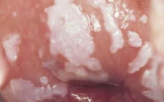
Diagnosed leukoplakia of the cervix (photo)
Extended colposcopy diagnoses both the listed abnormalities and more serious diseases:
- changes in tissue structure that precede cancer pathologies (dysplasia);
- the presence of part of the epithelium of the cervical canal on the surface of the cervix (pseudo-erosion or ectopia);
- the presence of growths (polyps);
- cancer of the cervix and vaginal walls;
- proliferation of epithelial cells (endometriosis);
- death (atrophy) of individual tissue fragments;
- keratinized fragments of the mucous membrane (leukoplakia).
The gynecologist reports conclusions on the diagnostic results immediately after the procedure. A full analysis of the obtained indicators may take several days. The colposcopic method is safe and informative. This is a real chance to detect malignant changes at the initial stage, preventing their further development. Women aged 35+ are recommended to undergo this examination annually.
Source: https://apkhleb.ru/endoskopiya/rashirnnaya-kolposkopiya
Diseases detected during colposcopy
Colposcopy is an examination of the cervix and mucous membrane of the genital tract using a magnifying optical device, a colposcope. A variation of the technique is video colposcopy, performed using modern optics that displays the image on a monitor.
The procedure can be as simple as a routine examination or as extensive as the area being examined is treated with a special dye that highlights the affected areas. If necessary, tissue samples are taken during the procedure for a biopsy (cellular analysis), which helps detect tumors.
Changes in the cervix detected during colposcopy
- True erosion is damage to the cervical mucosa caused by mechanical damage or irritating discharge. Without treatment, the disease develops into pseudo-erosion, requiring laser tissue removal.
- Pseudo-erosion is the spread of tissue from the cervical canal of the cervix to the cervical surface. Malignant processes may occur in the affected area, leading to cancer.
- Ectropion is an eversion of the cervix resulting from difficult childbirth and traumatic gynecological procedures. In this case, the tissues that should be inside the cervix, once on its surface, become inflamed and injured.
- Leukoplakia is a disease caused by keratinization of the cervical epithelium. It can be found on the walls of the genital tract and the mucous membrane of the intimate area. It is a precancerous pathology.
- Polyps are pedunculated tumors growing outside and inside the cervical canal. As they grow, they interfere with the outflow of menstrual blood and fertilization. May lead to bleeding.
- Condylomas are warty growths caused by papillomavirus. In addition to the cervix, they affect the entire genital area. May grow around the anus. Infection with the papilloma virus greatly increases the risk of cancer of the cervix, bladder, and rectum.
- Nabothian cysts are formations on the cervix containing fluid. They may rupture with the release of contents and the formation of ulcers. They provoke inflammatory processes – endocervicitis, endometritis. During childbirth, the full dilation of the cervix is prevented.
- Foci of endometriosis are cells that should normally be found in the inner uterine layer - the endometrium. These cellular structures respond to hormones and begin to fill with blood before menstruation. With cervical endometriosis, a woman notices brownish-brown discharge that appears 2-3 days before menstruation. Endometriotic lesions can degenerate into a malignant tumor - endometrioid stromal sarcoma.
- Consequences of cervical ruptures . In such women, scars are found that deform the cervix and narrowing of the internal uterine os, leading to impaired outflow of menstrual blood and problems with conception.
- Dysplasia is a disease in which whitish spots of altered tissue are visible on the neck. Sometimes the lesions may be red and resemble erosion. This pathology, as scientists have found, is caused by papillomavirus. Unlike other diseases in which tissue degeneration does not occur, with dysplasia, abnormally developed, ugly, giant cells are found in smears. This is the last step before cancer develops.
Cervical cancer
A tumor in this area is also often asymptomatic, especially in the early stages. Sometimes bleeding occurs after sexual intercourse and physical activity, and discharge with an unpleasant odor appears.
Upon examination, a tumor is visible growing towards the genital tract, resembling a bumpy formation or cauliflower. Some forms of malignant tumors look like ulcers with dense edges covered with a grayish coating.
Neoplasms growing towards the uterus are difficult to identify during examination, so patients are tested for oncocytology.
If malignant tumors are detected, the woman is referred to an oncologist for further diagnosis and treatment.
How to avoid cervical disease turning into cancer
Prevention of cancer pathologies consists of timely treatment of diseases in this area. Detected erosions are removed using a laser. This treatment is less traumatic compared to cauterization with electric current or surgery. A gynecologist removes polyps, foci of endometriosis and cysts.
Reliable prevention of cervical cancer is vaccination against the human papillomavirus. This measure allows you to avoid infection with viruses types 6, 11, 16 and 18, which cause cancer and genital warts. Vaccinations are given to teenage girls aged 12-15 years.
To prevent severe pathologies of the cervix, you need to regularly visit a gynecologist and undergo a PCR test that detects viruses and other microorganisms that provoke the formation of malignant tumors, erosions and dysplasias. Every year you need to do a colposcopy and take smears for atypical cells, even in the absence of complaints. If any suspicious symptoms occur, you should contact a gynecologist as soon as possible.
About doctors
Make an appointment with obstetrician-gynecologists of the highest category - Erkhan Karolina Pavlovna and Maisuradze Liana Georgievna today. We will do everything to accommodate you as quickly as possible. The Rainbow Clinic is located in the Vyborg district of St. Petersburg, just a few minutes walk from the Ozerki, Prospekt Prosveshcheniya and Parnas metro stations. See the directions map.
ILBI (laser blood irradiation)
800
Intravenous injection (excluding drug costs)
300
Intravenous injection (without the cost of drugs, dropper)
800
Intramuscular injection (without drug costs)
150
Opening a Bartholin gland cyst
6500
Intravaginal treatment (bath + medicine)
550-1000
Colposcopy
1300
Colposcopy (Videocolposcopy)
1300
Complex conservative treatment of STIs with medications
from 18 000
Consultation with a gynecologist (endocrinologist)
1300
Laser locally
400
Laser rectal/urethral
400
Laser vaporization of the cervix (depending on the size of the erosion)
5000-7000
Laser removal of cervical cyst (Ov. Naboti) – 1 unit.
1500
Laser removal of genital warts - 1 unit.
600
Laser removal of endometrioid cyst - 1 unit.
1000
Marsupialization
15000
Medical termination of pregnancy (Miropriston is a Russian drug) – up to 6 weeks
6000
Medical termination of pregnancy (Mifegin - a French drug) - up to 6 weeks
8800
Medical termination of pregnancy (Mifepristone - a Chinese drug) - up to 6 weeks
4500
Local anesthesia
1000
Multiple genital condylomatosis
up to 7000
Prescribing a course of therapy/Repeated consultation with prescribing treatment
1500
General anesthesia
3500
Initial consultation with a gynecologist on the selection of contraception
1000
Initial consultation and examination by a gynecologist
1000
Repeated consultation with a gynecologist
700
Reference
300
Removal of the intrauterine device (without technical difficulties)
1000
Removal of the intrauterine device (with technical difficulties)
from 3000
Removal of Bartholin gland cyst
15 000
Removal of lipoma on the genitals with general anesthesia
8000
Removal of a polyp of the cervical canal
5000
Installation of an intrauterine device (without the cost of the device)
1500
Installation of an intrauterine device (with the cost of the device)
5500
Chemical destruction of genital warts (Solcoderm)
1500 Return to list of articles
Source: https://www.raduga-clinic.ru/articles/chto-vyyavit-kolposkopiya/
Articles
Colposcopy and why is it performed?
Colposcopy is one of the leading methods of examining the cervix using an optical device - a colposcope (a binocular equipped with a strong light source). Simply put, an examination is similar to examining something under a microscope. Examination with a colposcope helps the practitioner in the early detection of cancer of the vaginal part of the cervix.
Colposcopy is indispensable for early diagnosis of cervical cancer and establishing its initial stages. Colposcopy also helps in the differential diagnosis of benign changes in the cervix, vagina and vulva.
The most minor tissue defects, such as small erosions, tiny tumors and microbleeds in the cervix, vagina and vulva, can only be diagnosed using a special optical device. In this regard, colposcopy has become an indispensable method of clinical research in the practice of gynecologists.
The colposcopy method opens up new opportunities for more effective treatment of cervical cancer. Colposcopy allows for differential diagnosis of cancerous tumors and other changes in the epithelium of living tissue, which is impossible to do without a colposcope. Today, the use of colposcopy has become widespread. Colposcopy requires some experience.
In addition, obtaining good photographic documents has become possible only recently. Therefore, compared with cytological examination, colposcopy remained on the sidelines.
However, it became clear to everyone that the use of colposcopy and cytological examination, especially when taking smears during a colposcopic examination, gives significantly better results during preventive examinations of women and especially with pathology of the cervix that is poorly visible through the eyes of a doctor. The use of colposcopy makes a great contribution to the prevention of gynecological pathology.
- Colposcopy Values:
- colposcopy must be used for every in-depth gynecological examination,
- colposcopy must be carried out along with taking a smear for cytological examination: only the parallel use of these two methods gives an optimal result,
- photographs during colposcopic examination improve diagnosis - they are necessary for long-term observation in case of deviations from the norm and for monitoring treatment,
- a biopsy using a colposcope is a reliable diagnostic method (tissue is taken from the cervix in the area that caused suspicion of the disease during colposcopy).
Positive experience with the use of colposcopy as a clinical examination method throughout the world has shown its great benefits in the early diagnosis of cancer.
However, colposcopy is necessary not only for the early diagnosis of precancer and early stages of cervical cancer, but also for the differential diagnosis of numerous benign changes in the cervix.
This possibility makes colposcopy mandatory in outpatient practice. A specialist who knows the method of colposcopy will never refuse to use it in everyday work.
The use of colposcopy has shown that true cancer is quite rare; changes in the epithelium are much more common in initially benign tumors. Thanks to colposcopy, it has become possible to monitor the treatment of various diseases of the cervix.
- Objectives of colposcopy:
- 1) assess the condition of the mucous membrane of the cervix and vagina
- 2) identify the lesion
- 3) differentiate benign changes from those suspicious for malignancy
- 4) carry out targeted smears and biopsies, which significantly increases the information content of the latter
- Colposcopy evaluates:
- 1) fabric color
- 2) the state of the vascular pattern
- 3) surface and level of the mucosa
- 4) presence and shape of glands
- 5) boundaries of identified formations (clear or blurred)
- Mixed formations detected during colposcopic examination:
- · Exophytic condylomas – occur as a result of human papillomavirus infection.
- · Inflammation – can be diffuse and local.
· Atrophy – usually the result of estrogen deficiency. Often observed in postmenopause.
· An ulcer (true erosion) is a defect of the epithelium, its local absence. Occurs against the background of inflammation, hormonal imbalance, injury, etc.
· Endometriosis – during colposcopy, foci of endometriosis may appear as bluish cysts or in the form of bleeding linear and dotted areas.
· Cervical polyps are growths that protrude into the lumen of the canal or beyond the external os. May be single or multiple.
The purpose of colposcopy is not only to determine them, but also to evaluate the condition of the surface according to the usual criteria. Colposcopy is mandatory for preventive examinations of women, because makes it possible to timely identify various diseases of the cervix, incl. precancer and early stages of cancer.
There are no contraindications to colposcopy, no complications are observed.
Colposcopy is performed by a gynecologist on an outpatient basis or in a hospital. No special preparation of the patient is required.
The subject is positioned on a gynecological chair, the cervix is exposed with vaginal speculum, the discharge from its surface is removed with a tampon, then the vaginal part of the cervix is examined using a colposcope installed near the gynecological chair.
Thus, extended colposcopy is one of the highly informative methods for diagnosing pathology of the cervix and vagina.
Source: http://aigerim.info/blog/show/rol-kolposkopii-v-ranney-diagnostike-zabolevaniy-sheyki-matki
Simple and extended colposcopy: all the details of the examination
Colposcopy is a classic method for diagnosing gynecological diseases. When examining the condition of the vagina, vulva and cervix, the gynecologist uses a special device called a colposcope. Colposcopy is an effective tool for detecting many female pathologies, including precancerous conditions.
CLICK TO SIGN UP
Many women experience great anxiety before a colposcopic examination. This happens due to simple ignorance. Colposcopic examination is a simple and painless, but extremely necessary gynecological procedure.
What is colposcopy
Colposcopy is a detailed examination of the internal female organs accessible to the gynecologist using a special microscope with built-in lighting - a colposcope. The procedure is similar to a regular gynecological examination - the vaginal walls are spread apart with a speculum, and the examination itself lasts no more than 20 minutes. Colposcopy is the choice of all modern medical centers.
To date, colposcopy is the only procedure that detects cancer in the early stages of development.
What does a colposcope show: why is colposcopy needed?
Using a colposcope, a gynecologist can examine the vaginal part of the cervix and the entire vagina - the organs most often suffering from infection, tumor processes and erosion.
Colposcopy reveals many diseases, even those at an early stage of development and occurring without any symptoms (latent, hidden), while providing extremely accurate research results.
The device sees all changes in the mucous membrane, and the magnification of the image can be varied.
In addition, problem areas can be removed and stored electronically, using the images later to compare the results of therapy, as well as for consultations with colleagues, without involving the patient.
The results obtained during colposcopy are already a reason to make a preliminary diagnosis. The gynecologist makes the final conclusion after receiving a response from the laboratory, where tissue samples taken during colposcopy are sent for cytological and histological examination. The diagnosis established during a complete examination is 100% reliable.
What diseases can be detected by colposcopy?
The main goal of colposcopy of the cervix and uterus is to identify the following diseases and pathologies:
- genital warts that can transform into cancerous tumors;
- cervical cancer;
- cervicitis (inflammation of the cervix);
- precancerous changes affecting the tissues of the vagina, cervix, vulva;
- vulvar cancer;
- vaginal cancer.
When is colposcopy performed?
The main goal of a colposcopic examination is the timely diagnosis of cervical pathologies. Colposcopy is definitely prescribed if:
- Cervical cytology results show dysplasia. Colposcopy of the uterus in this case will show the specific area of the cervix where the modified cells are located. If foci of pathological epithelium (covering tissue) are detected during colposcopy, the gynecologist immediately performs a biopsy.
- During a standard gynecological examination, the gynecologist detects suspicious areas on the surface of the cervix. The method reveals areas of modified epithelium (upper integumentary layer), which cannot be detected without the use of video equipment.
In addition, indications for the procedure are:
- Control of local treatment.
- Genital warts detected on the cervix.
- The woman is worried about itching, unpleasant odor, discharge and rashes in the vaginal area.
- During sexual intercourse, pain is felt, then a discharge mixed with blood or pus is noticeable.
- Prolonged nagging pain in the lower abdomen, not related to typical menstrual ailments.
- Bleeding outside the cycle.
- Swelling, growths, ulcers in the genital area.
- Decreased desire.
- Difficulty conceiving;
- Missed pregnancies , miscarriages .
Colposcopy falls into the category of preventive procedures that every woman must undergo annually.
How to prepare for the procedure
To obtain an accurate result, colposcopy is recommended to be performed in the first 2-4 days after the end of menstruation. It is not forbidden to carry it out on another day of the cycle, with the exception of menstruation, since the discharge will not allow a thorough examination of the organs.
An important point is that it is better to undergo colposcopy in the first phase of the cycle. After ovulation, a large amount of mucus accumulates in the cervical canal, so sometimes the results are distorted. In addition, in the second half of the menstrual cycle, tissue regenerates much more slowly, which increases the likelihood of discomfort during the procedure.
Preparation for colposcopy requires compliance with the following rules:
- Refusal of sexual contacts 2 days before the procedure;
- During this period, it is prohibited to use vaginal suppositories, sprays, tablets and creams, or douche. If a woman is undergoing therapy using them, she should consult a gynecologist about this.
- Do not take a bath, but temporarily switch to a shower.
- Personal hygiene should be carried out only by washing the genitals with water, without the use of special means.
How is the procedure performed?
This procedure is painless and can be completed in just 5-10 minutes. The doctor conducts an examination as usual, on a gynecological chair. First, the vaginal speculum is inserted, then the colposcope itself.
During the examination, the gynecologist takes swabs from the mucous membrane for study in the laboratory.
To avoid discomfort and unpleasant sensations, the patient should sit in it as comfortably as possible and, most importantly, relax.
During the examination, a colposcope is used, which includes a lighting device and a binocular. To make the cervix accessible for examination, the vaginal walls are spread apart using a speculum (gynecological instrument), as during a standard examination in a chair. The duration of the procedure is on average 10 - 15 minutes.
Using the device, the doctor can see ruptures, tumors, deformations, and inflamed areas.
If there are indications, additional tests may be used: the use of optical filters, treatment of the cervix with a solution of acetic acid, and so on. This is required to diagnose tissue areas where dysplasia is suspected. Samples reveal hidden defects in the mucosa - the initial stages of erosion and other pathologies.
Extended colposcopic examination
An extended examination is carried out as follows:
- The gynecologist inserts an ordinary dilator and a speculum into the woman’s vagina, and a colposcope is installed 10-15 cm from the entrance to the vagina.
- Using a cotton ball, a weak solution of acetic acid (3-5%) is applied to the surface of the mucosa. Using the device, the gynecologist monitors the condition of the blood vessels: healthy tissues contract, but no pathological changes occur.
- The surface of the mucous membrane is treated with Lugol's solution or iodine (the patient may feel a slight burning sensation). The gynecologist evaluates the reaction of the tissues: healthy ones should turn the color of the solution, affected ones should not change color.
- If pathological areas of the mucous membrane are detected, the doctor takes a biopsy test - “pinches off” a particle of the affected tissue.
Women should not worry: the colposcope is not inserted into the vagina, and the procedure itself, although not particularly pleasant, is practically painless. Lasts no more than 30 minutes.
Extended examination gives great opportunities to the gynecologist. The doctor can
- Assess the condition of the mucosal surface.
- Take photographs and enlarge affected or modified tissues.
- Determine the nature of the neoplasm (benign, malignant).
- Take material for biopsy.
- Take a smear from questionable areas of the mucosa.
Despite a number of advantages, colposcopy is not a panacea. An accurate diagnosis can be made and treatment can be prescribed only after a comprehensive examination including tests for oncology.
Fluorescent and color colposcopy
These methods differ only in the reagents used. During color colposcopy, the mucous membrane of the organ is treated with solutions that color it blue or green.
Fluorescent colposcopy is prescribed only if cancer is suspected.
It involves applying a fluorochrome, after which the condition of the mucous membrane is studied under the influence of ultraviolet rays: the cancer cells turn pink.
What diseases can be detected using extended colposcopy?
This procedure is indispensable when detecting the following pathologies:
- Chronic cervicitis is inflammation of the cervix.
- Cervical erosion – changes in the structure of mucous areas on the cervix. If the pathology is not treated, then there is a great threat of infection of the uterus, infertility, and subsequent development of oncology.
- Ectopia (pseudo-erosion) is the result of displacement of endocervix cells beyond the boundaries of the external os. Under the age of 25, this is often a physiological norm, but in older women it is a fact of the presence of an inflammatory process as a result of infection.
- Genital warts (papillomas, condylomas) are benign formations that can develop into malignant ones if left untreated.
- Leukoplakia is a lesion of the mucosal surface, manifested in keratinization of the epithelium. There is a risk of degeneration into a malignant tumor.
- Dysplasia is a precancerous change. Mild pathology rarely develops into a tumor, unlike grade 2 and 3 dysplasia.
- Oncological diseases.
Contraindications
The main factors in the presence of which the examination is prohibited:
- menstruation;
- inflammatory processes;
- 1-2 months after abortion or childbirth;
- allergy to the solutions used;
- operations on the uterus;
- atrophy of the ectocervix.
After colposcopy: complications
After a simple examination, no consequences or restrictions are expected, since the gynecologist did not come into any contact with the uterus. After dilation, a woman may experience dark brown discharge, but one should not be afraid of them: this is the remnants of Lugol’s solution. Also characteristic is nagging pain in the lower abdomen for 4-10 days.
There are cases when an infection gets into a woman’s vagina, resulting in inflammation and accompanying symptoms. But this fact does not directly relate to colposcopy; this occurs due to the negligence of the gynecologist who allowed the infection to occur. Or the infection may have been picked up before or after colposcopy, but until now it has not actively developed.
Some patients continue to bleed for 2 days after the procedure. In this case, only pads are allowed, tampons are prohibited. This happens quite rarely and is not a reason to contact a gynecologist. The exception is pregnancy.
As a rule, there are no other consequences after the procedure.
Colposcopy during pregnancy
Colposcopic examination is prescribed for pregnant women only in extreme cases. It is mainly carried out due to suspicion of cervical erosion, which is now common in young women.
In this case, the procedure should be performed by the most experienced gynecologist.
In addition to erosion, colposcopy will help identify many other diseases in a pregnant woman, the timely diagnosis of which is important both for the unborn child and for the woman herself.
The examination should be carried out as carefully as possible, otherwise there is a high probability of dire consequences - bleeding, miscarriage in the first trimester or premature birth at a later date. A biopsy during pregnancy may produce the same result. Without a threat to health, it is recommended to carry out it in the postpartum period (after 1.5-2 months).
Recommendations after the examination
After a simple colposcopy, you are expected to lead a normal lifestyle. But the extended one, especially in combination with a biopsy, requires compliance with the following rules for 1-2 weeks:
- personal hygiene using detergents is prohibited;
- refuse to visit the bathhouse, sauna, or take a bath;
- take a shower only;
- sexual activity is prohibited;
- do not use tampons (only pads), do not douche;
- limit yourself from heavy physical activity and sports;
- Do not take acetylsalicylic acid or medications containing it.
- You should immediately consult a doctor if:
- body temperature has risen above 38°C;
- heavy bleeding does not stop for more than a day;
- there was severe pain, chills, dizziness, the general condition deteriorated sharply;
- purulent discharge appeared;
- discharge with blood continues for more than 5 days.
Where to get a colposcopy in St. Petersburg
This diagnostic technique is classified as subjective, since the medical opinion is made based on what is seen, without confirmation by tests.
Therefore, in order to accurately assess the situation, the gynecologist must have extensive experience and high qualifications. These are the specialists who work at the Diana Clinic (St. Petersburg).
Specialists have a new colposcope with powerful magnification at their disposal. In the clinic you can undergo colposcopy at any time convenient for you.
Source: https://medcentr-diana-spb.ru/ginekologiya/kolposkopiya-metod-obsledovaniya/