Diagnostics of internal organs with image output on digital or paper media with determination of cellular structure was considered science fiction until the 50s of the twentieth century.
But scientists have developed a new method based on the work of computed tomography, positron emission tomography. The described type of examination is carried out in two stages and allows you to determine the condition of organs at the molecular level.
The procedure is characterized by the absence of pain and reveals the cause of the disease. Prescribed by the attending physician in accordance with medical indications.
Features of the diagnostic method
Positron emission tomography (PET-CT) is an examination using high-precision equipment that detects oncological or neuralgic diseases, pathologies of the cardiovascular system. Medicine is constantly evolving, new methods are being developed to study the body for the presence of dangerous pathologies.
This diagnostic method differs significantly from CT (computed tomography) and MRI (magnetic resonance imaging). A positron emission tomograph records the activity of the organs being studied, and after the study it is compared with the indicators of the normal functioning of the organ.
The device detects deviations at the genetic level that lead to serious diseases.
The operating principle of positron emission equipment is based on digital imaging of a diseased organ at the molecular level.
When the body is affected by a certain type of pathology, metabolic disturbances occur in the metabolism, which can be determined by the combined use of positron emission tomography and computed tomography. The combined study reduces the risk of complications.
The method replaces a number of types of diagnostics when examining the structures of the brain, cervical spine, upper and lower extremities, and other internal organs.
PET-CT, used in oncology, allows us to identify initial changes with fluorodeoxyglucose, which characterizes the oncological process in the body.
In medicine, a discovery has been made about the direct connection between the malignant development of a tumor in the tissues of the thyroid gland and the rate of accumulation of fluorodeoxyglucose (FDG). A study of the body with radioactive iodine is not able to determine oncology at the initial stage of formation.
The use of an advanced method helps to detect the disease at an early stage and begin timely treatment, increasing the chances of a full recovery.
PET-CT detected tumor in body by oxygen use of cancer cells
The abbreviation was obtained from the initial letters of the medical method - positron emission tomography, carried out in conjunction with computer scanning.
Indications for diagnostic use
PET-CT is prescribed for suspected serious pathologies that require treatment in the early stages of formation. This increases the patient's chance of recovery. Tomographs are able to detect deviations in the cellular structure at the initial stage of formation, so the doctor writes a referral for the study if the corresponding symptoms are present.
Indications for examination using PET-CT equipment:
- examination of an extraneous neoplasm for malignancy;
- study the characteristic structure of the tumor to determine the type of pathology;
- determine the area of spread of metastases;
- diagnostics of the body to detect melanoma or lymphoma;
- To clarify the diagnosis, a biopsy of biological material is required;
- for prevention and analysis of treatment for effectiveness, in order to adjust the prescription of chemotherapy courses or other medicinal techniques.
A medical examination shows the condition of any organ and helps to identify the following serious diseases:
- causes of short-term memory loss;
- a severe disease of the central nervous system that develops in adults - Alzheimer's disease;
- a disorder in the functioning of parts of the brain and nervous system that affects older people - Parkinson's disease;
- deviations in the functional activity of the gastrointestinal tract, enlarged lymph nodes in the abdominal cavity are observed;
- damaged structure of blood vessels caused by coronary heart disease;
- disturbed structure of arteries and veins after a stroke.
PET-CT is rarely used due to the high cost of the analysis and the risk of harming a weakened body. Typically, combined techniques are used to make a diagnosis - computer or magnetic resonance imaging, angiography, duplex or triplex scanning of blood vessels.
Positron emission tomography is recommended if there is a list of suspicions:
- there is a high risk of developing malignancy;
- it is necessary to clarify the diagnosis of suspected cancer;
- in oncology it is used to examine a tumor for the development of sarcoma;
- recommended for determining the effectiveness of therapeutic measures for epileptic seizures;
- severe damage to the circulatory system of the brain and neck was diagnosed;
- after surgery on the tissues of the head;
- high information content of PET-CT is required to confirm a serious disease;
- pain in the chest area of unknown origin;
- recommended for children with autism to determine the extent of the disease;
- A neuroendocrine tumor in the brain tissue was diagnosed.
The radiation dose after using the method may be harmful to the body, so the doctor carefully examines the medical indications and the patient’s well-being.
Contraindications for diagnosis
The accuracy and reliability of diagnosis helps to identify the presence of dangerous abnormalities in the structure of many organs, but the analysis reveals contraindications:
- the patient is in serious condition;
- women should not be diagnosed during pregnancy;
- while breastfeeding a newborn;
- a person has a dangerous infectious disease;
- after a recent laparoscopy or biopsy, the analysis is not prescribed;
- an inflammatory process in the body is diagnosed;
- in oncology at the stage of undergoing courses of chemotherapy and irradiation with gamma rays;
- individual intolerance to the incoming components of the contrast agent.
After chemotherapy, it is impossible to conduct a study using PET equipment for a month; after radiation exposure, it is recommended to abstain for 3 months. In these cases, combined options for diagnosing the body are used.
Advantages of the method
Positron emission tomography, when prescribed, performs several diagnostic methods simultaneously.
There is no need to carry out many instrumental tests - this will save time for making a diagnosis. Interpretation of results is carried out for a single examination, and not for many.
The advantages of the method are especially appreciated in the presence of a malignant tumor in a vital organ with aggressive development.
The method shows advantages over other analyses:
- The equipment is highly sensitive to detecting abnormalities at the cellular level.
- The diagnosis is 100% confirmed - the analysis error is 0.01%, which is extremely important in the presence of a life-threatening disease.
- The diagnostic method makes it possible to identify oncological neoplasms in parts of the brain at the initial stage of formation.
- A timely examination allows treatment to begin in the early stages of formation.
This method is characterized by high sensitivity to diseases of the heart muscle, brain, skeletal bones and soft tissues. There are known diseases that cannot be examined by traditional screening methods.
Types of PET-CT diagnostics
The examination of the body is carried out with a contrast agent. For diagnosis, certain radiopharmaceuticals (RPs) with high sensitivity to the radiation of malignant tumors are used. When conducting research, it is permissible to combine substances with two or three pharmaceuticals. The list of such solutions is small.
Preparation of the specimen for scintigraphy
The examination is carried out using contrast elements:
- With choline and radio particles it is used in the presence of an oncological tumor in the tissues of the liver, head or prostate gland. The drug is effective in the study of a dangerous type of cancer of liver tissue - hepatocellular carcinoma.
- With methionine and radio particles is required to confirm pathology of a neurological nature or oncology in the cerebral hemispheres. Also used in the presence of ischemic areas with suspected multiple sclerosis or epilepsy. The stage of development of Parkinson's disease, the degree of schizophrenia and other diseases are determined.
- With glucose containing fluorine and other radioactive elements, soft tissues, internal organs of the pancreas and prostate gland, tissues of the uterine body, and skin with lymph nodes are examined. Helps detect cancer of the gastrointestinal tract, genital organs, etc.
With contrast, the examination results are of high quality, the images are obtained in high resolution. The analysis is carried out with gallium, urografin and other elements that can reflect the radiation of the device. For increased diagnostic quality, it is necessary to pre-prepare the body.
Preparatory activities before the examination
Before carrying out the diagnosis, you will have to carefully prepare in order to increase the result and reliability of the results. The doctor gives recommendations to the patient before the procedure that need to be taken into account and followed. It’s not difficult to prepare for the test; you just need to live the day before the diagnosis correctly and adjust your diet.
Preparation for PET-CT includes the following steps:
- 2-3 days before the procedure you need to avoid alcohol and sweet sodas.
- It is recommended to test for the presence of an allergic reaction to the contrast agent.
- The patient is prescribed a low-carbohydrate diet containing foods with plant fiber.
- You should not eat smoked meats, fatty, salty and canned foods.
- Physical exercise is prohibited for 1-2 days.
- If you have diabetes, you need to notify your doctor in order to include data about the disease on the diagnostic sheet.
- Before PET-CT, people with diabetes undergo a glucose measurement, the results of which are recorded on a diagnostic sheet.
- In the morning you should not eat, drink water or smoke.
- Insulin will need to be eliminated before the procedure.
- Taking medications should be discussed with your doctor.
Before the examination, the doctor collects anamnesis and should answer questions honestly. If there are concomitant chronic diseases, the doctor will independently decide on the diagnosis.
Survey methodology
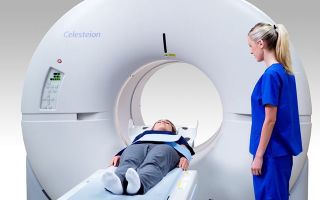
Positron emission tomography is considered a non-invasive diagnostic method. Any organ can be studied for the formation of pathology. The process is painless, but being stationary for a long time causes some discomfort.
To conduct the study, a contrast agent is injected. Diagnostics take place in a separate room, where outsiders are prohibited from entering. The patient is placed on a special couch, which is then moved into the scanner. If there is mild nervousness, the person is given a sedative medication. Accuracy and reliability require a calm state of the nervous system.
A person is injected with a contrast solution into an artery - using a catheter, inhalation or orally.
After this, the couch is placed inside the tomograph, where first a positron emission tomography scan is performed, and then a computed tomography scan. The slight noise produced by the tomograph may cause discomfort to the patient.
The procedure lasts up to 1 hour. The examination is recorded on digital and paper media, which is then transferred to the attending physician.
The study is carried out on an outpatient basis; hospitalization is not required. After the examination, it is not recommended to have contact with children and other people to avoid exposure to residual radiation. The volume of radiation is low in concentration - it is not harmful to human health.
For a child, pregnant women and nursing mothers, the harmfulness of such a dose increases, so diagnostics should be abandoned. The contrast solution is excreted naturally through the kidneys.
A large amount of liquid will help you recover after the analysis - you can drink plain still water, tea, fruit juice or compote.
Transcript of the study
Interpretation of the results after PET-CT is carried out by the attending physician, radiologist and nuclear physicist. The images are analyzed for any abnormalities in the structure of the organ.
A special table has been developed that includes data on the norm of indicators. After the examination, the difference between the obtained images and the table data is assessed.
The conclusion is issued to the patient when the doctor can decipher all the images.
Gradation that determines the result of the examination:
- Levels 1 and 2 indicate an inflammatory process in the body, characterized by varying degrees.
- Level 3 marks the presence of cancer in the initial stage.
- Level 4 is established with extensive metastasis of the body.
The result of the analysis may be known on this day or on the second - it depends on the area of the body area being examined and the complexity of the disease. After decoding, a decision is made on therapeutic measures.
Errors during the examination may occur if the following actions are present:
- the scanner has a low resolution;
- the pathology is characterized by a chaotic manifestation;
- the patient did not follow all the doctor’s recommendations in preparation for diagnosis;
- during the examination, metal objects remained on the body or extraneous movement was made, so sometimes hyperfixation is required;
- blood glucose level is increased;
- After a course of chemotherapy or radiation exposure, little time has passed - residual traces of the toxin are present.
The study is carried out by a doctor of the highest category, so erroneous tests are rare.
Possible complications
The method is characterized by low radioactive exposure, which is easily tolerated by the body. Unpleasant consequences may occur if the dose of contrast agent is exceeded.
Side effects from the procedure are diagnosed:
- sensation of heat and burning with contrasting water;
- feeling of itching;
- attacks of nausea with slight dizziness;
- short-term manifestation of claustrophobia.
These symptoms do not require medical attention. Signs of discomfort usually disappear within 1-2 hours. It is only recommended to drink more fluid - this will help relieve discomfort faster.
Medical centers using PET-CT
In Russia there are a small number of clinics capable of performing positron emission tomography. The equipment is quite expensive, the cost of analysis varies between 40,000-50,000 rubles. Not every clinic can afford an expensive device, which complicates the diagnosis.
In Western countries, Israel and the USA, each cancer center is equipped with a positron emission tomograph and CT scanner. In Moscow, a number of clinics can perform examinations:
- National Medical Research Center of Oncology named after N.N. Blokhin (RONC).
- EMC Cancer Institute.
- Clinical Hospital No. 1 of the Administration of the President of the Russian Federation.
- Scientific Center for Cardiovascular Surgery named after. A.N. Bakuleva, PET center.
- National Medical Research Center for Neurosurgery named after Academician N. N. Burdenko.
- Federal Scientific and Clinical Center for Pediatric Hematology, Oncology and Immunology named after Dmitry Rogachev and others.
Since 2016, this method has been included in the list of services provided free of charge under the compulsory medical insurance policy. To undergo a free examination, a referral from your attending physician is required. A person signs up and undergoes a preliminary examination of the body.
Source: https://onko.guru/medic/pet-kt.html
Positron emission tomography
Until recently, the ability to visualize pathological processes occurring at the level of body cells was considered science fiction.
Now positron emission tomography makes it possible to actually carry out this diagnosis.
It refers to a completely painless, rapid research method that reveals the root causes of the disease, even when the lesion affects a small number of cells.
What is PET
PET first became known half a century ago. Then nuclear energy took a step forward. At this time, the first scanners and pharmaceuticals for this examination were created. Next, the safe dosage of radiation was refined, and installations began to be produced that would allow large-scale research to be carried out by anyone who needs ultra-precise diagnostics at the molecular level.
The essence of tomography is based on the use of radioactive particles that are contained in a drug administered before the procedure. After penetration into the human body, they produce energy radiation of a given frequency. Moreover, the radiation in healthy and damaged tissues is different.
The research principle is based on the following:
- With the help of radioactive substances, the natural radiation of human cells is increased.
- The device's sensors are able to pick up signals from tissues and transmit them to a computer.
- A special program processes the received signals into a three-dimensional image.
Positron emission tomography does not exceed the average radiation dose and does not cause negative consequences in the form of radiation sickness or cancer progression. This diagnosis reveals oncological, cardiovascular, and neurological diseases.
A radioactive substance is used to conduct research
Positron emission tomography is a difficult research method that allows you to see the presence of tumors that do not manifest the slightest symptoms. It reveals a pathological focus up to a hundredth of a millimeter in size and metastases that can spread to neighboring organs. This diagnostic can evaluate therapy and monitor changes in tumors.
Types of PET
Positron emission scanning has several types, based on the type of drug used, for a unique highlighting of the areas under study. Today there is no universal substance that will be equally effective for diagnosing different organs. Combined tomography is performed with the following types of tools:
- With choline and radioactive substances. This type is indicated for the study of the liver, head structures, and prostate gland.
- With methionine and radioactive elements. Recommended for use in neurology, ischemia, epilepsy, myocardial infarction.
- With fluorodeoxyglucose and radioactive substances. Used to examine the uterus, skin, intestines, lymph nodes, mammary gland, thyroid gland.
The solutions used are distributed among organs and systems as they are absorbed by the body.
Modern equipment is used to carry out the procedure, increasing its productivity.
The advantages of the technique include: increased research accuracy, the ability to replace several different diagnostics, non-invasiveness, absence of pain, the ability to examine several organs and systems in one procedure, the absence of harmful factors. The main task of the diagnostic method is to obtain an image in color. This makes it possible to clearly see any change.
When is the study indicated?
Most often, doctors recommend undergoing CT and MRI. Positron emission tomography is prescribed in more complex situations. The reasons for its passage are:
- multiple sclerosis of the brain;
- Alzheimer's syndrome;
- schizophrenia;
- Parkinson's disease;
- epilepsy;
- malignant neoplasms of the lungs, intestines, head structures, prostate;
- cardiac ischemia;
- myocardial infarction;
- search for metastases;
- assessment of blood supply to the heart muscle;
- clarification of the disease before and after surgery.
Using emission tomography, pathologies can be detected in several directions
This procedure records blood flow in tissues and determines the process of oxygen and glucose consumption by organs. This method is used to diagnose:
- In oncology. It detects cancer formations at the primary stage of development, while cancer cells have not yet begun to progress. Identifies chains of tissues and organs affected by tumors. This is possible due to the greater absorption of glucose by cancer-affected tissues. Tomography allows us to determine the effectiveness of treatment.
- In cardiology. Detects impaired blood circulation in individual cardiac areas. As a result, even old heart attacks and ischemia are clearly diagnosed. By using positron emission scanning, it is possible to distinguish a dead part of the heart muscle from a living one, but poorly contracting. Thanks to this, it is possible to more accurately determine the indications for surgical intervention.
- In neurology. The procedure at the initial stage identifies pathologies that are fraught with the development of senile dementia. Thus, a predisposition to the disease is revealed. It most accurately determines the brain area where the epileptic focus is located. Allows you to effectively plan surgery. Using this diagnosis, the post-stroke state is assessed and schizophrenia is detected.
What you can explore
Using positron emission scanning, any changes in the following structures can be examined:
Tomography with contrast
- esophagus;
- lungs;
- heads;
- neck;
- lymphatic system;
- intestines;
- thyroid gland;
- hearts;
- mammary gland;
- genitals;
- skin.
This method can detect melanoma that is difficult to diagnose with other methods. Since it can only be determined by a biopsy, which traumatizes the tumor and leads to its progression.
With positron computed tomography, the patient does not have to risk his health.
If you repeatedly scan for oncology, you can track the dynamics of the tumor and the effectiveness of treatment.
Preparation
To undergo a positron emission examination, no special preparatory measures are required. If the patient has diabetes, the doctor must be notified.
A recommendation may be made to reduce the dosage of medications.
Since insulin affects the course of metabolic processes, as a result of which the results of the examination can be seriously distorted.
There are also minor restrictions on food intake; it is advisable to come to the procedure on an empty stomach. If the scan is scheduled for the morning, the last appointment should be in the evening. When the examination is scheduled for the daytime, at least 5 hours must pass between the diagnosis and the last meal.
Smoking is prohibited on the day of the procedure
You should not drink coffee or strong tea during the day. It is recommended to avoid taking products that contain caffeine. Any alcoholic drinks should be avoided the day before the examination.
If the positron emission procedure is required by a nursing mother, then after it it is necessary not to breastfeed the baby for two days.
You must come to the procedure in comfortable clothing that does not contain metallic inclusions; women do not need to use cosmetics or hairspray. Since these substances often contain metal particles.
How the research is carried out
The procedure is carried out in a specialized room. The patient is injected with a solution into a vein. It is also possible to administer it by inhalation; this method is used to diagnose the lungs.
After which the patient lies more comfortably on the couch and is secured with soft straps. Scanning can be performed on different types of tomographs.
Based on this, the couch moves inside the device or along it.
The installation has built-in sensors that capture radiation and transmit it to the computer. Using a special program, the image is transferred to the monitor in the form of a three-dimensional image. How long the procedure will take depends on the extent of the examination and the complexity of the area being diagnosed. It usually takes about 30–40 minutes.
Contraindications
Despite the high information content, this research method has some contraindications. Among which, pregnancy and breastfeeding are considered absolute. Since this diagnosis belongs to the category of radiation and can cause significant damage to an unformed fetus and a newborn child.
The patient should not be diagnosed in a serious condition. Since he needs to remain motionless for a fairly long period of time. If the procedure is performed for people suffering from diabetes, then the procedure with glucose is considered a contraindication. In this case, consultation with your doctor is required. It will help to carry out a preliminary correction of the patient’s blood sugar.
Diabetes mellitus is a contraindication for studies with glucose components. Then you need to consult your doctor and pre-correct your blood sugar level.
Diagnostics should be used with caution in the presence of renal failure. Since due to the possible delay of radiological drugs, data distortion may occur.
For your information, it is prohibited to carry out diagnostics on persons who have implanted devices with metallic inclusions, crowns, or tattoos.
If the patient is hypersensitive to the injected drug, antiallergic drugs may be administered during the examination.
The following conditions are relative contraindications:
- fear of confined spaces;
- undergoing radiation treatment, chemotherapy;
- breastfeeding period;
- exacerbation of a chronic disease;
- purulent infections;
- viral diseases;
- sarcoidosis
Such conditions may have a negative impact on the examination result.
Possible complications
If you follow all the doctor’s recommendations, there are usually no complications after positron emission diagnostics. The doctor calculates the individual dose of the radioactive substance. However, during scanning the following side effects may occur: burning in the area where the component was inserted, nausea, fear of confined spaces.
Radioactive components leave the body on their own throughout the day. To remove them as quickly as possible, you should drink as much fluid as possible. Using positron emission scanning, it is possible to detect pathology at the molecular level. As a result, the diagnostic quality increases and, consequently, the chance of obtaining a favorable prognosis during treatment increases.
Source: https://apkhleb.ru/kt/pozitronno-emissionnaya-tomografiya
PET-CT - what kind of study
Views: 1081
PET or Positron Emission Tomography is a modern diagnostic study of human internal organs, which makes it possible to identify the disease at the earliest stages of its development . This scanning is based on special radioactive tracers that are introduced into the patient’s body before the procedure. This may be an injection into a vein, a capsule to be swallowed, or an aerosol. The method of delivering indicators to the body depends on the part of the body being examined using PET.
A PET scanner most often always works in conjunction with CT or MRI scanners, hence the double name PET-CT (MRI).
Radioactive tracers are tracked by the PET scanner during scanning and help the doctor determine the condition of the organ being examined. They are concentrated in tissues with the greatest chemical activity, and certain diseases provoke a surge in such chemical activity during their development. And on the scanner, areas with increased activity will appear as bright spots.
In addition, PET scans can measure blood flow, oxygen consumption, how the body uses sugar and many other important parameters to track health.
PET scanning is an outpatient procedure, i.e. After the examination, the patient can go home or go about his other business.
In Russia, about 2,000 diagnostic PET-CT studies are performed annually, despite the fact that the actual need is 200,000.
This “deficit” is due to the fact that in our country there are only 4 positron emission tomography centers, which are located in Moscow and St. Petersburg.
And most of these scans are performed on a commercial basis, and not on referrals from local clinics. And prices start from 30,000 rubles.
According to modern medical standards, 1 PET scanner should serve 1 million people, but in our country there are 4 for 150 million people.
Why is a PET scan done, indications?
Most often, PET-CT examination is prescribed in case of suspicion of:
- cancer;
- cardiovascular diseases;
- brain disorders, including problems with the central nervous system (CNS).
Because PET testing works at the molecular level, so it will also be prescribed for other diseases that are difficult to classically diagnose. The scope of its application is very wide.
Cancer
Cancer cells have a very high metabolism compared to normal cells. Due to their high chemical activity, they are clearly visible during PET-CT examination and they appear on the tomograph screen as bright spots. Such diagnostics are prescribed not only to detect the presence of a cancerous tumor, but also for:
- determining the presence of metastases in other organs;
- determining the effectiveness of the treatment;
- checking for a possible relapse of the disease after a certain amount of time.
Despite all the effectiveness and accuracy of PET-CT examination, the doctor should read the results with caution, because there is a possibility that some benign neoplasms may be mistaken for cancer. Also, solid tumors are often not diagnosed with PET scans.
Cardiovascular diseases
PET scans identify areas in the heart with low blood flow. Healthy heart tissue absorbs more radioactive tracers compared to weakened tissue. The scan results will show areas of the heart with different brightness, showing the different condition of the tissues and heart muscles, which will help your doctor decide on the further course of treatment.
Brain disorders
Glucose is the main fuel for the brain. During a PET scan, radioactive tracers are “attached” to compounds such as glucose. Thus, the scanner determines which areas of the brain actively absorb glucose. Using this picture, an experienced doctor will be able to determine the state of brain function and identify hidden disorders.
PET-CT brain
PET can diagnose a variety of CNS abnormalities, including:
- Alzheimer's disease;
- depression;
- epilepsy;
- head injuries;
- Parkinson's disease.
Comparison of PET examination with other diagnostic procedures
PET scans show metabolic changes in organs or tissues at the cellular level. This is an important feature of this study, because A large number of dangerous diseases begin at the cellular level. When comparing PET with CT or MRI, the latter two studies do not show cratin at the cellular level.
A PET scan can detect changes in cells at a very early stage. MRI and CT scans show changes that have already occurred in the structure of tissues or organs.
Diagnosis of diseases at the cellular level shows the doctor a complete picture of such complex systemic diseases as:
- IHD (coronary heart disease);
- brain tumors;
- memory disorders;
- seizures.
As mentioned earlier, PET examination is now combined with CT or MRI.
- Computed Tomography (CT) itself uses X-rays to produce a picture of the body.
- MRI uses a magnetic field and radiofrequency pulsations to create images of internal organs, tissues, and bones.
When a CT or MRI scan is combined with a PET scan, a so-called fusion is obtained. A computer combines images from two scanners to create a 3-dimensional image that gives a more complete picture of the patient's condition, thereby increasing diagnostic accuracy.
Gallium scanning is somewhat similar to a PET scan. It uses gallium citrate as a radioactive tracer. For patients, the main difference with gallium scanning is that it lasts several days, because... gallium citrate needs to penetrate the tissue. This procedure is also not used to diagnose cancer.
Possible risks during PET-CT examination
A PET scan (no matter with CT or MRI) involves the introduction of radioactive tracers into the body. But in this case their radioactive influence is small. They begin to disintegrate already during the diagnostic process and are completely eliminated from the body within 2 days. When using the radiopharmaceutical 18F-FDG, which is now the standard in this field, the exposure will be 14 mSv.
For comparison: during fluorography, a person receives a dose of 0.02 mSv, and CT of the chest - 8 mSv. The maximum permissible radiation dose for nuclear energy workers is 50 mSv. So everything here is within normal limits. But with PET-CT, the radiation dose will be higher and will be up to 26 mSv. If this bothers you greatly, please consult your doctor about this issue first.
But all the risks of radiation exposure are negated in comparison with the benefits of PET studies, especially when it comes to cancer.
Allergies and other medical conditions
An allergic reaction to radioactive tracers is possible. If you are allergic to iodine, aspartame or saccharin, be sure to tell your doctor. For those patients who cannot tolerate iodine-based tracers, barium-based tracers are used.
Most often, an allergic reaction to iodine indicators occurs in people suffering from such diseases as:
- asthma;
- heart diseases;
- dehydration;
- polycythemia vera;
- multiple myeloma;
- kidney diseases.
For pregnant women
Radiation in any dose is not safe for fetal development. So if you are pregnant or think you are pregnant, you should refuse the PET study.
Other possible risks
Other risks include discomfort during the procedure, claustrophobia, or fear of needles. In addition, injection of a radiopharmaceutical can lead to symptoms such as bleeding, swelling, and bruising.
How to prepare for a PET scan
Your doctor should give you comprehensive information about preparing for a PET diagnostic study. Tell your doctor about any prescribed drugs you take and those you take without a prescription.
A few days before
You should avoid increased physical activity. Avoid any physical exercise 24-48 hours before the test.
One day before
During the last 24 hours before diagnosis, you should follow a low-carb diet and completely eliminate sugar from your diet. Here's what you shouldn't eat during this time:
- porridge;
- paste;
- bread;
- rice;
- milk, yogurt and other dairy products;
- fruits and fruit juices;
- alcohol;
- caffeine (tea, coffee);
- candies, including chewing gum and mints, etc.
You can eat meat, tofu, nuts and vegetables without starch.
A few hours before
If you will be given anesthesia for the procedure, you should not eat or drink anything the entire morning before the PET scan. If you are taking any essential medications, take them with just a few sips of water.
But even if you are not prescribed anesthesia during the scan, you should avoid eating any food six hours before the examination. Chewing gum and any candy, including, are also prohibited. and cough medicine, mint tablets or candy. You can really drink, but not a lot.
Immediately before the procedure, you will be changed into hospital clothes, and you will also be asked to remove all jewelry, incl. and piercings.
If you are having a PET CT scan, medical devices such as a pacemaker or artificial joints will not affect the results of the test. But you cannot undergo a PET-MRI without prior approval of any medical devices or metal implants.
Other factors
- If you are pregnant or think you may be pregnant , be sure to tell your doctor because... PET is not safe for the baby.
- If you are breastfeeding , then you need to suck breast milk 24 hours before the procedure and prepare it for the baby, because You should not breastfeed for the next 24 hours after the test.
- If you have diabetes , any fasting from eating can cause your blood sugar levels to rise. You will likely be allowed to take your standard dose of insulin and have a light snack 4 hours before your scan.
How is a PET-CT study performed?
Before the scan itself, a radiopharmaceutical drug will be injected into your body, i.e. radioactive tracers. This can be given by injection into a vein, orally, or as an aerosol that you inhale. Your body then needs time to absorb the drug, so the actual scan will begin in about another hour.
How long the indicators are absorbed depends on what part of the body is being examined. While you wait, try to minimize any movement, relax and rest. If you are undergoing a PET scan of the brain, you will not be able to watch TV, listen to music or read.
The scanning process itself takes from 30 to 45 minutes. All this time you will lie on a flat table, which will be located inside a PET machine, which is somewhat similar to a giant letter “O”. The table on which you will lie will periodically move.
You need to lie still and not move throughout the scan. You may be asked several times to hold your breath for a few seconds. You will hear buzzing and clicking sounds throughout the scan.
Once all the necessary images have been recorded, the table will leave the PET machine. The research is completed.
After PET examination
After the procedure, you can go about your business unless your doctor, of course, gives other instructions.
The radioactive drug will remain in your body for another 12 hours, so you are advised to limit contact with pregnant women and young children.
Drink more water after PET-CT scan, because... water promotes the rapid removal of radioactive substances from the body. Typically, radioactive tracers are completely eliminated from the human body after 48 hours.
At this time, a specialist radiologist will interpret the results of the study and transmit this information to your doctor. Such decryption usually takes no more than 2 business days. Look forward to your next appointment with your doctor to discuss your results and whether or not to prescribe any necessary treatment.
Additional Information:
Source: https://doktorlaser.ru/diagnostika/pet-kt.html
Positron emission tomography is a breakthrough in the diagnosis of cancer
Computed and magnetic resonance imaging, ultrasound and endoscopic research methods... The arsenal of modern medicine is wide and allows us to identify many health- and life-threatening ailments at the earliest stages.
At the same time, science does not stand still. In European countries and the USA, along with the now traditional diagnostics, the positron emission tomography method is actively used. In our country it is not yet so widespread. What sets it apart from other modern medical imaging methods? In terms of diagnostics, what can PET do that CT and MRI cannot?
- Let's consider these and other questions.
- What is positron emission tomography and what is the essence of this study?
- Positron emission tomography (or, otherwise, PET) is a modern method of medical imaging, which is based on its own special principle.
The essence of PET is as follows. Before the study, a pharmaceutical preparation containing a radioactive component is injected into the body - a radiopharmaceutical (this can be, for example, chemically modified glucose). In the body it decays with the release of positrons.
The latter begin to interact with electrons of organs and tissues, resulting in special electromagnetic radiation that is recorded by special sensors. Based on its characteristics, a conclusion is drawn about the state of the observed structures at the molecular and cellular level.
PET scan shows not only the structure of the area under study, but also its function. Glucose from the administered radiopharmaceutical is “included” in the metabolism, and with the help of the device it is possible to record how it “behaves” in a particular tissue or organ.
For example, it is known that malignant tumor cells grow rapidly. This happens for a reason: rapid growth requires, in particular, energy.
Thus, the administered glucose is more intensively consumed by the cells of the tumor tissue, which makes it possible to “see” them on tomograms.
- What is PET/CT (or PET-CT)?
- This is a study that combines data obtained using both methods, which makes it possible to accurately identify the presence and location of a pathological formation.
- What are the indications for positron emission tomography?
First of all, these are many oncological diseases, including those with the formation of metastases. This method will also help with neurological diseases - in particular, with cerebral vascular pathology, epilepsy, memory impairment, Alzheimer's disease and other types of dementia, degenerative brain lesions (Parkinson's disease, Huntington's disease).
- What role does MRI play in diagnosing epilepsy? Read here
- In addition, PET is used to assess blood circulation and vitality of the heart muscle (myocardium).
- Are there any contraindications to PET CT?
- Yes, they are.
- — The study is not prescribed during pregnancy or its potential. If it is necessary for nursing mothers to carry it out after the procedure, resumption of feeding is allowed no less than 24 hours later;
- — Kidney and liver failure;
- — The use of the method in patients with diabetes mellitus is limited;
- — Allergy to components of the radiopharmaceutical and contrast agent;
- — Diseases of the thyroid gland (when planning to use contrast in CT);
- — General serious condition of a person and some others.
- MRI has its own contraindications, which you can read about here
All details regarding possible contraindications should be discussed with your attending physician and the doctor who will perform the diagnostic procedure. Give them only reliable and as detailed information as possible.
How to prepare for the examination and undergo a PET scan correctly? Is it necessary to follow a diet before the examination?
There are different types of PET, each of which has its own characteristics in preparation. To understand the basic principles, consider the option for diagnosing tumors.
- In order for the diagnosis to be as informative as possible, the following recommendations must be followed:
- - be sure to inform the doctor who will conduct the study about any allergies or reactions to medications you have, or any existing or suspected pregnancy;
- — bring with you a list of medications you are taking;
- - 48 hours before the test, stop intense physical activity and avoid massage;
- — 36 hours before the test, avoid hypothermia. If the procedure is carried out in the cold season, then dress warmly on the day of the examination;
- - 24 hours before the test: follow a very low carbohydrate diet, completely eliminating sugar. Do not use chewing gum or mints;
- - 6 hours before the test: stop eating, drink only non-carbonated unsweetened water in large quantities; continue taking prescribed medications unless otherwise advised by your doctor;
- — Patients with diabetes should consult their doctor about any changes in diet and medication.
- How is positron emission tomography performed?
First, a radiopharmaceutical is injected into the patient's body. In this case, a person usually does not feel anything; occasionally there is a feeling of heat. After administration, large amounts of water should be taken as this will affect the quality of subsequent images.
Within an hour, the product is distributed throughout the organs and tissues, including the pathological focus. All this time you need to lie still and avoid talking. This contributes to the fact that during the study the neoplasm is more clearly identified, where more of the radiopharmaceutical is concentrated.
Then the scanning procedure itself is carried out. First, a CT scan is performed (sometimes, if indicated, with contrast enhancement), and then a PET scan is performed. Both studies take place in the same tomograph and, like MRI, CT and ultrasound, are completely painless.
When are CT, MRI and ultrasound prescribed? Find out here
What to do after undergoing positron emission tomography?
In the absence of contraindications, it is recommended to consume at least 2 liters of fluid per day and limit salty and smoked foods. Avoid contact with pregnant women and children for 24 hours. Breastfeeding women should refrain from feeding for 24 hours. During this time, express all milk produced; Other family members need to care for the baby during this period.
If any health symptoms occur, immediately notify medical personnel. If complaints appear after you have left the clinic, contact the doctor who conducted the study or call an ambulance.
What are the advantages and disadvantages of positron emission tomography?
Among the advantages of PET are the non-invasive and painless procedure, high diagnostic accuracy, detection of pathologies in the early stages, the ability to examine all organs at once, and monitoring the effectiveness of the therapy.
The disadvantages of the method include the presence of a certain radiation exposure provided by a radiopharmaceutical, and when PET is combined with CT, by x-ray radiation. Examination of patients with diabetes mellitus is problematic or impossible.
The reliability of diagnosis strongly depends on the quality of preliminary preparation. PET alone is not always sufficient to make a definitive diagnosis, so it is usually combined with CT or MRI.
Also, the disadvantages of the method include its relative high cost.
- Enver Aliyev
- ***********************************************************************************
- Realizing that people’s awareness of this method is high, for an expert opinion we turned to Andrei Vladimirovich Korobov, Candidate of Medical Sciences, a specialist in the field of radiation diagnostics, a member of the board of the Expert group of medical companies, and Director of the Expert Institute.
- - Andrey Vladimirovich, when is it necessary to do without PET CT?
Taking into account the unique ability to determine and visualize this combined method of cellular and molecular metabolism, the use of such an expensive method as PET CT can be considered fully justified in the diagnosis of any oncological diseases with expected high metabolic activity.
This allows you to monitor the patient’s entire body for the presence of a primary tumor and metastases in one session, measure functional changes in biochemical processes, distinguish benign from malignant formations with high accuracy, promptly determine the effectiveness of the treatment used, and conduct early diagnosis of an impressive list of oncological diseases.
- Is there an alternative to PET CT?
All medical imaging methods are, to one degree or another, alternative to each other.
At the same time, if you ask the question: “what is better to do...” and start listing ultrasound, radiography, computed tomography, MRI, SPECT-CT, PET-CT, and other methods, then with a high degree of probability you will get the answer which is better and more correct in a full-fledged diagnostic process, use several different methods.
In this case, the choice is largely determined by the doctor based on the expected type of disease. There are various legends extolling certain individual methods of radiation diagnostics. Legends that exaggerate the identification of one or another unique information that is inaccessible to identification by other methods.
At the same time, as a rule, the fact is kept silent that in the example given, the patient has already undergone a lot of other studies, which excluded a great many missing changes and significantly narrowed the direction for the search. Of course, in the search for small lesions in suspected cancer with high metabolic activity, PET, and even more PET CT, has undeniable advantages.
A research method such as single photon emission computed tomography (SPECT) has similar properties to PET. Both methods belong to the field of nuclear medicine.
A full, correct comparison of these methods in terms of their value for the diagnostic process is difficult to achieve due to both the relative scarcity of both methods and the difficulty of justifying their simultaneous use in the same patients.
That is why discussions about the comparative advantages of these diagnostic methods, which are similar in physical meaning, are still ongoing. Authors of scientific publications point out the advantages of PET CT in sensitivity, specificity and diagnostic accuracy.
At the same time, these SPECT CT indicators are also quite high, reaching values close to 90%, which, despite the many times higher cost of PET, gives grounds for the wider use of SPECT.
- Where can I get positron emission tomography in our country?
These studies are currently available mainly in large cities. The data varies, but today PET can be performed in Moscow (PET Technology, Sofia Cancer Center), St. Petersburg (LDC MIBS), Voronezh (MMCRDiLOZ). The Federal Network of PET Technology also has its branches in Ufa, Lipetsk, Yekaterinburg, Belgorod, Tambov, Orel, and Kursk. There are also centers in Tyumen, Chelyabinsk, Kazan.
- If you need to undergo such research, I recommend checking the relevance of the information, since the list of cities is constantly expanding.
- For reference:
- Andrey Vladimirovich Korobov
- Candidate of Medical Sciences, specialist in the field of radiation diagnostics, member of the board of the Expert group of medical companies, director of the Expert Institute.
Source: https://www.mrtexpert.ru/articles/540