Actinomycosis is an infectious disease caused by radiant fungi (actinomycetes) and has a primary chronic course with the formation of dense granulomas, fistulas and abscesses.
Actinomycosis can affect not only the skin, but also internal organs.
Diagnosis of the disease is based on the detection of characteristic fungal mycelium in the discharge and detection of the growth of specific colonies when inoculated on nutrient media. In the treatment of actinomycosis, the administration of actinolysate is used in combination with antibiotic therapy, ultraviolet irradiation of the skin, and iodine electrophoresis. According to indications, abscesses are opened, fistulas are surgically treated, and the abdominal and pleural cavities are drained.
Actinomycosis is an infectious disease caused by radiant fungi (actinomycetes) and has a primary chronic course with the formation of dense granulomas, fistulas and abscesses.
Actinomycosis can affect not only the skin, but also internal organs.
Diagnosis of the disease is based on the detection of characteristic fungal mycelium in the discharge and detection of the growth of specific colonies when inoculated on nutrient media.
The causative agents of actinomycosis are fungi of the genus Actinomyces, which are often found in nature. They can be found on soil, plants, hay or straw. Actinomycetes enter the human body through damaged skin, inhalation or through food.
In most cases, they do not cause disease, but live on the mucous membranes of the eyes or mouth as saprophytic flora. Inflammatory processes in the mouth, gastrointestinal tract or respiratory organs can lead to the transition of actinomycetes to a parasitic state with the development of actinomycosis.
Actinomycosis also occurs in farm animals. However, human infection does not occur from animals or people with actinomycosis.
Actinomycosis of the skin can occur primarily when actinomycetes penetrate through wounds and other lesions on the skin. Secondary skin lesions develop from the inside, when the infection spreads from the underlying tissues (tonsils, teeth, lymph nodes, muscles, mammary gland) and internal organs.
Depending on the localization of the pathological process in actinomycosis, the following forms are distinguished:
- cervical-maxillofacial;
- thoracic;
- abdominal;
- cutaneous;
- genitourinary;
- actinomycosis of joints and bones;
- actinomycosis of the central nervous system;
- actinomycosis of the foot (mycetoma, Madura foot)
The duration of the incubation period for actinomycosis is not precisely known. The disease is characterized by a long and progressive course and can last 10-20 years. In the initial period, the patient remains in normal health, but when internal organs are damaged, the condition becomes severe and cachexia occurs.
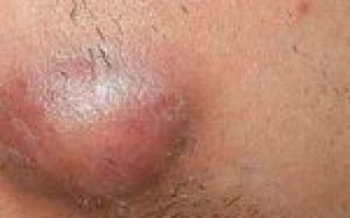
Actinomycosis of the skin most often affects the submandibular, sacral region and buttocks. It is characterized by the appearance of compactions in the subcutaneous tissue and a bluish-purple coloration of the skin above them.
The seals are spherical in shape and practically do not cause pain. At first they are very dense, then they soften and open with the formation of poorly healing fistulas. There may be an admixture of blood in the purulent discharge of the fistula.
Sometimes it contains yellow grains - drusen of actinomycetes.
There are 4 types of skin actinomycosis. In the atheromatous variant, which occurs mainly in children, infiltrates resemble atheromas.
Tuberculate-pustular actinomycosis begins with the formation of tubercles in the skin, turning into deep pustules, and then fistulas. The gummous-nodular variant is characterized by the formation of nodes of cartilaginous density.
Ulcerative actinomycosis usually develops in weakened patients. With it, the stage of suppuration of the infiltrate ends with tissue necrosis and the formation of an ulcer.
Cervical-maxillofacial actinomycosis is more common than others and occurs in several forms: with damage to intermuscular tissue (muscular form), subcutaneous tissue or skin.
The process can spread across the face and neck, involving the lips, tongue, penetrating the larynx, trachea and orbit.
In the muscular form, a characteristic infiltrate most often forms in the area of the masticatory muscles, causing trismus and leading to facial asymmetry.
Thoracic actinomycosis begins with cold symptoms: general weakness, low-grade fever, dry cough. Then the cough becomes wet, mucopurulent sputum is released, which has the taste of copper and the smell of earth.
Gradually, the actinomycotic infiltrate spreads from the center to the chest wall and reaches the skin, forming fistulas coming from the bronchi.
Such fistulas can open not only on the surface of the chest, but also in the lumbar region and even the thigh.
Abdominal actinomycosis often mimics acute surgical pathology (intestinal obstruction, appendicitis, etc.).
It spreads to the intestines, liver, kidneys, spine and can reach the anterior wall of the abdomen with the formation of intestinal fistulas opening on the skin. Actinomycosis of the rectum occurs with the clinical picture of paraproctitis.
Actinomycosis of the genitourinary organs is a rare disease that often occurs secondary to infection from the abdominal cavity.
Actinomycosis of joints and bones usually occurs when the process spreads from other organs. Damage to the joints is not accompanied by a significant impairment of their function, and actinomycosis of bones occurs as osteomyelitis. The spread of infiltrate to the surface of the skin leads to the formation of fistulas.
Mycetoma begins with the appearance of several dense “peas” on the sole, the skin over which gradually acquires a brownish-violet color. The number of compactions increases, swelling occurs, the shape of the foot changes and purulent fistulas form.
The process may involve the tendons, muscles and bones of the foot.
With the development of a characteristic clinical picture of actinomycosis, diagnosis does not cause difficulties. However, it is important to make the correct diagnosis in the initial period of actinomycosis. Detection of actinomycetes in sputum, throat or nasal swabs has no diagnostic value, since it is also observed in healthy people.
Therefore, for examination, fistula discharge is taken or a percutaneous puncture of the affected organ is performed. Conventional microscopy of the test material can reveal drusen of actinomycetes, which allows a quick preliminary diagnosis of actinomycosis.
Subsequent immunofluorescence reaction (RIF) with specific antigens is aimed at determining the type of actinomycetes.
Difficulties arise in those cases of actinomycosis in which there are no drusen in the studied material, which is observed in 75% of the disease. In such cases, the only reliable diagnostic method is to culture pus or biopsy material on Sabour's medium.
A complete and reliable culture test for actinomycosis may take more than 2 weeks. But after 2-3 days, microscopy can detect colonies characteristic of actinomycosis.
When studying culture, the growth of accompanying microflora and its sensitivity to antibiotics must be taken into account.
Serological diagnosis of actinomycosis, unfortunately, is not specific enough. And PCR research methods for this disease are still under development.
Treatment of actinomycosis is carried out by intramuscular and subcutaneous administration of actinolysate. Along with this, antibiotic therapy is carried out, aimed at suppressing the accompanying flora and preventing secondary infection. Like any chronic infection, actinomycosis requires additional detoxification and restorative therapy.
Physiotherapeutic treatment used for actinomycosis includes ultraviolet irradiation of the skin in the affected area, local electrophoresis of actinolysate and iodine. When abscesses form, it is necessary to open them. Surgical treatment of fistulas, drainage of the pleural cavity or abdominal cavity may also be required. In some cases, for large lung lesions, a lobectomy is performed.
In the absence of specific treatment, actinomycosis of internal organs can be fatal. The mildest form is considered to be cervical-maxillofacial actinomycosis. After patients recover, relapses may develop.
There is no specific prevention of actinomycosis. Nonspecific prevention includes maintaining hygiene, preventing skin injuries, timely treatment of teeth, inflammatory diseases of the oral cavity, tonsils, respiratory organs and gastrointestinal tract.
Source: https://www.KrasotaiMedicina.ru/diseases/zabolevanija_dermatologia/actinomycosis
Actinomycosis in cows (cattle): symptoms and treatment, control measures
Cows, along with many other farm animals, are susceptible to an infectious disease such as actinomycosis. It poses a great danger because humans can also become infected. In this article we will consider information about symptoms, routes of infection, methods of prevention and treatment of actinomycosis.
What is actinomycosis
Actinomycosis is a chronic fungal infection of various organs and tissues of animals and humans, which is characterized by the formation of granulomatous foci - actinomycosis.
The disease affects all representatives of cattle, sheep, pigs, and horses. It is observed in animals of different ages. Recorded all over the world. It can occur at any time of the year, but is most often diagnosed in the spring and winter.
Historical reference
The bacterium that causes actinomycosis was first isolated and described in 1877 as a microbe in the jaw tissue of a cow. The disease was then called dixomycete. In 1878, 2 cases of the disease detected in humans were described. They were registered in Germany. That same year, German veterinarian Otto von Bollinger named the disease actinomycosis.
The causative agent was named radiant fungus. Until 1940, it was believed that the causative agent of the infectious disease was identical to Actinomyces israelii. However, in the same year, D. Erickson proved that these are 2 separate organisms.
Pathogen
The causative agent of actinomycosis is the gram-positive bacterium Actinomyces bovis, which is classified in the order Actinomyces.
It is susceptible to hot temperatures - it is destroyed in 5 minutes when heated to +70...+80 °C. It is resistant to drying. At low temperatures it lives for 1–2 years.
Actinomyces bovis can be killed by exposure to a 3% formaldehyde solution for 5-7 minutes.
Sources and routes of infection
The pathogen enters the animal's body through wounds in the oral mucosa when eating roughage (straw, cereal awns, mouse barley) or teething in calves, through damage to the skin, and nipples of the mammary glands.
Infection can also occur anaerobically.
The active growth of bacteria and the rapid development of the disease are facilitated by the presence in the body of purulent inflammation, injuries, and disturbances in the functioning of the immune system.
The mechanism of the onset and development of the disease
After penetration into the body, the pathogenic bacterium provokes the development of inflammation, in which a granuloma is formed. Most often, lesions are localized in the bones, tissues of the lower jaw, and lymph nodes. Subsequently, a tumor and fistula form with yellowish purulent discharge containing gray drusen. Afterwards, blood and remains of dead tissue are recorded in the purulent discharge.
Clinical signs
Clinical signs may vary depending on where the pathogen is localized. A characteristic symptom is the presence of a dense tumor, which, as it develops, opens and pus flows from it.
With actinomycosis of the head, neck, jaw, and tongue, the following symptoms are present:
- pain when touching the area with actinomyoma;
- pain when eating food;
- increased body temperature (rare);
- leakage of pus from the actinomyc;
- change in head shape;
- tooth loss;
- complications of breathing and swallowing as a result of the presence of actinomics in the respiratory tract, mouth, throat, and tongue;
- exhaustion;
- enlargement and prolapse of the tongue.
If the pathogen has settled in the intestines or abdominal cavity, the following clinical symptoms develop:
- temperature increase;
- pain in the peritoneum;
- vomit;
- diarrhea or constipation;
- partial intestinal obstruction.
Important! If tumors and fistulas are detected, you should not treat the cow yourself. This may lead to undesirable consequences. At the first manifestations of the disease, you should seek veterinary help.
With actinomycosis of the udder the following are observed:
- necrosis of the skin;
- abscesses and fistulas in the mammary glands.
If the disease has affected the lymph nodes, the animal will experience the following symptoms:
- tissue swelling;
- depressed state;
- hyperthermia;
- redness or bluish discoloration of the skin at the site of inflammation.
Diagnostics
The disease is diagnosed through a veterinary examination and history taking. If it is necessary to exclude other diseases with similar symptoms (actinobacillosis, streptotrichosis, lymphangitis), then material is taken from the actinomyoma for microscopy in the laboratory.
How to treat
When actinomycosis is diagnosed, treatment of the disease should begin immediately. You need to start by placing sick individuals in quarantine and treating the room where they were kept with disinfectants.
Disinfection of the premises
Disinfection is carried out with a 2–3% solution of caustic alkali or freshly slaked lime. The person who performs disinfection must work in a special suit and observe personal safety measures.
Find out how to treat infectious rhinotracheitis in cattle.
Iodine or potassium iodide solution
One of the treatment methods is intravenous administration of iodine solution. It is prepared by mixing 1 g of iodine, 2 g of potassium iodide, 0.5 l of distilled water.
Antibiotics
Antibiotic therapy is also used against the pathogen. Possible introduction:
- Penicillin - 100–400 thousand units into the tumor for 4–5 days;
- Oxytetracycline - 200 thousand units + 5-10 ml of saline solution for calves up to one year old, 400 thousand units for adults in healthy tissue surrounding actinomyoma for 10-14 days with simultaneous suction of pus from the tumor;
- Clindamycin;
- Erythromycin;
- a complex of antibiotic drugs with sulfonamides.
In parallel with antibiotic therapy, autohemotherapy is performed - blood transfusion from the jugular vein into the affected area.
Surgical intervention
If conservative treatment methods do not lead to an improvement in the cow’s condition and recovery, then surgical intervention is performed - the actinomyomas are opened, cleared of pus and disinfected. After this, antibiotic therapy and painkillers are administered.
After treatment, scars remain on the animal's body. If the inflammatory process affects only soft tissues, the cow can be completely cured. It is unknown whether the cow acquires immunity against the disease after recovery. If bones, joints, and vital organs are affected, the prognosis is unfavorable.
Important! If treatment is not started on time, the disease will become generalized and affect vital internal organs - liver, lungs, kidneys, brain, heart. The meat of such animals is prohibited for consumption.
Prevention
In order to prevent this serious disease from entering the herd, it is necessary to adhere to certain rules for keeping and feeding animals:
- Do not release cattle onto pastures located in swampy lowlands.
- Before feeding roughage, heat it to soften and disinfect it.
- Before putting straw in the feeder, it must be calcined.
- Regularly disinfect the premises where cows are kept and equipment used to care for them.
- Periodically inspect animals for scratches and minor wounds for their timely treatment and treatment.
Thus, actinomycosis is a serious infectious disease of cattle that affects animals, regardless of their age. It is characterized by the appearance on the body, most often in the head and neck area, of painful tumors that develop into fistulas with purulent contents. When the disease is detected in the early stages and proper therapy is provided, it can be effectively treated.
Source: https://fermer.blog/bok/zhivotnye/krupnyy-rogatyy-skot-krs/bolezni-krs/infekcionnye-bolezni-krs/283-chem-lechit-aktinomikoz-u-korovy.html
Timely treatment of actinomycosis of the oral cavity
April 18, 2018 Last revised: December 18, 2019 Oral diseases
What to do if a patient is suddenly diagnosed with actinomycosis of the oral cavity? On medical websites you can find information that some forms of actinomycosis can be fatal for patients.
is it so bad? What health problems are meant by this term, why does it occur, by what signs can this disease be suspected, and is it really possible to completely cure maxillofacial actinomycosis? How serious and inevitable are the complications of actinomycosis, and what measures with the greatest effect will help protect against this chronic disease, prone to relapses? We invite you to learn about all this in our article.
Description of the disease
Actinomycosis is an infectious disease that can affect the skin and internal organs of a person. This disease has a chronic course with a tendency to relapse and occurs due to the entry of actinomycetes into the body.
With this disease, granulomatous lesions appear in various organs. In most cases, actinomycosis primarily affects the maxillofacial area.
The causative agent of actinomycosis is well known - it is the “human radiant fungus”. Actinomycetes are quite common in nature; they can also often be found in the body of healthy people.
Often these microorganisms are found in a person’s mouth and gastrointestinal tract.
How does oral actinomycosis appear?
Most often in the body, radiant fungus is found in gum pockets, carious cavities, dental calculus, tonsil lacunae, damaged or inflamed tissues of the oral cavity, nose, salivary glands, and gastric mucosa.
The fungus can also enter the body from the outside, from the environment. Microorganisms can stay in the body for a long time without causing any harm until the mucous membrane is damaged.
Actinomycetes spread inside the human body mainly through the blood (hematogenously), and occasionally they talk about the possibility of entering various organs through the lymph (lymphogenously).
In order for the disease to develop, it is necessary to repeatedly enter the body with radiant fungi. Most often, the patient himself becomes infected with the fungus from his own body (autoinfection).
Before the first symptoms of the disease appear, it usually takes 1 to 3 weeks after actinomycetes enter the body. In general, the incubation period can vary from a couple of weeks to 2-3 years.
Radiant mushrooms are also common in nature (in soil, hay, straw or on plants). People of mature age, schoolchildren and preschoolers suffer from actinomycosis more often. The cervicofacial region and lower jaw are most often affected by infection.
The impetus for infection with actinomycosis can be injections, injuries or surgical operations. For actinomycosis to develop, it is enough to injure the mucous membrane with a fish bone, damage it with a fragment of a carious tooth, bite the cheek, or have gums damaged by inflammation.
Symptoms
The cervical-maxillofacial form of the disease (genitourinary, skin, abdominal, osteoarticular, thoracic and other forms are also found) occurs most often, in almost 80% of cases of actinomycosis.
At the beginning of infection or self-infection, the patient’s well-being does not suffer in any way. The cervical-maxillofacial form occurs in several forms with damage:
- skin (cutaneous form);
- mucous (mucous form);
- subcutaneous tissue and muscles (subcutaneous muscle form);
- jaw bones (jaw shape).
The disease has a very long course and can last 10-20 years. Gradually, the pathological process can spread throughout the face, reaching the lips, tongue, larynx, involving the eye sockets and penetrating the neck area.
The muscular form most often affects the masticatory muscles.
Most often, actinomycosis of the oral cavity affects the tongue, salivary glands or tonsils. Actinomycosis of the upper jaw is less common.
Actinomycosis of the tongue
Actinomycosis of the tongue usually develops with chronic injuries to this organ with poorly fitting prostheses, sharp broken edges of teeth, damage to the lingual tissues by sharp bones, after tongue piercing, etc.
The clinical picture of the disease depends on the location of the source of infection. If the tip of the tongue or its back is affected, then these areas will not have any external changes for a long time and will not hurt.
However, after some time, the actinomycosis node becomes fused with the mucous membrane of the tongue or becomes abscessed, resulting in the formation of a fistula in the node.
Actinomycosis of the tonsils
Actinomycosis of the tonsils is less common than other forms. With this form, the tonsils are enlarged and compacted, resembling cartilage in density. Subsequently, the pathological process spreads to neighboring tissues. The main symptom of this form of the disease is the feeling of a foreign body in the pharynx.
Actinomycosis of the salivary glands
Actinomycosis of the salivary glands can be primary or secondary. More often, the radiant fungus enters the salivary gland through the duct. There are several forms of this type of actinomycosis:
- exudative limited and diffuse disease;
- limited productive and diffuse disease;
- lesions of the deep lymph nodes in the area of the ear gland.
Actinomycosis of the maxillary sinus
Actinomycosis of the maxillary sinus is rare. In this form, the infection enters the jaw from the affected tooth (odontogenic pathway) or from the nose (rhinogenic pathway). Manifestations of actinomycosis of the upper jaw resemble sinusitis: nasal breathing is impaired, and purulent discharge may come out of the nose. During exacerbation, swelling of the zygomatic, buccal and infraorbital areas is noticeable.
- What manifestations should alert the patient and force him to see a doctor for actinomycosis?
- At the onset of the disease, it is difficult to detect due to the normal temperature and good health of the patient.
- Although the mucous membrane is already damaged and lesions in the form of nodes begin to form in the tissues, the pathological process can proceed deeply and not be noticeable externally.
- If actinomycosis affects the oral mucosa, the patient will experience symptoms such as:
- swelling on the oral mucosa;
- inflammation and pain in the mouth;
- formation of fistulas in the oral cavity with the release of purulent fluid.
The local course of the disease and the absence of general signs of intoxication are characteristic of all forms of actinomycosis.
Actinomycomas or granuloma nodules can be found in tissues or mucosa. And only at the stage of disintegration of these nodes, pus is released from them and intoxication processes occur in the body (poisoning with tissue decay products).
Symptoms appear in the patient only at the stage of node disintegration. In this case, pus is released from fistulas (channels in the node).
At the stage of nodular decay, the patient is concerned about manifestations in the form of:
- headache;
- temperature (usually 38-38.5 degrees);
- general weakness;
- poor appetite.
When the nodes are located on the lower jaw, there may be spasms of the mouth in the form of convulsions (trismus), which interfere with eating.
Forms of actinomycosis
With actinomycosis of the oral cavity, infectious foci develop in the tissues of the oral mucosa.
Actinomycosis of the oral cavity can also occur in three forms:
- Gummy-nodular, with characteristic red or brown infiltrates. In this case, the skin in the affected areas softens, fistulas form on it, from which purulent fluid with inclusions is released.
- Tuberous-pustular, in which deeper ulcers are formed, combined with the formation of pustules and fistulous canals. This form is characterized by the appearance of purulent crusts on the skin.
- Ulcerative, in which there are skin defects of varying depths. Skin ulcers subsequently lead to tissue scarring and cicatricial atrophy.
Actinomycosis of the oral cavity in children
In pediatrics, actinomycosis is not so rare and usually occurs more often in children with carious teeth, frequent inflammation of the tonsils, or with the habit of putting or chewing various spikelets and stems of plants (especially barley, rye, oats, sorrel) in their mouths.
After damage or microtrauma to the mucous membrane, the fungus enters the tissue and causes a red swelling to appear in the mouth, which grows slowly. In some cases, actinomycosis may manifest itself in the form of acute phlegmon (spread inflammation). In this case, the swelling suppurates, but no outflow of pus occurs.
Phlegmon occurs in a child with local pain and spastic clenching of the jaws due to tissue swelling (if the neck and chin area become inflamed), which prevents the baby from opening his mouth.
After suppuration, the purulent formation can open on its own and form fistulas. With actinomycosis of the tongue, a visible infiltrate (swelling) usually forms on its front part.
Clinical manifestations of the disease in children
Just like in adults, actinomycosis in children begins in the form of local (limited) inflammation, which does not affect the general well-being of the patient. The course of the disease becomes chronic, with the temperature being low or high.
If a child's disease is not treated, it can become generalized (with multiple lesions) and lead the child to complete exhaustion (cachexia). This is due to constant intoxication, poor appetite and difficulty chewing food.
Diagnostics
Actinomycosis has similar manifestations with many infectious and inflammatory processes in the skin (phlegmon, abscess) or bone (osteomyelitis, periostitis), some infectious diseases (syphilis, tuberculosis) and tumor diseases.
In the diagnosis of actinomycosis, in addition to examining and collecting anamnesis from the patient, the use of diagnostic research methods is also used in the form of:
- blood and urine tests (general, biochemical) to determine the nature of the existing infectious inflammation;
- microbiological, to determine the colony of actinomycetes in the infiltrate;
- immunological, most often in the form of a skin allergic reaction in the form of subcutaneous administration of actinolysate;
- pathohistological, with examination of granulation tissue under a microscope.
- X-ray (when the process moves to the jaw bones) with determination of the bone cavity;
- biopsy with examination of granuloma tissue to exclude oncology.
Treatment
Treatment of actinomycosis of the jaw and oral cavity is always carried out with the simultaneous use of general and local therapeutic measures.
The main types of therapeutic measures for actinomycosis are the following methods:
- Surgical treatment. This method includes removal of carious teeth, tonsils, opening of foci of actinomycosis, curettage of pathology inside the bone, and removal of affected lymph nodes. Actinomycosis lesions and purulent wounds are also surgically treated.
- Anti-inflammatory treatment. Includes antibiotic therapy in the form of intravenous Penicillin for up to 6 weeks and a transition to intramuscular administration of it or Amoxicillin for 6-12 months. Amoxiclav, Doxycycline, Phenoxymethyl, and Erythromycin are often used orally. This also includes the use of antifungal agents: Nystatin, Levorin, Miconazole, Ketoconazole, Decamine, potassium iodide preparations.
- Methods for developing specific immunity. To do this, patients undergo intradermal administration of actinolysate or actinomycetic polyvalent vaccine (course of 20-25 injections) or long-term course use of Ftivazid (from 3 to 8 months).
- Increases general immunity. Methods of vitamin therapy (complexes of vitamins and microelements), the use of adaptogens (Dibazol, ginseng, Eleutherococcus, pantocrine, Manchurian aralia, etc.), autohemotherapy, intravenous administration of blood or blood substitutes are used.
- Treatment by staying in a pressure chamber in the amount of 10-15 procedures per course.
- X-ray therapy in the form of the use of X-rays according to an individually selected scheme.
- Physiotherapy using ultrasound, electrophoresis (with lidase, dimexide, calcium chloride), laser therapy, iontophoresis with hydrocortisone, ultraviolet radiation.
Complications and prognosis
The prognosis for this disease is quite serious. It is important to identify and treat actinomycosis as early as possible.
Some forms of actinomycosis of internal organs can be fatal in the absence of specific therapy. For example, mortality in abdominal and pulmonary forms of this disease can reach up to 50%.
The maxillofacial form of actinomycosis is considered the mildest and is more treatable than other forms. However, even after successful treatment, the patient must be registered at the dispensary for 6 to 12 months.
A prolonged course of the disease in the maxillofacial area can lead to dangerous complications in the form of metastasis to internal organs: the meninges, the lung area, and the gastrointestinal tract. In some cases, actinomycosis can lead to complications in internal organs in the form of amyloidosis (a disease leading to atrophic changes and hardening of internal organs).
The most serious complication of actinomycosis is the occurrence of actinomycosis sepsis.
The prognosis for timely treatment of actinomycosis is generally favorable if therapy is started in a timely manner and carried out correctly. However, this disease can have a long course (as with tuberculosis), leading to repeated relapses.
Prevention
There are no 100% methods for preventing actinomycosis, since actinomycetes in many cases live in the human body constantly.
There is no specific prophylaxis to prevent this disease yet.
To prevent actinomycosis, it is important to take care of maintaining good general and local human immunity: eat right, avoid stress and hypothermia, and avoid bad habits.
Important methods of protection against actinomycosis are timely treatment of diseases of the oral cavity, carious teeth and inflammation of the gums, chronic tonsillitis, respiratory and digestive diseases.
For long-term and difficult-to-treat processes in the mouth or maxillofacial area, it is important to examine patients for actinomycosis.
Actinomycosis is a serious disease that lasts a long time and has the potential for serious complications. If treatment is delayed, the disease can have dangerous complications.
To prevent the occurrence of actinomycosis, it is important to monitor your own health, promptly treat diseases of the tonsils, teeth, and oral cavity and maintain the general immunity of your body.
Good health to you and your children!
Sources used:
- Prof. A. I. Arutyunov, Candidate of Medical Sciences N. Ya. Vasin and V. L. Anzimirov. Handbook of Clinical Surgery / Prof. IN AND. Struchkova. - Moscow
- Codman, E. A. (August 11, 1898). "A Case of Actinomycosis". The Boston Medical and Surgical Journal .
- "A Case of Actinomycosis in a Heifer". The Boston Medical and Surgical Journal
Source: https://CreateSmile.ru/aktinomikoz-polosti-rta/
Actinomycosis
Actinomycosis is a disease of an infectious nature. The causative agents of the disease are radiant fungi, actinomycetes. If they penetrate the human body, the latter develops suppurations (abscesses), convex round granulomas and fistulas on the skin. In this article we will tell you what actinomycosis is, the causes of its occurrence and possible risk factors.
Causes and risk factors
The causative agent of actinomycosis is microorganisms of the genus Actinomyces. In the environment, they are found in contaminated soil, old hay/straw or plants. Radiant mushrooms can enter the human body in one of the following ways:
- with low-quality or poorly processed food;
- by inhalation;
- through small wounds and damage to the skin.
Once ingested, actinomycetes do not always cause inflammation. In most cases, they remain on the mucous membrane of the mouth or eyes and feed on dead cells. Actinomycetes are caused by previous inflammatory processes in the respiratory system, oral cavity or gastrointestinal tract.
Skin infection can be primary or secondary. In the first case, actinomycetes enter the body through wounds and other damage. With secondary infection, skin infection occurs from the tonsils, mammary glands, lymph nodes, etc.
Types of actinomycosis
The disease can develop on any part of the skin or internal tissues. Depending on this, there are several types of disease:
- actinomycosis of the skin. Fungi penetrate the skin through various lesions and cause pronounced changes in the integument. External changes occurring in the epidermis are visible to the naked eye: the body is covered with convex nodules, which then become deep-lying infiltrates. As the disease progresses, the latter form a fistula and open. Most often the cervicofacial region is affected. In this case, pathogenic microorganisms attack the subcutaneous fat layer, the skin becomes bluish-red, dense and shiny. Over time, the fistulas soften and liquid pus is released. Actinomycosis of the skin becomes chronic, affects internal organs, and can be fatal;
- actinomycosis of the oral cavity. This is a chronic disease that mainly affects middle-aged and older people. The cause is often primary pathologies that weaken local immunity. These include caries, tartar, plaque, gum disease, and actinomycosis of the tonsils. Damage occurs when the oral mucosa is damaged. In this case, actinomycetes invade the tissues and provoke actinomycetes in the maxillofacial cavity. The inflammatory process spreads to the bone tissue, and it begins to die. Symptoms of the disease develop gradually: first, granulomas are located deep in the mucous membrane, then purulent fistulas appear in the oral cavity. Then signs of intoxication appear: a slight increase in temperature, general weakness, headache;
- actinomycosis of the lungs. Affects the lower part of the lungs. The clinical course and symptoms are similar to pneumonia. A person experiences severe hyperthermia (increased body temperature), a wet cough with blood, loss of strength, and increased sweating. The breathing of a sick person becomes heavy; when listening, doctors detect wheezing. In the future, actinomycosis of the lungs progresses, so it is recommended to start treatment at the 1st stage of the disease;
- actinomycosis of the liver. The damage to this organ is secondary. First of all, actinomycetes penetrate the intestines, appendix and rectum. After this, microorganisms reach the liver through the veins and cause liver damage. The lesions appear as grayish-white masses and resemble metastases of cancerous tumors in appearance. Associated symptoms: high fever, sweating, headache, exhaustion. When diagnosed, an enlarged liver is observed.
Experts recommend going to the hospital at the first signs of illness. This way you can ease the course of actinomycosis and prevent serious consequences.
Diagnosis of actinomycosis
The most common method for diagnosing fungal diseases is microscopic examination of scrapings or mucous discharge.
However, in the case of actinomycosis, the use of this method is not always justified, since actinomycetes are found in the body of healthy people. For examination, discharge is taken from fistulas in case of skin lesions.
If a patient is admitted to the hospital with an infection of internal organs, the diagnosis is made using a puncture through the skin.
After the initial diagnosis, doctors identify the type of actinomycetes. For this purpose, a special procedure is performed - an immunofluorescence reaction (RIF). Everything happens in several stages:
- Biological material is scraped from the genitourinary tract, after which a smear is prepared. In some cases, the patient's mucous discharge or blood is examined.
- The smear is treated with special antibodies that help detect the antigen.
- If antigens are present in the smear, antibodies bind to them. This gives a specific glow, which is detected through a fluorescent microscope. As soon as this glow is detected, the doctor makes a diagnosis of “actinomycosis”.
The advantage of RIF is the high speed of research. Disadvantages include relative inaccuracy (80%), so immunofluorescence reaction alone is not enough. To final confirm the diagnosis, bacteriological culture is prescribed.
Microorganisms taken from biological material are placed in a special environment. Within 2 weeks, the growth rate of actinomycete colonies and their sensitivity to antibiotics are observed. Final results are usually ready in 14 days.
Treatment of actinomycosis
Actinomycosis must be treated comprehensively. The process includes taking etiotropic antibiotics, immunomodulators and other drugs. In advanced cases, surgical intervention is permissible. When treating actinomycosis, the following principles are observed:
- when prescribing etiotropic drugs, the sensitivity of the radiant fungus and the bacterial flora that has joined during the disease is taken into account. If antibiotics are prescribed without taking these factors into account, bacteria become resistant to the prescribed drugs. The natural microflora is suppressed, which weakens the patient’s immunity even more;
- administration of antibacterial drugs should be parenteral. This is a method in which the gastrointestinal tract is bypassed, which distinguishes this use of drugs from their traditional use. This is done by injection or inhalation. In advanced and severe cases of the disease, a combination of different drugs is used;
- An additional aspect in the treatment of actinomycosis is immunotherapy. This therapeutic method helps to increase the effectiveness of the use of antibiotics and reduce the severity of side effects. In the process of immunotherapy, Actinolysate is used - a cultural liquid of self-lysing (dissolving) actinomycetes. It is used directly in the treatment of actinomycosis and is considered one of the most effective medicines.
During the treatment process, it is recommended to take foods rich in iodine: seaweed, seafood, onions, beef and iodized salt. Natural antibiotics are suitable to maintain immunity: onions, garlic, honey, mint, rosemary. You can also use onion juice topically to lubricate fistulas.
Macropreparations in the treatment of actinomycosis
Tetracycline drugs, benzylpenicillin, fluoroquinols and macrolides are used to combat radiant fungus and bacteria. The selection of the appropriate agent is determined by the type of actinomycosis and various factors. Medicines to combat the disease:
- actinomycosis of the facial and cervical zone is treated with Flemoklav or Amoxiclav in combination with penicillins. The doctor prescribes 2.4 g of the drug 3 times a day. The course of treatment is up to 7 days, after which the dose is reduced by 2 times. With the new dosage, the drugs must be taken for another 1 week. If the pathology affects the chest and lungs, the therapeutic course is at least 3 weeks;
- Actinolysate is used to increase cellular immunity. After its administration, the severity of the inflammatory reaction decreases, and the body’s defenses become significantly higher. Actinolysate is injected into the patient's body intramuscularly 3 times a week. The permissible dose is 3 milliliters. The course of treatment is determined by the patient's condition and can be 10, 20 or 25 injections. If necessary, the therapeutic course is repeated after a month;
- as auxiliary drugs, doctors prescribe vitamin complexes, detoxification solutions, potassium iodide;
- to neutralize allergic reactions, Suprastin, Tavegil, Diazolin and other antihistamines (antiallergic) drugs are prescribed;
- Stimulation of the immune response is carried out using injections of aloe extract or autohemotherapy - subcutaneous injection of the patient’s own blood, previously taken from a vein. Course - 4 procedures. Conducted once a week;
- fistula tracts are washed with antiseptic solutions. Local preparations (creams and ointments) containing antifungal and antibacterial active ingredients are also used. For rapid healing of wounds in the postoperative period, the following are prescribed: Vinilin, Methyluracil, Solcoseryl. The lamp used for quartz treatment also has bactericidal properties. It is recommended to use this method during periods of acute inflammation;
- the accumulation of dead cells, blood and lymph is eliminated by electrophoresis using Lidase and Iodine. To enhance the effect, exposure to ultrasound is recommended. The doctor prescribes a course of 25 daily procedures.
Surgical treatment is carried out along with conservative therapy. The surgeon's help consists of opening abscesses and phlegmons with further washing and pumping out the purulent contents. In advanced cases, excision of adhesions is used.
Source: https://GribokTela.ru/mikoz/actinomycosis.html
Actinomycosis - symptoms, routes of transmission and treatment
Actinomycosis is a chronic, slowly progressive infectious disease of humans and animals; caused by radiant fungi - actinomycetes; characterized by granulomatous damage to tissues and organs, the development of dense, often stringy infiltrates, the formation of abscesses, fistulas and scars.
The main route of infection is considered to be endogenous - due to the activation of actinomycetes - common inhabitants of the skin and mucous membranes.
The pathogen can penetrate through the mucous membrane of the oral cavity (including tonsils), gastrointestinal tract (in particular, the intestines, for example, the ileocecal region), lungs, skin; less often - by other routes (urethra, eyes, cervix).
Its penetration deep into the surrounding tissue causes the development of a primary lesion such as an infectious granuloma. Hematogenous dissemination of actinomycetes from foci of mycosis already existing in the body is possible.
In healthy people, as noted, actinomycetes can be found in a saprophytic state - in the mouth, carious teeth, dental granulomas, tonsil crypts (including in local inflammatory processes - odontogenic, rhinotonsillar and other various diseases), as well as the respiratory tract, intestines.
It should be noted that in nature there are many actinomycetes (more than 300 species), including soil, but not all of them and only under certain conditions can be pathogenic. It is figuratively noted that “actinomycetes are not yet actinomycosis.” In a healthy body, the fungus is in unfavorable conditions and is usually lysed.
Damage to the oral mucosa.
Its transformation from a saprophytic to a pathogenic state is facilitated by:
- helminthic infestation.
- exposure to accompanying microflora, especially in an immunosuppressive organism.
- diseases of the nervous system and blood vessels.
- sensitization, repeated exposure to the fungus and even nutritional errors (fatty foods).
- Trauma (damage to the mucous membrane in the mouth, worms in the intestines) is of particular importance in the occurrence of the disease.
And at present, an exogenous route of infection cannot be ruled out.
The wide distribution of actinomycetes in the air, soil, and on plants can be one of the factors of exogenous infection (for example, when they come into contact with an open wound surface, or are pricked by plants containing fungi).
It was believed that infection of humans (and animals) could occur through the introduction of actinomycetes into damaged mucous membranes, for example, by chewing cereals infected with actinomycetes.
There is very demonstrative evidence of the alleged transmission of actinomycosis from a sick person or animal to healthy individuals (but these cases represent rare exceptions). Despite the significant distribution of actinomycetes in nature, actinomycosis is relatively rare and is not noticeably contagious.
Symptoms of actinomycosis:
It is assumed that the incubation period for actinomycosis varies widely and ranges from 9-20 days to 11-22 years (usually in the range from 1-2 years to 10 years). The clinical manifestations of actinomycosis are highly variable.
In this case, the mycotic process can affect all organs and tissues - skin, mucous membranes, bones, joints, internal organs, nervous system.
There are stages of actinomycosis: initial, “woody infiltrate”, abscesses and fistulas, metastases.
Actinomycosis of the maxillofacial region:
Actinomycosis of the maxillofacial region.
The most common and characteristic actinomycosis occurs in the maxillofacial region (including the maxillary sinuses) and neck (cervicofacial actinomycosis occurs in up to 80% of cases). With actimomycosis of the head and neck, painful dense infiltrates (immobile or inactive, fused with surrounding tissues) appear in places where the radiant fungus is introduced; the nodes are dense, bluish-red in color, followed by softening, opening and the formation of long-term non-healing fistulas (with purulent-bloody discharge). In place of the suppurating infiltrates, ulcers and scars form. The abscess form of actinomycosis is also known (proceeds as phlegmon, abscesses).
In many patients, actinomycosis does not cause significant subjective sensations (including pain); however, sharp, burning, “fiery” pain in the area of the fistula occurs upon palpation. Lesions in the oral cavity may be observed (incl.
on the tongue), salivary glands, masticatory muscles (trismus, facial asymmetry develops), bones; later the process sometimes spreads to the accessory cavities, the skull area, surrounding areas of the skin and subcutaneous tissue.
Lymph nodes are usually not involved; however, when they are affected, the course of actinomycosis can be protracted.
With thoracic actinomycosis (about 13-15% of cases), the process involves the organs of the chest cavity and chest wall - with fistulas emerging on the skin of the chest, destruction of intercostal muscles, ribs, vertebrae (periostitis or destructive osteomyelitis with sequestration).
With actinomycosis of the lungs, weakness, loss of appetite, and weight loss increase; patients are bothered by a painful cough with scanty sputum and hemoptysis. Less commonly, the disease begins acutely, like simple pneumonia, later taking a chronic course. Sometimes pulmonary abscesses form.
Characteristic is the involvement of the pleura in the process - in the form of exudative pleurisy or empyema. There are known forms of actinomycosis in the form of bronchiectasis.
Abdominal actinomycosis (about 3% of cases) affects the abdominal organs and abdominal wall tissue. The process usually spreads throughout the retroperitoneal tissue.
The first manifestations are often noted in the gastrointestinal tract (usually in the ileocecal region). Less commonly, other parts of the intestine and stomach are affected. The formation of fistulas is typical.
Differential diagnosis should be carried out with neoplasms, abscesses of various etiologies, echinococcosis, etc.
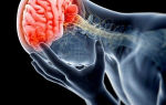
There are also actinomycosis of the pelvic organs and genitourinary areas, pararectal (paraproctitis), sacrococcygeal (with bone damage), and gluteal. Cases of generalization of infection have been described - with the development of actinomycotic brain abscesses and meningoencephalitis; in rare cases - lesions of the cornea, etc.
More on the topic of fungal diseases:
- Toenail fungus.
- Aspergillosis.
The clinical diagnosis of actinomycosis should be confirmed:
1) bacterioscopic studies; In this case, the detection of drusen of radiata fungus is decisive for diagnosis.
The materials for the study are: punctates, sputum, biopsy samples and especially the discharge of dense infiltrates, fistulous tracts and pus.
For research, white or yellowish dense grains (“grains”) are extracted from the material and crushed - for maceration, a 15-20% solution of sodium or potassium hydroxide is added, the glass is slightly heated, and a cover glass is applied.
Microscopy of unstained preparations is carried out under high magnification of a dry system.
At the same time, characteristic drusen are visible - densely intertwined thin threads of mycelium in the center of the conglomerate; along the periphery - flask-shaped formations are located radially, sharply refracting light (representing the final “swellings” of the mycelium).
When stained according to Gram, the mycelium of the fungus is purple (stained with gentian violet), and the flask-shaped “swellings” are red (discolored and perceive magenta staining).
However, even with a typical clinical picture, drusen are not always detected, but thin branching threads of mycelium (possessing acid resistance) are detected - the so-called. atypical Berestnev actinomycosis (differs from the typical one in the absence of drusen in the pus - without any clinical differences). 2) Microscopic studies are supplemented by cultural ones ("grains" containing elements of the fungus are inoculated).
3) Research is also recommended: purulent discharge using a direct test of fluorescent antibodies; ultrasound scanning; CT scan; radioisotope research (can help in detecting “silent” abdominal abscesses).
4) In the diagnosis of actinomycosis, great importance is attached to histopathological studies; in this case, it is possible to detect drusen of radiata fungus in the affected tissue.
Actinomycosis is differentiated - with tuberculous ulcers (scrofuloderma, lupus), syphilitic gummas, chronic deep pyoderma, tumors, deep mycoses, osteomyelitis of other etiologies and other suppurative processes.
In this case, one should take into account the most characteristic clinical signs of actinomycosis (a very high density of nodes and infiltrates, their tendency to open and form fistulas), and most importantly, the detection of drusen of radiata fungus (the detection of drusen was considered a mandatory criterion for diagnosing actinomycosis).
Treatment of actinomycosis
Treatment of actinomycosis includes: specific immunotherapy, antibiotics, sulfonamides, restoratives and stimulants, vitamins, surgical and physical methods. Complex treatment of actinomycosis can be carried out sequentially:
Stage 1 - combined use of actinolysate and antibiotics. The main specific immunodrug for actinomycosis is actinolysate; is entered according to 2 schemes:
- IM 3 ml 2 times a week, for a course of 20-25 injections; after 1-1.5 months the treatment is repeated;
- intravenously, starting from 0.5 ml to 2 ml 2 times a week, in courses of 3 months with an interval of 1-1.5 months.
After clinical recovery, 2-3 courses of anti-relapse therapy are carried out. It is noted that the intradermal method of administering actinolysate is more effective and economical than the intramuscular one. Actinolysate is considered one of the most effective means of treating actinomycosis (various clinical forms and localizations).
The use of antibiotics occupies one of the leading places in the treatment of actinomycosis; Tetracyclines are prescribed (Unidox-solutab, doxibene, vibromycin, oxytetracycline, etc.
); penicillins (long-term and in high doses: penicillin G 10-20 million units/day intravenously, for 4-6 weeks; then switch to phenoxymethylpenicillin orally, 2-4 g/day, 6-12 months); you can use ampicillin intravenously at 50 mg/kg/day (4-6 weeks) - followed by replacing it with oral forms - amoxicillin 0.5 g/day orally, 6 months.
It is possible to use other antibiotics (erythromycin, streptomycin, clindamycin, ristocetin, etc.). 3rd generation cephalosporins (ceftriaxone) are recommended. Sometimes treatment is combined with isoniazid, course dose is 70-120 g.
The 2nd stage includes the administration of sulfonamides (course, dose 60-100 mg), incl. combined agents are used (bactrim, groseptol, berlocid, etc.). Sulfadimezin is prescribed at a dose of 4-6 g/day (course 1-5 weeks). During this period, physical methods of treatment are used (phonophoresis, potassium iodide electrophoresis, UHF), autohemotherapy.
Stage 3 - use of iodine preparations - potassium iodide orally in the form of a 25% solution (in milk or meat broth); inhalation - for actinomycosis of the lungs. At all stages, general strengthening and stimulating therapy is carried out (vitamins C, gr.
B, biostimulants; according to indications - immunocorrectors, gamma globulin, interferon inducers). Food should be rich in proteins and vitamins. In severe cases, detoxification therapy is carried out; blood transfusions of 200 ml are used once a week.
According to indications, surgical intervention is performed (opening and draining abscesses, excision of fibrous tissue).
The course of the disease is usually 1-3 years; without treatment, the process progresses, causing destructive changes.
[blockquote align=»center»]After clinical recovery, patients with actinomycosis should be monitored for at least 2 years (due to possible relapse of the disease).
[/blockquote] Prevention of actinomycosis consists of sanitation of the oral cavity, combating injuries and timely treatment of microtraumas (ioddicerin, 5% alcoholic iodine solution) - especially in rural areas.
Source: http://med-brand.ru/aktinomikoz-simptomy-puti-peredachi-i-lechenie/