Lymph nodes indicate the general condition of a person. Ultrasound of the lymph nodes of the neck is an accurate, safe research method that can detect dangerous pathology at the very beginning of its development. This article will tell you what an ultrasound scan of the lymph nodes of the neck shows when this examination is indicated.
Characteristic
Lymph nodes belong to the organs of the protective system. These are some kind of filters. Their function is to retain pathogenic substances circulating in the blood.
Such “pests” include bacteria, cancer cells, various viruses, and toxic substances.
Any change in the functioning of the lymph nodes causes their transformation, which is manifested by an increased size, compaction, irregular shape, excessive mobility, and a malfunction in the tissue ratio.
Moreover, the changes do not have an equal relationship with the degree of the disease. This allows you to do an ultrasound of the lymph nodes of the neck earlier. This makes it possible to identify pathology at an early stage.
Ultrasound diagnostics during the procedure records the acoustic resistance of tissues. Due to the fact that different types of cells block ultrasonic propagation to different degrees.
Next, the differences are transferred to the device monitor.
This allows you to record the changed parameters of the lymph nodes:
- shape;
- size;
- length;
- width;
- echogenic character.
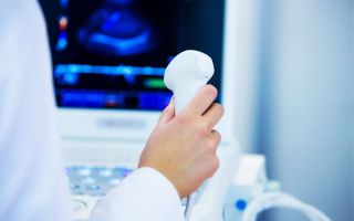
Ultrasound of the cervical lymph nodes does not require special preparation. To carry out the procedure, the patient must lie on the couch. Next, the doctor treats the area to be examined with gel and presses the sensor tightly to the neck area. The ultrasound machine monitor projects the image.
The only condition for diagnosis is the patient's calm state. Because of this, he should avoid stressful situations before the procedure. The session is not performed if the skin of the neck is damaged. You also do not need to wear high-collared clothing for the examination. For women, it is better to put their hair in a ponytail and remove the chains from the neck area.
Each of the presented lymph nodes can be involved in the pathological process
What pathologies does it reveal?
During an ultrasound examination, a number of diseases are detected in the neck area that are associated with the presence of an inflammatory process in the tissues.
Enlarged lymph nodes signal a violation of such organs as: the thyroid gland, palate, salivary glands, hearing organs, sinuses, hearing organs.
Some parasites also cause inflammation of the cervical lymph nodes.
However, diagnosing oncology is of primary importance. Ultrasound allows you to determine the presence of metastases, which indicate a malignant pathology of a nearby organ.
The technique also diagnoses:
- lymphoma of any nature;
- mycoses;
- leukemia;
- lymphadenitis;
- leukemia;
- abscesses;
- lymphosarcoma;
- cancerous tumor;
- inflammatory processes of the eyes and ears.
Diagnostics are often used to monitor prescribed treatment. It allows you to monitor the dynamics of the disease.
Children's Study
As a result of the immature immune system, children may experience frequent inflammation of the lymph nodes. Moreover, their cervical nodes have some peculiarities. It is common for a child to have a lymph node size within 1 cm. If it increases to 1.5 cm, then an ultrasound examination should be performed.
An increase of more than 2 cm requires immediate action to avoid serious complications. When inflammation appears after an infectious disease, there is no reason to worry. Because as a result of complete recovery, this phenomenon disappears.
Ultrasound is safe for children
When is the study indicated?
Indications for ultrasound examination are local and general. Ultrasound is recommended in the following cases: if enlarged lymph nodes persist for 14 days after complete cure of the infectious disease, when the lymph nodes have enlarged on their own, with difficulty breathing, with difficulty swallowing, with painful palpation.
Can ultrasound be wrong?
In addition to the increased size, the doctor may recommend an ultrasound in the following cases:
- joint pain;
- with severe headaches;
- fever;
- redness of the neck area;
- disturbed sleep;
- decreased appetite;
- weakness of the body;
- reduced ability to work.
The listed symptoms require immediate consultation with a specialist and an ultrasound scan.
After the procedure
After the ultrasound, the result is given to the person being examined. Sometimes, to determine the severity, they may recommend undergoing additional diagnostics.
The most accurate information can be obtained when undergoing the procedure on a more modern device. By checking the results obtained with the average parameters, the doctor prescribes treatment if necessary.
The norm for a healthy person is represented by the size of the lymph node in the range of 8-10 mm. However, we should not forget about the individuality of the body.
If the person being studied has suffered many infectious diseases during his life, then his indicators may be slightly increased.
In addition, overestimated parameters of the cervical lymph nodes are found in chronic patients, people suffering from frequent manifestations of herpes. Based on this, minor swelling is within the normal range.
In this case, the patient may be recommended to undergo an ultrasound annually to monitor the behavior of the nodes.
It is important to diagnose at the first symptoms of diseases
If an ultrasound shows an increase in the diameter of the node by 2 cm with clear, even boundaries, then the patient may have non-purulent serous lymphadenitis. When the node has slightly increased in width, its density is reduced, the boundaries are blurred, and the shape is uneven, this indicates purulent lymphadenitis.
Significantly increased density, irregular shape, large size of the node indicate metastatic damage. Ultrasound diagnosis of cervical lymph nodes as a preventative measure should be performed once a year.
These measures will allow you to monitor the functioning of the thyroid gland and identify many pathologies.
Useful article? Share on social networks:
Source: https://medpath.ru/uzi-sheynyh-limfouzlov/
Ultrasound of regional lymph nodes - what is it?
Ultrasound of the lymph nodes in the abdominal cavity is necessary to detect mesadenitis, which is most often observed in children
Ultrasound of the lymph nodes is a procedure aimed at examining the condition of the organ using ultrasound. This simple and painless examination can tell a lot about a person’s overall health.
Ultrasound of the lymph nodes of the neck, as well as the axillary nodes, is often prescribed for young children. This is due to the fact that the nodes of these zones can suddenly increase in size in response to latent infectious processes in the body.
Ultrasound of the lymph nodes in the groin area is often prescribed for women with gynecological diseases, including uterine cancer. In addition, an ultrasound examination of the inguinal lymph nodes can be prescribed if this area is enlarged and inflamed due to furunculosis, syphilis, gonorrhea and other infectious diseases, including those that are sexually transmitted.
Ultrasound of the lymph nodes in the abdominal cavity is necessary to detect mesadenitis. The pathology is accompanied by inflammation of the lymph nodes in this area and is most often observed in children. Since specific symptoms of the disease are not enough to establish mesadenitis, the doctor may refer the patient for an ultrasound examination of the retroperitoneal lymph nodes.
An examination is prescribed if there is a sudden enlargement of the lymph nodes or their inflammation. This procedure is necessary if there is discomfort in the affected lymph nodes, and there are no symptoms of the disease that could provoke the disorder.
In general, for lymphadenopathy (enlarged lymph nodes), this examination method is the main and quite informative.
An ultrasound examination of the cervical lymph nodes is prescribed when a tubercle or lump appears on the side of the neck - this is how an enlarged lymph node manifests itself.
As a rule, this occurs against the background of infections and inflammations of the ENT organs, so additional examinations may be prescribed to a child or adult simultaneously with an ultrasound of the lymph nodes in the neck.
The main indication for ultrasound examination of axillary lymph nodes in women is mastopathy or pain in the mammary glands. This is due to the fact that lymphoid organs often react to neoplasms in the breast, and we are talking not only about cancer, but also about benign processes.
An ultrasound of all peripheral lymph nodes can be prescribed to a patient with complaints of general malaise and swelling. In this case, an ultrasound examination of the lymphatic system allows us to identify a number of pathologies that could potentially lead to impaired lymph outflow.
Alarming symptoms indicating lymphadenopathy, which should alert a person and force him to consult a specialist as soon as possible:
- enlarged lymph nodes;
- redness of the skin around the lymph nodes;
- general malaise;
- pain syndrome;
- night sweats.
Specific symptoms depend on the location of the enlarged lymph nodes. Thus, ultrasound of the lymph nodes under the arm is a mandatory test for suspected breast cancer. Moreover, this study can be prescribed even with an unconfirmed diagnosis of cancer, to study the dynamics of changes in the tissues of the lymphatic system.
Ultrasound of the inguinal lymph nodes is prescribed for enlargement or inflammation of the nodes located in the inguinal folds.
Examination of regional lymph nodes is an important diagnostic procedure for identifying cancer pathologies.
Ultrasound of the lymph nodes of the neck, chest, abdomen, armpits and groin area allows us to detect both the onset of malignant processes and to determine progressing cancer. In addition, with a long asymptomatic course of cancer, metastases can spread to the lymph nodes, which is also visible from the results of ultrasound.
Many people do not know what exactly an ultrasound of the lymph nodes shows. This examination allows you to assess your general health according to several criteria:
- size of lymph nodes;
- clarity of their contours;
- structure of lymph nodes.
For various pathologies in the lymph nodes, ultrasound shows deviations from the norm, on the basis of which an accurate diagnosis is made.
Contraindications
Ultrasound of inflamed lymph nodes during pregnancy should be done only after consultation with the attending physician - gynecologist
Ultrasound examination of internal organs is one of the safest diagnostic procedures, therefore it has practically no contraindications. The procedure should be abandoned if there are wounds or rashes in the area of the lymph node being examined. Moreover, in this case, the ultrasound is simply postponed until the damage to the epidermis heals.
Ultrasound diagnosis of lymph nodes in the armpit is not carried out in the presence of cuts, wounds, inflammations, or ulcers on the skin.
Lymph nodes in the groin area are often enlarged due to sexually transmitted diseases. In this case, the patient must first consult a venereologist.
Pregnant women should undergo ultrasound of lymph nodes only as prescribed by a gynecologist.
Ultrasound does not require special preparation, except when examining the lymph nodes of the abdomen, mesentery and abdominal region. Typically, this procedure is prescribed for suspected mesadenitis.
In this case, the patient is recommended to refuse fatty and heavy foods the day before the examination, and immediately before the procedure, do a cleansing enema.
These measures are necessary to ensure that intestinal contents do not distort the results of ultrasound diagnostics.
The examination is carried out in three stages and lasts about 10 minutes. The patient lies down on the couch if the lymph nodes of the kidneys, chest, abdominal area, groin area, and lower extremities are examined. Examination of the nodes of the neck, submandibular region, armpits and arms is carried out in a sitting position.
The doctor applies a special gel to the area under study, which ensures the passage of ultrasound pulses to the tissues of the organ. Then, using a special sensor, the specialist scans the area of interest, and the image is displayed on the monitor.
Sometimes examination of several regional groups of lymph nodes is prescribed. In this case, the patient must lie on the couch during the procedure, as the examination takes about half an hour.
After an ultrasound examination of the lymph nodes, the diagnostician draws up a protocol, which includes all the nuances of the procedure. Based on this protocol, the attending physician will subsequently establish an accurate diagnosis.
Ultrasound examinations for children are carried out in the same way as for adults without any restrictions.
Ultrasound has no age-related contraindications or restrictions, so it can be performed on all patients without exception.
Preparing children for a kidney ultrasound if they suspect enlarged lymph nodes includes avoiding fatty, fried and heavy foods three days before the procedure. Kidney ultrasound in children is interpreted by a doctor.
If the procedure was carried out with a full intestine, there is a possibility of obtaining unreliable results.
An ultrasound of the lymph nodes in the baby’s neck is prescribed when they are enlarged. Often this symptom indicates hidden infectious processes, so the doctor will definitely prescribe additional examinations. In general, ultrasound for children is performed in the same way as for adults.
Lymphadenopathy on ultrasound
Lymphadenopathy is an increase in size, as well as a change in the shape of one or a group of lymph nodes. This is a symptom of a wide variety of viral and bacterial infections, but can also be a sign of a malignant process.
When fighting the infection, the lymph nodes become inflamed. Inflamed lymph nodes rapidly “grow” at the beginning of the disease and quickly “deflate” during recovery.
On ultrasound, the lymph node is enlarged due to the cortical and pericortical zone, hypoechoic in the periphery and hyperechoic in the center, oval in shape, clear contour, blood flow only in the hilum or absent.
If the inflammation extends into the surrounding tissue (periadenitis), an abscess may form.
Drawing. In children with ARVI, ultrasound shows enlarged cervical lymph nodes with preserved architecture - oval in shape, the contour is clear and even, hypoechoic along the periphery with a hyperechoic center. Conclusion: Lymphadenopathy of the cervical lymph nodes.
Drawing. A 6-month-old boy with severe dermatitis. On ultrasound, the cervical (1) and submandibular (2) lymph nodes are enlarged, elongated, hypoechoic along the periphery with a hyperechoic linear structure in the center. Note the submandibular lymph node with a wavy outline. Conclusion: Lymphadenopathy of the cervical and submandibular lymph nodes.
Drawing. On ultrasound, the lymph nodes are enlarged, oval in shape, with a clear and even contour, reduced echogenicity, the cortical zone is somewhat expanded, the central scar is clearly visible; the blood flow in the hilum is increased, the vessels are located correctly - they diverge radially, the subcapsular blood flow is not determined. Conclusion: Lymphadenopathy with signs of a high degree of activity.
Lymphadenopathy with signs of a high degree of activity. With cat scratch disease, small pustules form at the site of the bite or scratch and at the same time nearby lymph nodes become inflamed. One or a group of lymph nodes increase in size to 5-10 cm, become painful, and become denser. Self-healing occurs after 2-4 weeks. Sometimes abscesses and fistulas form.
Tuberculosis most often affects the lymph nodes of the neck, axillary and groin areas. Typically, tuberculous lymphadenitis develops slowly, the lymph nodes are painless, the average size is 3 cm, but sometimes can reach 10 cm. On ultrasound, the affected lymph nodes are enlarged, hypoechoic, with an unclear contour; pronounced periadenitis and fused lymph node packages can often be seen.
Drawing. An ultrasound scan of the neck reveals a group of enlarged lymph nodes of irregular shape; echogenicity is reduced, there is no central scar; heterogeneous due to anechoic avascular zones - foci of necrosis; blood flow is increased, the course of blood vessels is irregular, subcapsular blood flow is pronounced. Conclusion based on the biopsy results: Tuberculosis of the lymph nodes.
Drawing. Ultrasound shows enlarged lymph nodes, irregular in shape with blurred boundaries; echogenicity is reduced, there is no central scar; heterogeneous due to small cystic cavities and hyperechoic inclusions with an acoustic shadow behind (calcifications). Conclusion based on the results of the biopsy: Damage to the lymph nodes by atypical mycobacteria.
"God is in the details"
Decoding
The health of the lymphatic system can be assessed by studying the diameter, structure and echogenicity of the lymph nodes. Normally, they range in size from 3 to 10 mm, depending on location. The thoracic lymph nodes are smaller (3-5 mm); axillary, cervical and inguinal can reach 10 mm in diameter. An enlarged lymph node compared to normal indicates lymphadenopathy.
The mere fact of lymphadenopathy does not indicate a specific disease. In this case, the doctor will prescribe a number of additional examinations to identify the cause of the disorder.
Violation of the structure of the lymph node and unclear contours may indicate lymphadenitis. This is an inflammatory disease of an infectious nature.
If the main part of the ultrasound indicators of the lymph nodes corresponds to the norm, but areas of decreased or increased echogenicity in the nodes are observed, it is necessary to undergo a detailed examination by an oncologist, since such a picture may indicate malignant processes.
The norm for cervical lymph nodes is considered to be up to 10 mm in diameter, but if they are larger on ultrasound, we are talking about lymphadenopathy.
Axillary nodes do not exceed 5 mm in length, but their diameter on ultrasound may be larger, and here it is important to be able to distinguish the normal size of the node from pathology. Since the size of the lymph nodes is a physiological feature of each person, only a doctor can make an accurate diagnosis based on ultrasound results.
10 signs of a malignant lymph node on ultrasound
- Large sizes, more than 10 mm;
- Round shape, long/short size ratio (L/S) {amp}lt;2;
- Echogenicity is diffusely or locally reduced down to anechoic;
- Concentric or eccentric expansion of the cortex;
- The hyperechoic central scar is thinned or absent;
- Heterogeneous echostructure due to hyperechoic calcifications and/or anechoic zones of necrosis;
- An uneven and blurred outline when tumor cells grow into the capsule;
- Often form large conglomerates;
- Defective blood flow - the vessels are displaced, chaotically organized, the diameter does not decrease towards the capsule, pronounced subcapsular blood flow, avascular zones, etc.;
- High resistance index (RI {amp}gt;0.8) and pulsation (PI {amp}gt;1.5).
"God is in the details"
Drawing. On ultrasound, lymphadenopathy with echo signs of a malignant process: enlarged (28x16 mm) lymph node, round in shape (D/C {amp}lt;2), hypoechoic without a central scar; subcapsular blood flow is determined, the diameter of the vessels does not decrease towards the capsule, RI 0.88.
Drawing. A 63-year-old man discovered a “tumor” on his neck: it is growing slowly without pain or fever. Ultrasound in the area of the sternocleidomastoid muscle reveals a group of sharply hypoechoic lymph nodes without a central scar, size 10-20 mm; some lymph nodes are round; There are lymph nodes with sharply increased blood flow. Conclusion based on the biopsy results: Lymphoma.
Drawing. A 32-year-old woman with a “tumor” on her neck. Ultrasound in the left supraclavicular region reveals one large and several small hypoechoic lymph nodes, round in shape, with a thin central hyperechoic scar; the blood flow is noticeably increased, the vessels are located chaotically, the diameter does not decrease towards the capsule, subcapsular blood flow is pronounced.
Drawing. A 50-year-old man complains of hoarseness and a “tumor” on the left side of his neck. Ultrasound reveals a round formation on the neck with a large anechoic cavity in the center - a zone of necrosis.
CT scan shows a large tumor in the supraglottic region on the left. Conclusion based on the biopsy results: Enlarged lymph node with metastases of squamous cell carcinoma.
Central necrosis of the lymph nodes is typical for squamous cell carcinoma.
Drawing. Ultrasound of a lymph node with metastases of papillary thyroid cancer: heterogeneous echostructure - small anechoic cavities and microcalcifications; the central scar is not defined; subcapsular blood flow is visible.
Drawing. Ultrasound shows a group of enlarged round lymph nodes in the neck: hypoechoic, heterogeneous due to small and large anechoic, avascular zones - foci of necrosis. Conclusion based on the biopsy results: Lymph nodes with metastases of adenocarcinoma. The primary tumor could not be found.
Drawing. On ultrasound, metastases of lung adenocarcinoma destroyed the normal architecture of the lymph node: heterogeneous due to the alternation of hyper- and hypoechoic areas, the central scar is absent, the shape of the lymph node is uncertain, the contour is unclear, which indicates infiltrative growth into the surrounding tissues.
Drawing. Ultrasound shows lymphoma (1,2) between the angle of the lower jaw and the submandibular salivary gland, as well as a lymph node (3) with metastases.
Lymphogranulomatosis or Hodgkin's lymphoma is a malignant hyperplasia of lymphoid tissue. The tumor develops from a single focus, most often in the cervical, supraclavicular, and mediastinal lymph nodes. Ultrasound shows a pack of enlarged lymph nodes, clearly demarcated, not growing into the capsule and not merging with each other.
Drawing. Massive lymph nodes in the neck were biopsied and turned out to be Hodgkin's lymphoma.
Take care of yourself, Your Diagnosticer!
Source: https://ladyless.ru/uzi-regionalnykh-limfouzlov-chto-eto/
Ultrasound of lymph nodes
Ultrasound of lymph nodes is a popular and accurate way to examine patients. It is practiced all over the world. The method is harmless and informative. Ultrasound diagnostics are carried out on: infants, children, adolescents, middle-aged people, elderly, weakened patients, and even expectant mothers who are pregnant and carrying babies.
Lymph nodes are examined by ultrasound
In the peripheral system responsible for immunity, there are lymph nodes. They purify the blood from various viruses and bacteria, and remove toxins from atypical cells. Thanks to them, immune antibodies are produced.
If you are not sick and your body is functioning normally, they do not manifest themselves in any way. When this or that pathology develops in the body, they become denser, change shape, and become larger. This is what the ultrasound machine will show on the screen.
Ultrasound waves, reflected from the tissues of the lymph nodes, show their structure on the screen. A specialist in size, structure, echogenicity will understand whether the lymphatic system functions normally or if there are pathologies?
If the pathology is obvious on the screenshots. The doctor will prescribe the necessary treatment course. You will pass it and significantly improve your general condition of the body, since the lymph nodes cleanse the blood.
Who is this procedure prescribed for?
Ultrasound of the lymph nodes is prescribed when it concerns pathologies occurring directly in them or if another organ is sick, but you need to know the condition of the lymphatic system, and therefore how clean the blood is. Her examination is carried out both to clarify the diagnosis and control the severity of the disease. You will be prescribed a test if your lymph nodes:
- Pathologically enlarged after some infectious disease, for example, an acute respiratory infection or flu, sore throat, etc. Most often they become enlarged in the groin and neck. They become inflamed during illness and become bad if they do not return to their previous size. when you will be discharged;
- They have increased on their own, but the doctors do not note any chronic disease in you;
- When the lymph nodes, for example, in the throat are noticeably enlarged and it hurts you to swallow. If you measure your temperature, it will be elevated;
- If in the groin, on the neck or under the armpits, you can feel them yourself or a doctor will do it, feel them with your fingers. This indicates an inflammatory process.
The doctor will prescribe an ultrasound if, for example, one of the lymph nodes in the neck has become larger and has a different consistency, which can be felt even with your fingers. The doctor may suspect the development of lymphoma or tuberculosis, lymphosarcoma or syphilis, or unpleasant leprosy. "Advice. Be prepared that in addition to tests of the lymph nodes located in the neck, you will undergo a biopsy. They will take some tissue from the lymph node and conduct a study.”
In addition, the doctor will examine the lymph nodes located under the jaw.
How is the ultrasound procedure performed?
It doesn’t matter whether they do an ultrasound of the lymph nodes in the groin area or the cervical area, under the jaw; no special preparation is required from the patient. It is enough to take a shower and put on fresh clothes shortly before visiting the doctor.
If they say that they will do an ultrasound of peripheral lymph nodes, these are those located in the abdomen and groin. Most often, the cervical ones are examined, where one system includes nodes located in the armpits and jaw.
If the lymph nodes are normal, then when you feel the places where they are located, they will not protrude noticeably. And if they are palpable, it means that inflammation is occurring in them.
If you need to examine lymph nodes located in the abdomen or groin, the doctor will ask you to take a deep breath. If they are enlarged, then you need to look for inflammation in the gastrointestinal tract, liver or uterus with ovaries.
"Advice. If you need to examine the lymph nodes located in the armpit area, sit or stand quietly. The doctor will ask you to raise your hand and will palpate.”
What are the errors in the examination and are there other methods?
Ultrasound of the lymph nodes is not enough to establish the correct diagnosis. A doctor, feeling them on the neck or under the armpits, may not understand whether there is simple inflammation there or a cyst formed and slowly growing? The echogenicity will be very similar and even an experienced specialist will not understand what is happening in your body? Therefore, the doctor will refer you for a biopsy or duplex echosonography.
If an ultrasound is performed in the groin area, then the doctor may not understand whether a hematoma or hernia has formed there? Therefore, the doctor will additionally prescribe a biopsy and an MRI examination.
The good thing about ultrasound is that the method is very simple and accessible to everyone. The examination is safe and very information-rich.
"Advice. Do not neglect an ultrasound examination; thanks to it, lymph node disease can be detected at an early stage. If the doctor has made some preliminary diagnosis, then it can be confirmed or refuted.”
During an ultrasound scan of the axillary lymph nodes or other lymph nodes, the patient feels comfortable and does not experience discomfort. The main thing is to wear clean and loose clothes. so that the desired area of the body can be exposed.
the doctor will apply a special gel to it, move a sensor over it and see the examination results on the screen. Will take screenshots. It doesn't matter how old the patient is. This procedure is completely painless and safe.
This is how the lymphatic system is examined even in infants.
Source: https://uzibook.ru/golova/uzi_limfouzlov.html
Everything you need to know about ultrasound of neck lymph nodes
The lymph node system in the human body plays the most important role of a protective barrier against the spread of attacking harmful microorganisms, rods, helminths, viruses, cancer cells, and toxic substances in the body.
Lymph nodes are among the organs of the immune system that are the first to take the hit and change due to infection.
Therefore, the occurrence of the slightest painful symptoms in places where immune filters are concentrated requires their detailed examination to draw up a complete clinical picture of the disease.
The cost of ultrasound of lymph nodes in Moscow at the Men's and Women's Health Clinic is affordable and designed for patients of any income. Qualified doctors will conduct diagnostics, and doctors will assess your health based on the results obtained.
Ultrasound as a method for diagnosing cervical lymph nodes
Ultrasound examination of the lymph nodes of the neck is carried out with a special device using high-frequency waves. The method is based on the ability of ultrasound to penetrate soft organs without consequences, be reflected from them and return. The scanner picks up the returning waves and converts the information onto the screen in the form of a “picture” of the internal organs.
Ultrasound of the soft tissues of the neck and lymph nodes will reveal pathologies, among which are often found:
- inflammatory changes;
- metastases;
- cysts;
- tumors, etc.
An ultrasound of the lymph nodes is completely safe and painless.
The method does not cause discomfort or pain to the patient during the study, does not cause harmful effects to the body, and therefore is prescribed to patients regardless of condition, age and suspected disease. An exception may be damaged skin in the diagnosed area or the postoperative period.
When is it necessary to perform an ultrasound examination of lymph nodes?
A qualified doctor can make an accurate diagnosis only based on the results of diagnosing the patient’s condition. Having the conclusions of a set of diagnostic procedures, the doctor will correctly determine the source and degree of development of the pathology and prescribe a course of effective treatment.
One of the informative instrumental methods for studying the health of the body is ultrasound of the lymph nodes. Using ultrasound equipment, you can determine any deviations from the norm in the condition of the lymph nodes:
- changes in shape and consistency;
- increase in contours and sizes;
- change of localization;
- compaction of the structure;
- the relationship with surrounding soft tissues changes;
- redness of the surface, etc.
These characteristics make it possible to identify and determine the nature of lymphadenopathy (deviations from the normal state).
Since the course of pathological processes leads to changes in the lymph nodes, the occurrence of any of these symptoms is a mandatory reason to sign up for an ultrasound scan of the lymph nodes.
An accurate study using modern premium equipment is carried out by specialists from the Clinic for Men’s and Women’s Health. Here you can do an ultrasound of the lymph nodes without queues or fuss, the price for services is affordable, and the results are reliable. Pre-registration is made by telephone based on a doctor’s referral.
Indications for the ultrasound procedure
The patient’s complaints of malaise are a reason to pay attention to the condition of the lymphatic vessels. Indications for prescribing an ultrasound of the lymph nodes of the neck, which shows the presence of minimal deviations from the norm, may include a number of accompanying symptoms:
- the appearance of a lump, bump or redness at the location of the cervical lymph nodes;
- pain in the area of clusters of lymphatic vessels;
- night sweats;
- general weakness;
- temperature, etc.
The presence of one or a complex of these disorders requires immediate contact with a specialist. At the Men's and Women's Health Clinic, distinguished doctors of the highest category will provide qualified medical care with care and understanding.
What diseases can cause changes in the lymph nodes of the cervical region?
The cervical region is a place of accumulation of lymph nodes, which doctors especially often pay attention to. If they are soft, the body fights dangerous pathogens; if they become dense, this is a sign of inflammation, which is called lymphadenitis.
After palpation, the doctor prescribes an ultrasound of the cervical lymph nodes and a number of additional tests that will help clarify the cause of the ailment. Since the nodes in the neck are located very close to the mouth, nose, nasopharynx, throat and trachea, diseases of the lymphatic system can occur with the following diseases:
- caries;
- sinusitis;
- tonsillitis;
- pharyngitis;
- tracheitis;
- sinusitis, etc.
An ultrasound of the lymph nodes should be performed if there is slight compaction that persists for a long period. This may be a signal of a hidden inflammatory process. Therefore, ultrasound of the lymph nodes of the neck can identify a number of diseases that occur unnoticed in the body:
- HIV/AIDS;
- diabetes;
- monoculosis;
- tuberculosis;
- Sezary's disease;
- syphilis;
- leprosy;
- oncological diseases;
- diseases affecting the lymphatic system.
If external changes or pain symptoms occur, it is recommended to consult a doctor without taking self-medication measures. If necessary, the doctor will order an ultrasound of the lymph nodes of the neck and additional tests.
Ultrasound of lymph nodes in Moscow can be performed at the Clinic for Men's and Women's Health, where certified physicians, using innovative scanners, will make a diagnosis and coordinate further treatment tactics with clients.
How to prepare for an ultrasound procedure
Depending on the symptoms, the doctor gives a referral for ultrasound diagnostics of one of the groups of nodes. The choice falls on the lymphatic vessels that are as close as possible to the location of the disease. In case of severe condition, it is possible to diagnose several groups of nodes.
For effective diagnosis, the doctor may ask you to prepare for the examination in advance. Ultrasound of the lymph nodes in the cervical region is performed without special preparation, with the exception of cases when the patient needs to remove hair growing on the neck, special bandages or bandages.
You must arrive for the examination on time in agreement with the diagnostician. Before the procedure, you need to inform your doctor about taking medications, since a number of medications provoke changes in the lymph nodes.
Age threshold for ultrasound
There are no age restrictions in using the ultrasound method. Examinations can be carried out at any age. The meaning of the results is equally important for the doctor, who will interpret the results according to the age group and the characteristics of the person’s constitution.
Since the procedure is completely painless, the child can be diagnosed without prior psychological preparation.
Survey methodology
The diagnostic room has a specialist workplace, and a couch for the patient is required.
Examinations of the cervical spine are performed while sitting. The area being diagnosed is exposed. The doctor applies a gel to the area, which promotes soft gliding and effective passage of ultrasound pulses towards the tissues and back. The information received is displayed on the monitor and recorded in the protocol.
An ultrasound of the lymph nodes, which shows the slightest deviation from the norm, can diagnose the disease at an early stage and prevent death in the future.
The procedure lasts from 10 minutes to half an hour, depending on the complexity of the placement of the nodes and the thoroughness of the diagnosis.
The diagnostician enters the results obtained on a form and gives them to the patient. A specialized doctor deciphers the data and makes a diagnosis.
Are additional tests needed?
The accuracy of the diagnosis depends on the completeness of the information received about the patient’s condition. In addition to ultrasound, the doctor must prescribe additional tests that will complement the clinical picture of the disease. Current research methods:
- general blood analysis;
- blood chemistry;
- tests for tuberculosis;
- blood test for HIV/AIDS;
- X-ray;
- MRI;
- CT scan.
The functionality of the tomograph in our clinic allows us to most accurately perform ultrasound, which eliminates the need for many additional tests.
Inaccuracies and preliminary diagnoses based on ultrasound diagnostics
Correct diagnosis and interpretation of ultrasound results determines the further outcome of treatment. Conducting ultrasound examinations by inexperienced specialists or under conditions of turmoil and nervous tension in public health facilities shows low effectiveness. In stressful environments, human error may occur more often than usual.
For an error-free diagnosis, it is recommended to contact a private diagnostic and treatment institution, the Women’s and Men’s Health Clinic, which offers ultrasound of the neck lymph nodes at a low price. The center does not create queues outside the office door, accepts patients by appointment, and pays due attention to patients.
How much does an ultrasound scan of neck lymph nodes cost in Moscow?
The price of an ultrasound scan of lymph nodes depends on the area being examined. The men's and women's health clinic has a loyal pricing policy. The cost of medical care is acceptable for residents of Moscow and the regions. Specific figures for all types of services can be found on the website in the “Cost of Services” section.
Price of ultrasound of lymph nodes in the clinic The difference in cost is insignificant; it differs from the cost of the procedure in city multidisciplinary clinics. I can compare with the comfort, efficiency of examination and treatment provided in our center.
Source: https://neurourologist.ru/vse-chto-nuzhno-znat-ob-uzi-limfouzlov-shei/
What is the value of ultrasound examination of lymph nodes?
Why is ultrasound of lymph nodes performed?
Lymph nodes are organs of the immune system that play the role of a kind of protective filter that retains harmful substances circulating in the blood - viruses, bacteria, toxins, tumor cells, etc.
Almost any pathological process leads to transformations in the structure of the lymph nodes in the form of its enlargement, compaction, acquisition of an unnatural shape, mobility, and the ratio of tissue components.
These changes are not always adequate to the degree of clinical manifestations of pathologies (lymph nodes can react before the appearance of serious symptoms), which makes it possible to carry out early diagnosis of a number of diseases, in particular, using ultrasound. The method is based on recording differences in the acoustic resistance of tissues - the ability of different tissues to prevent the propagation of ultrasound in the body to varying degrees.
These differences are projected on the monitor screen in the form of a specific image, allowing the diagnostician to detect changes in a number of parameters of the lymph nodes:
- size
- length/width ratio
- nature of echogenicity
- forms.
IMPORTANT! Ultrasound has limited capabilities, and its data without additional studies cannot be the basis for making a diagnosis.
How is a lymph node examination done?
Ultrasound of lymph nodes does not require special preparation and is extremely simple.
The sensor of the device is lubricated with a gel-like substance and pressed tightly against the area of the body being examined that is not covered by clothing. An image is projected on the monitor of the ultrasound unit, formed by sound waves sent deep into the body and reflected back.
An exception is the ultrasound procedure of the lymph nodes in the groin. Since changes in the lymph nodes of this area are predominantly of a venereal nature, it is necessary to undergo an examination by a venereologist before carrying out the procedure.
In what cases is ultrasound of the lymph nodes of the neck prescribed?
The study of various groups of lymph nodes can be carried out both for local and general indications as a clarifying, differential or control study.
General nuances of ultrasound examination of soft tissues
https://www.youtube.com/watch?v=84BMTO3JNEY
Ultrasound of the lymph nodes in the neck is most often performed when there is suspicion of:
- benign and malignant lymphomas
- cancer
- lymphosarcoma
- Sézary's disease
- with metastases from other organs - lungs, larynx, throat, tongue, trachea
- actinomycosis
- tuberculosis
- syphilis
- leprosy
In the chronic asymptomatic course of these diseases, ultrasound of the cervical lymph nodes is of a differential diagnostic nature before obtaining biopsy data. The presence of symptoms (primary syphilide, woody density, isolated Koch bacillus or lepromatous cells) is the basis for refusing an ultrasound.
When is an ultrasound of the submandibular lymph nodes prescribed?
Submandibular lymph nodes are examined in an ultrasound diagnostic room when:
- deformations of the bone structures of the dental system and soft tissues without pain
- bleeding gums, their overgrowth and increased night sweats, i.e. with signs of leukemia.
Ultrasound of axillary lymph nodes
Axillary lymph nodes are examined in cases where:
- the doctor suspected the presence of benign or malignant neoplasms of the chest organs and/or mammary glands
- in women after mastectomy (complete or partial removal of the mammary gland), it is carried out routinely to monitor the presence/absence of metastases
- with enlargement of the cervical and inguinal lymph nodes against the background of relative health (suspicion of HIV infection or oncology).
Ultrasound of the axillary lymph nodes does not make sense to perform earlier than 2 months after suffering toxoplasmosis and mastitis.
Ultrasound examination of the groin area
The lymph nodes of the groin area are the first to take the hit in infectious and inflammatory diseases of the pelvic organs. Changes in them are often accompanied by a pronounced clinical picture, and diagnosis is not difficult.
On what day of the cycle is it necessary to do an ultrasound of the mammary glands?
Oncological processes and a number of sexually transmitted diseases (HIV-AIDS, syphilis, etc.) often manifest themselves only as lymphadenitis. Therefore, an ultrasound scan of the inguinal lymph nodes is appropriate and justified, especially with polyadenitis (simultaneous enlargement of several groups of lymph nodes) and previously diagnosed oncologies in remission (to control the spread of metastases).
Watch a short video about lymph nodes.
When performing an ultrasound scan of the lymph nodes of the neck, interpretation errors are possible in the presence of cysts (congenital lateral and median) and abscesses that have the same echogenicity. Differential diagnosis in this case consists of analyzing the clinical picture and performing CDE (color duplex echosonography) and biopsy.
Ultrasound of the lymph nodes of the groin area may provide inaccurate data in the presence of hematomas, hernias and aneurysms. To clarify, CDE, MRI, palpation examination of the inguinal canal, and biopsy are also performed.
The cost of an examination, for example, in Moscow, is from 700 rubles. In regions, prices may vary up or down. In any case, the study is justified because it allows you to make the correct diagnosis.
Ultrasound of lymph nodes is a painless, non-invasive, easily accessible and fairly inexpensive research method that allows you to clarify a possible diagnosis, effectively monitor the dynamics of benign tumor diseases in remission, and also identify the first signs of metastases in malignant processes. The method does not require special preparation and does not cause discomfort to the patient.
ATTENTION! The information on the site is for reference or popular information only. Correct treatment and prescription of medications can only be carried out by a qualified specialist, taking into account the diagnosis and medical history.
Successful diagnosis and treatment, health and well-being! Your uzilab.ru.
04/29/2015 UziLab
Source: https://uzilab.ru/soedinitelnyie-tkani/uzi-limfaticheskih-uzlov.html
Ultrasound of retroperitoneal lymph nodes
Ultrasound of the retroperitoneal lymph nodes is an ultrasound method that allows you to diagnose the pathological process in the lymph nodes of the retroperitoneal space.
The content of the article:
The abdominal cavity of the human body contains the internal organs of the digestive tract. It is separated from other areas by the peritoneum, a thin epithelial membrane. The retroperitoneal space is soft tissue and fiber located behind the posterior wall of the abdominal cavity.
The retroperitoneal space also contains kidneys, ureters, large vessels (abdominal aorta, inferior vena cava), lymphatic vessels and nerves.
It contains groups of lymphatic nodes that filter tissue fluid flowing from the lower part of the body, abdominal organs, and kidneys.
Infectious-inflammatory, hematological and oncological processes in these areas lead to lymphadenopathy - enlargement of the lymph nodes, changes in their normal structure. The location of the retroperitoneal lymph nodes makes the diagnosis of these changes difficult.
Computed tomography and nuclear magnetic tomography make it possible to accurately determine lymphadenopathy of the retroperitoneal lymph nodes. However, these methods are very expensive, and their use for diagnosis and routine treatment control is not economically profitable for the clinic and the patient.
Lymphography - the introduction of an X-ray contrast agent into the lymphatic ducts - is a rather complex procedure that also allows for the diagnosis of pathologies of this group of lymph nodes.
Ultrasound of the retroperitoneal lymph nodes is the optimal method for diagnosing lymphadenopathy and monitoring the effects of treatment. This is an inexpensive, painless and accurate technique that is widely used in medicine.
Indications for ultrasound of retroperitoneal lymph nodes
- pain in the abdomen and lower back, of unknown origin;
- specific infections (tuberculosis, syphilis, AIDS, etc.);
- persistent increase in body temperature for no apparent reason;
- changes in blood tests that allow one to suspect a hemato-oncological disease (increased ESR, appearance of atypical lymphocytes, leukocytes, other blood cells, anemia of unknown origin);
- diagnosis of metastases in oncological diseases of the genitourinary system and abdominal organs;
- control over the dynamics of the disease and treatment results.
Minimal preparation is required before the test. A few days before the examination, it is necessary to refrain from eating large meals, especially those that cause gas formation in the intestines. These are vegetables, carbonated drinks, legumes, dairy, sweets and baked goods, bread, alcohol, pickles and delicacies. Increased gas formation leads to bloating of the intestines, the formation of pockets of gas in it. Ultrasound does not penetrate gases. For this reason, failure to follow the diet may distort the results or make the study impossible.
Patients suffering from flatulence are prescribed carminative drugs - espumizan, Maalox, simethicone, fennel tincture. Sometimes it is recommended to take laxatives, cleansing enemas, and activated charcoal.
The study is carried out while lying on a couch. A sound-conducting gel is applied to the skin of the abdomen to improve the passage of ultrasound. During the procedure, the doctor may ask you to hold your breath while inhaling or exhaling, roll over on your side, and strain your abdominal muscles.
The examination is painless and takes 10-20 minutes.
During the study, the density, structure, size, and contours of the lymph nodes are assessed. Normally, they are small in size, oval in shape, and not fused to each other. The contours are clear and even.
The structure is homogeneous, without areas of decreased or increased echogenicity. The sizes are small, do not exceed 1-3 cm.
Enlarged lymph nodes may be a sign of infection, autoimmune pathology, inflammation, cancer or hematological disease.
Conclusion Ultrasound of the retroperitoneal lymph nodes is not a definitive diagnosis. It can only be determined by a doctor based on all the results of tests and studies!
Source: https://www.mosmedportal.ru/article/uzi-zabryushinnykh-limfouzlov/