Magnetic resonance imaging (MRI) of the spine is a method for studying soft tissues and vertebrae, based on the ability to record changes in their own magnetic fields, which change in various pathologies.
Since all tissues contain hydrogen, the behavior of the atoms of this element is studied.
During MRI, the patient is exposed to the external magnetic field of the machine, as a result of which hydrogen atoms briefly adjust their magnetic moment to it.
After the scanner is turned off, they return to their original state, releasing energy. This process is captured by special devices and recorded as a digital image.
What parts of the spine are examined on MRI?
When examining the spine, MRI makes it possible to study the condition of not only all bone parts (from the cervical to the coccyx), but also other important structures:
- intervertebral discs;
- spinal cord and its membranes;
- vessels and nerves;
- ligaments;
- cartilage tissue;
- intervertebral joints;
- adjacent muscles and fiber.
Magnetic resonance imaging accurately determines the location of herniated protrusions of intervertebral discs, their size, the degree of degenerative changes, and impaired circulation of the cerebrospinal fluid. With its help, adhesions in the epidural tissue are differentiated from inflammatory ones, as well as from tumor and vascular neoplasms.
Spinal sections:
- cervical;
- chest;
- lumbar;
- sacral;
- coccyx.
All departments can be studied in detail during magnetic resonance scanning.
MRI diagnosis of the spine: indications and contraindications
Magnetic resonance imaging is used in the diagnosis of almost all spinal pathologies. Researched:
- injuries;
- developmental anomalies;
- inflammatory diseases (spondylitis, osteomyelitis, discitis);
- degenerative processes (osteochondrosis, spondyloarthrosis, protrusion of intervertebral discs);
- malignant neoplasms and metastases.
Osteochondrosis
This is a degenerative-dystrophic chronic disease that begins with the nucleus pulposus of the intervertebral disc and spreads further to all elements of the vertebral motor segment.
It makes sense to do an MRI for any persistent back pain, since making an accurate diagnosis during an initial examination by a neurologist based only on clinical signs is not an easy task. Difficulties may be due to the fact that;
- there are individual differences in the structure of the spine, the level of nerve exit from the spinal cord and the innervation of the lower extremities;
- There are a number of diseases that cause similar symptoms: pathologies of the hip joints, ischemic neuritis of the lower extremities, vascular diseases, abdominal and pelvic organs.
Therefore, a neurological examination can only suggest the localization of the process. To make a final diagnosis, it is necessary to visualize the process on MRI, which will allow you to immediately choose the right treatment tactics.
Spondylitis
An inflammatory disease in which early diagnosis and adequate treatment tactics make it possible to avoid severe disabling consequences.
Most often it is the result of the introduction of microorganisms with the bloodstream into the adjacent space of the spinal disc (there are no vessels in the disc itself). This disease is quite rare and is often mistaken for degenerative, leading to inappropriate treatment.
X-rays and CT scans do not detect spondylitis at an early stage. Therefore, in case of persistent back pain that is not amenable to conservative treatment, increased inflammatory markers in the blood or fever, it is necessary to do an MRI of the spine.
If the diagnosis of spondylitis is delayed, spinal cord damage and sepsis may develop.
The key signs of spondylitis on MRI are:
- inability to visualize the boundaries of the intervertebral disc and the adjacent vertebral body;
- increased signal intensity from the disc itself and the vertebral body.
MRI with intravenous contrast, in this case, reveals the spread of the inflammatory process and the formation of an epidural abscess. Standard MRI without contrast makes it possible to distinguish spondylitis from degenerative and tumor processes.
Dynamic studies (repeated) are prescribed for patients with a confirmed diagnosis of spondylitis and who have little improvement after conservative treatment. This is due to the fact that changes in inflammatory markers in the blood are not always indicative of assessing the effectiveness of treatment. Repeated MRIs may reveal abscess formation that requires surgical drainage.
Spinal injuries
According to the state of neurovascular formations, they can be:
- uncomplicated (without damage to the spinal cord and spinal nerves);
- complicated (with their damage).
Spinal cord injury occurs:
- acute - directly at the time of injury by foreign bodies or bone fragments;
- early - in the next 10 days, as a result of compression by hematoma, edema, secondary displacement of the vertebrae;
- later - after several weeks, and sometimes even years, due to the scar-adhesive process or the formation of cysts.
MRI makes it possible to establish a diagnosis in 98% of cases, as it visualizes:
- ligaments;
- intervertebral discs;
- membranes of the spinal cord;
- spinal cord and its pathologies (hemorrhages, swelling, ischemia);
- violation of the structure of the vertebral bodies.
MRI is the main method for diagnosing post-traumatic disc herniations.
In a certain group of patients with spinal trauma in the acute period, there are no signs of vertebral damage on the radiograph. But after 3-8 months. sudden sharp pain occurs at the site of the previous injury. Repeated examination reveals a compression fracture of the spine.
This is explained by the fact that in the acute period hemorrhage occurred in the vertebral body. After its resorption, aseptic inflammation and destruction of bone structures developed. The action of axial load on the weakened vertebral body led to its compression fracture.
MRI allows you to visualize these hemorrhages in the acute period and take timely measures, which significantly affects the patient’s future health.
Metastatic lesions of the spine
The appearance of pain in the spine, an increase in existing pain in the spine and lower extremities in patients with cancer should be the reason for urgent MRI of the entire spinal column with intravenous contrast.
However, the scope of primary diagnostic measures depends on the severity of the patient’s problems at the time of examination. Asymptomatic lesions in the spine of a metastatic nature, identified on CT or scintigraphy, should be confirmed by MRI with contrast.
The appearance of threatening symptoms in the form of weakness in the limbs, dysfunction of the pelvic organs or sensitivity is the reason for performing an emergency MRI of the entire spine with contrast, even if the patient has a history of degenerative disease of the spine.
Magnetic resonance imaging is contraindicated in patients:
- having implants or foreign bodies made of ferromagnets in the body;
- wearing non-removable electronic devices (pacemakers, cochlear implants, insulin pumps and others);
- with vascular clips installed on the arteries of the brain;
- pregnant women in the first trimester;
- if it is impossible to remain still due to claustrophobia, pain or other reasons.
Our clinic does not examine children under 5 years of age or adults with a body weight of more than 130 kg or a waist circumference of more than 150 cm.
Preparing for magnetic resonance imaging of the spine
No special preparation is required for this procedure. If you plan to administer contrast, then it is advisable to refrain from eating for 4-5 hours.
Before scanning, you must provide the doctor with the results of all previous studies and documents for implants (if any).
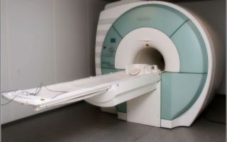
MRI of the spine: how the procedure works
Before the examination, you must remove all metal accessories, watches, and place bank or other magnetic cards. The patient is placed on a special table that will be located inside the scanner. During the procedure, you need to maintain a stationary position for better image quality.
The doctor retires to the next office, but maintains contact with the patient via two-way communication and sees him on the monitor. The scanner maintains a comfortable temperature, good air circulation and lighting. You can use headphones with pleasant music.
After the procedure, you can relax and drink coffee while waiting for a description of the images. This will take 15-20 minutes.
Normal MRI findings of the spine
When assessing MRI of the spine, they take into account the fact that there are individual structural variations that may be similar to certain pathological conditions.
- Cleft arch of the 1st cervical vertebra is considered normal in the absence of neurological disorders and is often discovered by chance.
- Non-fusion of the posterior part of the lumbar vertebrae - in the absence of complaints, can also be considered an individual feature.
- A fatty conglomerate in the bone marrow, which is a normal variant, sometimes looks like a vertebral hemangioma on imaging.
These and other types of spinal structure do not affect the patient’s health in any way and are easily recognized by competent doctors. Our clinic employs specialists with more than 7 years of experience.
For correct diagnosis, it is necessary to take into account the existence of structural features of the spine.
Source: https://mrtdon.ru/mrt/info/mrt-pozvonochnika-chto-pokazyvaet/
How is an MRI examination of the spine performed: how to prepare for the examination, contraindications and cost
MRI of the spine, or magnetic resonance imaging of the spine, is currently considered one of the best and safest methods for diagnostic examination of osteoarticular and muscular diseases of the spinal cord and spine in general.
What it is?
Magnetic resonance imaging of the spine, or MRI of the back, is a diagnostic study of an anatomical object, a layer-by-layer image of which is obtained in a three-dimensional projection.
Tomography of the spine or its parts is carried out using nuclear magnetic resonance - a physical measurement of the electromagnetic response of hydrogen atoms to the resonant excitation of the object under study.
The year of the founding of magnetic resonance imaging is considered to be 1973, when American doctors led by Paul Christian Lauterbourg discovered to the world this unique method of magnetic resonance induced imaging of the structural interaction of anatomical objects of the human body.
The first magnetic resonance imaging scanner, for which an American scientist received the Nobel Prize in 2003, has been stored at New York University in Stony Brook for many years.
However, the sluggishness of the Soviet government bodies in charge of inventions did not allow the Russian scientist Vladislav Aleksandrovich Ivanov to claim primacy in the invention of magnetic resonance imaging, which he sent to the USSR State Committee back in 1960.
Why do it?
The MRI procedure of the spine is performed to identify various diseases of the musculoskeletal system.
The MRI conclusion is deciphered on the basis of the results obtained after saturation of the structural tissues of the osteoarticular elements of the spine with nuclear resonance of the hydrogen proton.
Spinal tomography is not the only diagnostic use of magnetic resonance scanning.
First of all, it should be noted that the MRI procedure of the back determines the following clinical conditions or pathological manifestations in the osteo-articular organization of the spinal column:
- Conduct a study for the presence of congenital pathologies of the cervical, thoracic or lumbosacral region (i.e., lower back).
- Determine the condition of the intervertebral discs.
- Allows you to detect spinal stenosis, when narrowing of the spinal canals occurs.
- Magnetic resonance imaging is used to determine the degree of compression pressure on the nerve roots in the intervertebral segments.
- The monitor shows the effect of harmful components and other substances on bone tissue or nerve fibers, in the event of the appearance of a tumor-like neoplasm, cyst or bone abscess.
- The diagnostic use of resonance tomography allows one to determine circulatory insufficiency in the vessels in a certain area of the osteoarticular structure of the spine.
- Finally, the examination results provide a complete diagnostic picture in the case of an infected lesion of the osteoarticular and muscular components of the musculoskeletal system.
Kinds
There are other types of magnetic tomography of the spine that can detect pathological abnormalities in the human body:
- spinal cord.
- head (pituitary gland).
- any parts of the spine.
- abdominal organs and so on.
Compared to ultrasound (ultrasound), MRI of the back or other structural components of the body is a more sensitive, accurate and informative method of laboratory instrumental diagnostics.
Magnetic resonance imaging of the spine (ridge) will show projection scanning of various angles and planes of individual elements of the musculoskeletal system of the spinal column.
The data obtained by tomography, or MRI images of the entire spine, will help with the reconstruction of the supporting system in the future, when other painful symptoms and signs suddenly appear that require therapeutic or surgical treatment.
Contraindications
Contraindications to MRI of the spine may include various chronic diseases and conditions, neuropsychic abnormalities of the patient, and hormonal imbalance in women during menstruation.
Thus, we determine who should not have an MRI:
- Diagnostic examinations are not carried out on patients with obvious mental disorders.
- People with epilepsy, or with frequent seizures of the upper or lower extremities.
- People with orthopedic support metals in the osteoarticular segments cannot pass.
- A pacemaker is also a contraindication.
Also, when administering a contrast agent, it is necessary to make sure that the patient does not have an allergic dependence on the proposed medicinal drug, and in case of pregnancy, it is necessary to notify the diagnostician about this. If the menstrual cycle is disrupted, then the doctor should also know about this.
How the procedure goes and preparation for it
Before doing an MRI, it should be borne in mind that the diagnostic procedure involves the influence of a magnetic field on the human body. Therefore, precautions and safety precautions must be observed. All preparation for an MRI of the spine consists of removing various jewelry and objects attracted by a magnet, for example, coins, keys, magnetic cards, etc.
You can eat and drink, but it is better to do this 1.5-2 hours before the appointment of the diagnostic procedure. Such restrictions correspond to certain physiological needs of a person, when at the most inopportune moment one wants to leave the diagnostic capsule. The patient should be patient, since the entire process (procedure duration) usually takes from 15–20 to 45–60 minutes.
Examination using contrast
Slightly different conditions exist when preparing for an MRI with contrast. After conducting a biochemical laboratory test (tests), the attending physician, for specific medical indications, before performing a diagnostic procedure, prescribes the injection of a special dye into the venous artery in order to get a clear picture of the abnormal zone.
The person is invited to familiarize himself with the conditions of contrasting and sign the appropriate agreement on the possible risks of prescribing magnetic resonance imaging with a contrast agent.
Using prepared dyes, you can detect an increase or decrease in the diagnosed musculoskeletal joint or other vital organ, as well as determine the degree of its damage, or lack thereof. Coloring drugs passing through the muscle tissue, changing color, determine the clear boundaries of a particular clinical pathology. The rate of staining depends on the intensity of the main blood flow.
- Thanks to this diagnostic action, the accuracy of the tomogram improves significantly.
- To carry out diagnostics using magnetic resonance scanning, gadolinium salt is used, which differs from other substances in its low toxicity, rapid solubility and high efficiency. Currently, the Ministry of Health of the Russian Federation has approved other contrast agents containing the main medicinal component - gadolinium:
- Omniksan;
- let's finish;
- gadovist;
- premovist;
- Magnevist.
Other coloring substances for magnetic resonance imaging that have not passed the expert certification of the Russian Ministry of Health are prohibited.
Absolute contraindications:
- woman's pregnancy up to 2 weeks;
- individual intolerance to the active substance;
- children under 15 years old.
Relative contraindications:
- dehydration of the body;
- allergic reactions to any types of dosage forms;
- renal and liver failure;
- severe cardiac and/or vascular pathologies;
- chronic diseases of the bronchopulmonary system;
- anemia of vital organs;
- myeloma, polycythemia, etc.
Possible risks and consequences of tomography with contrast
To conduct a diagnostic examination, take a minimum medicinal dose of the coloring component in order to reduce the risk of possible complications:
- skin itching;
- allergic reaction;
- coughing, shortness of breath, or sneezing;
- painful discomfort in the eyes.
As you can see, the list of contraindications and negative consequences is negligible, and amounts to approximately 1% of the total number of possible complications, so you should not be afraid of diagnostic procedures, you should and can undergo them with confidence.
How long does it last?
The duration of a magnetic resonance scan is determined by medical indications, depending on the complexity of the clinical pathology and other factors. An MRI can be done in 15-20 minutes, or it can take more than an hour.
How do they do it?
The diagnostic method is based on local and constant electromagnetic fields, as well as high-frequency power current energy. During a diagnostic examination, the patient is in a pressure chamber, or scanner.
The powerful force of the magnetic field aligns hydrogen protons in the tissue structures of the human musculoskeletal system.
Using radio frequencies, signals from subatomic particles of tissue fibers are transmitted to the tomograph receiver.
A diagnostic doctor, on a powerful tomograph monitor with a fairly decent expansion, sees an image of individual particles of the body being examined, and thanks to a huge number of radio frequency signals, begins processing them on diagnostic equipment.
This method of clinical diagnosis of the condition of vertebral structures allows one to determine possible traumatic injuries to osteoarticular elements, identify vascular pathology, bleeding, infection processes of bone and articular segments, and also prevent the possible development of future clinical complications or pathological formations.
In addition, interpretation of the MRI of the spine (tomogram) allows you to confirm or exclude clinical conditions that were detected during diagnosis by computed tomography (CT), ultrasound scanning (US), or x-ray examination of bone or articular segments of the spine.
How often can I do it?
The undeniable advantage of magnetic resonance imaging compared to radiography is the absence of ionizing radiation and a more informative diagnostic picture. There is no substantiated evidence of the negative effects of magnetic fields on the human body yet.
There were also no complicating factors for deterioration of well-being after magnetic resonance exposure. Therefore, it is believed that an MRI procedure, if necessary, can be prescribed several times a year.
A repeat examination is a clarifying diagnostic procedure that allows you to determine the effectiveness of the treatment process or confirm the recovery of the vital organ being examined. However, medical experts are inclined to believe that re-examination for children under 15 years of age should be carried out no more than 2 times a year.
How is a photograph decrypted?
Deciphering MRI data requires a high level of training from a specialist:
- knowledge of the topographic location of the articular and bone segments of the skeletal frame;
- knowledge of pathological anatomy;
- understanding of morphological, histological, biochemical and physiological processes in the human body.
Based on this knowledge, the diagnostician determines the relationship between the shape and relative position of the organs. Literally translated, the word tomography from the ancient Greek language means section. The specialist’s task is to calculate the time during which the hydrogen protons, after being exposed to them, will take their original position.
Each pixel on an MRI monitor is a signal of frequency, density of hydrogen atoms and phase vector, which have their own color shades. Tomographic images are a layer-by-layer image in transverse and sagittal projection. The quality of the diagnostic test depends on the power of the equipment.
High-field magnetic resonance imaging scanners produce a magnetic field voltage of 5–7 Tesla, which allows one to obtain a more accurate picture of the location of the pathological anomaly. To establish an appropriate diagnosis, information for a specialist is:
- size, shape and number of inflammation foci;
- color shade of the pathological deviation;
- other indirect indicators.
What is the name of the doctor who does an MRI?
A radiologist interprets the MRI results of the back.
What is the price?
An MRI can be done free of charge as part of your compulsory health insurance policy.
Since this is one of the most expensive methods of diagnostic examination of the human musculoskeletal system, there is a long queue for free testing with a waiting period of several months. This is due to the fact that limited quotas are allocated for free examinations.
In different regions of the country, the cost of the procedure can vary from 3.5 to 5 thousand rubles or more.
It all depends on the severity of the osteoarticular disease, location (upper or lower part of the spine), type of MRI equipment and cost of the contrast component.
Of no small importance is the popularity rating of a particular medical clinic, as well as the time of the diagnostic procedure. Some diagnostic centers perform MRI of the spine in the evening and at night.
Where can I do it?
- As a rule, most regional and less often district medical institutions are equipped with diagnostic MRI equipment. In large cities of the Russian Federation and CIS countries, there are special city services for making telephone appointments for magnetic resonance and computer diagnostics:
- in St. Petersburg – 8 (812) 313-26-79; in Moscow – 8 (495) 230-78-55, 8 (499) 519-32-95; in Minsk – 8 (017) 270-21-42, 8 (017) 509-72-42; in Almaty – 7 (727) 331-33-31; in Kyiv – 8 (044) 259-78-00, 8 (044) 321-09-33.
- By making an appointment at night or on weekends, you can significantly save your budget.
Source: https://MoiPozvonochnik.ru/otdely-pozvonochnika/pozvonochnik/mrt-pozvonochnika
Magnetic resonance imaging of the spine
Magnetic resonance imaging is widely used in various areas of medicine. Neurology and orthopedics are no exception. MRI of the spine has great diagnostic value in examining the musculoskeletal system.
When performing an MRI, the patient is not exposed to radiation, since the operating principle of the tomograph is based on the effects of nuclear magnetic resonance. When performing an MRI, the entire spine (the area of three sections) can be examined, or individual parts of it can be examined.
Magnetic resonance imaging centers perform the following MRI examinations:
- cervical region;
- thoracic region;
- lumbosacral region;
- sacroiliac joints;
- sacrococcygeal region.
Patients who are prescribed such a diagnostic procedure are very interested in what an MRI of the spine shows, what types of MRI there are, and how the MRI procedure of the spine is performed.
In addition, despite the fact that many have heard about the greater safety of MRI compared to CT, patients are still somewhat wary of such a diagnosis.
And they also want to know if there are any contraindications for conducting such a study.
Indications
Magnetic resonance imaging of the spine is performed if the patient has the following symptoms:
- severe pain in the neck, thoracic spine, chest, lower back, sacrum, pelvis, lower and upper extremities;
- radicular syndrome and nerve root lesions;
- serious reasons to suspect an intervertebral hernia;
- signs of cancer pathology;
- spinal cord diseases;
- weakness and numbness of the upper or lower extremities;
- inflammatory and neoplastic processes in the meninges;
- acute or chronic circulatory disorder of the spinal cord;
- destruction of the myelin sheath of neurons of the central or peripheral nervous system;
- experienced injuries to the spine and spinal cord;
- congenital anomalies of skeletal development.
Various diseases of the spine can produce similar symptoms and signs, but at the same time require fundamentally different approaches to treatment.
Thus, back pain can be caused not only by hernias, but also by overload of the joint complexes with accompanying degenerative changes in them, inflammatory processes in the muscles and ligaments, joint capsules, acquired deformities of the musculoskeletal system, non-inflammatory lesions of the nervous system, radicular syndrome, neoplasms and other reasons.
A type of examination of the upper spine is MRI of the neck vessels
To make a correct diagnosis, you cannot do without magnetic tomography of the spine. MRI of the back can solve the following problems:
- assess the anatomy of the vertebrae and identify the presence of deformity;
- identify discopathy (destructive processes in intervertebral discs);
- identify areas of congenital skeletal anomalies;
- evaluate the results of surgery;
- diagnose spinal cord compression;
- identify deformations of the intervertebral disc without its rupture (protrusion) and with rupture of the fibrous ring (hernia);
- prepare the patient for surgery;
- determine the effectiveness of steroid therapy;
- identify the source of back pain that cannot be diagnosed by other means;
- determine the presence of infectious pathologies of the spine;
- confirm or identify malignant tumors.
Back pain is not always associated with the spine. Referred pain can occur as a result of pathology of internal organs (tumors of the abdominal or thoracic organs, intestinal dyskinesias, colitis, enteritis, cholecystitis, nephritis).
Contraindications
Diagnosis of the musculoskeletal system using magnetic resonance imaging is considered a safe procedure for the body. However, there are some contraindications to MRI of the spine:
MRI of the sacral spine
- patients with high weight;
- people suffering from claustrophobia;
- if it is impossible for the patient to remain motionless in a horizontal position for a long time;
- the presence of metal structures and electronic devices in the patient’s body;
- patients with severe mental disorders;
- patients suffering from epileptic attacks or convulsive syndrome;
- during pregnancy, especially in the 2nd trimester.
If scanning with contrast is used, the main contraindications are possible allergic reactions or hypersensitivity to the contrast agent. It is also harmful to carry out such diagnostics for patients with severe renal failure.
Preparation
An MRI of the spine does not require any special preparation. The patient may not violate his usual daily routine and diet, but doctors still insist that it is better to refuse food 3-4 hours before the procedure. If a spinal examination is performed on a woman, you should make sure that she is not pregnant.
If the patient plans to remain in his clothes during the examination, then they should be loose and not have metal fittings. Also, there should be nothing unnecessary on the patient’s body (jewelry, removable dentures).
If an MRI of the sacrolumbar region with the introduction of contrast is planned, then you will need to prepare more carefully. Additionally, it is recommended to take blood tests that will help prevent the development of an allergic reaction.
In mentally unstable patients and young patients, mild sedatives may be offered before an MRI.
Gadolinium is mainly used as a contrast, which causes fewer side effects, but does not completely eliminate them
To prevent the patient from using his imagination, the attending physician or diagnostician must explain how an MRI of the spine is done and how long the procedure lasts.
The patient will not have to experience anything terrible during this diagnostic method. Arriving at the office where the examination will take place, he will face the following.
After removing all unnecessary items and changing into hospital clothes, the patient must lie down on a special movable table.
It will be fixed on it with belts and, depending on which sections will be examined, cushions will be placed in the necessary places.
Before turning on the tomograph, medical personnel go to an adjacent room. The table slides into a massive cylindrical pipe. The tomography device makes loud noises during the scanning process.
You can communicate with medical staff using a special microphone.
The duration of the examination, depending on the indications and diagnostic tasks, can take from 5 to 30 minutes (up to 90 with contrast).
At the end of the study, the doctor must make a transcript of the results and give it to the patient on the same day. Before performing an MRI with contrast, a complete MRI examination of the problem area without contrast is performed.
After this, a contrast agent is administered and a scan is performed in three areas.
If the patient is not allergic and is not registered with a nephrologist, then the procedure turns out to be completely safe and does not cause negative consequences.
Computed tomography (CT) is an x-ray diagnostic method that provides three-dimensional images of the area being examined. And this is exactly how it differs from classic x-rays. Not one picture is taken, but a whole series, from different points and at different angles. Then all these images undergo computer processing and a 3D model is created.
CT is most often prescribed in the following cases:
- to study damage to bone and joint structures;
- diagnosis and study of traumatic brain injuries;
- identifying various diseases of the spinal column (hernias, increased fragility of bones due to decreased bone density, spinal deformities);
- studying the condition of blood vessels (aneurysm, atherosclerosis);
- examination of the chest organs, hollow abdominal organs and pelvic organs.
A CT scan clearly shows stones, hollow tumors filled with fluid, and cancerous tumors. MRI is usually prescribed to examine soft tissues,
- movable joints of skeletal bones and blood vessels:
- confirmation or detection of neoplasms in soft tissues;
- examination of cranial nerves and brain structures;
- study of the meninges;
- after experiencing an acute cerebrovascular accident;
- examination of patients with various neurological diseases;
- studies of connective tissue cords that connect bones to each other or hold internal organs in a certain position, and muscles;
- diagnosis of joint and bone diseases.
It is quite difficult to objectively say which method is better: in some cases, one or another method turns out to be more or less informative
But it is known for sure that CT is more universal for lesions of bone structures and pathologies of internal organs. And MRI shows its best side when studying the condition of muscles, tendons, synovial membranes, fibrous connective tissues, blood vessels and nerves.
Of course, when performing a CT scan, the patient is exposed to radiation exposure, but with modern equipment it is reduced to a minimum. In addition, direct contact of X-rays with the patient's body is short-lived, even if the entire scan takes 60 to 90 minutes. At the same time, MRI is considered absolutely safe and does not have any negative effects on the body.
Reviews
People with spinal diseases usually leave positive reviews about MRI.
Lydia, 28 years old: I decided to get an MRI of the vertebra because I had been suffering from lower back pain for a long time, and I was planning to become a mother.
After visiting the neurosurgeon, I was sent to have my lower spine examined. In the process, I felt claustrophobic, but I closed my eyes and started counting. My procedure took half an hour.
The result was given to me within 15 minutes. I turned out to have a vertebral hernia (8.8 mm) and osteochondrosis.
Veronica, 30 years old: I have already had to undergo an MRI of the brain before. So then only the head and neck were in the capsule of the device. And when they did an MRI of my spine, they brought me completely into the tomograph capsule. I started to panic at first, but then the panic subsided and everything went smoothly.
Evgenia, 38 years old: Once a year I have to do a control examination of the vessels of the neck and brain. I can say for sure that for people without nervous disorders there is nothing critical in this procedure. But for someone as impressionable as me, this is a quiet horror. But I have no choice, so I cry, but I go.
The human spine provides stability to the entire organism. At the slightest painful symptoms, you should immediately seek medical help. To establish the causes of the disease, many specialists now prefer MRI of the spine, which helps to do without additional diagnostic methods due to its high information content.
Source: https://apkhleb.ru/mrt/magnitno-rezonansnaya-tomografiya-pozvonochnika
MRI of the lumbosacral spine when a video is prescribed
The human spine is the foundation and support of the entire body. Not only the well-being of each of us, but also the proper functioning of internal organs and their systems, including human immunity, depends on his health.
Diseases of the back and lower back (with the latter, problems arise much more often), usually caused by pathologies of the spine, require correct diagnosis and appropriate treatment. MRI of the lumbosacral spine is prescribed to detect and detail them.
What kind of diagnostics is this, how is it performed and what will it tell you about?
The human spine is the foundation and support of the entire body
Indications for MRI of the lower back
The magnetic resonance imaging procedure performed in the sacrum covers the lumbar region. It consists of five vertebrae, called lumbar vertebrae, connected to each other by soft cartilage tissue - the intervertebral disc. The lumbar vertebra is the largest and strongest in the human body, because it bears the bulk of the load of the human body.
This diagnostic method allows you to detect pathologies such as:
- benign tumors of the ridge;
- cancerous tumors of the spine;
- injuries and damage to the integrity of the sacrum.
IMPORTANT!!! The magnetic tomograph used for the procedure allows you to examine every part of the lower back and every vertebra of the spine, without causing any harm to the patient’s internal organs.
The magnetic tomography scanner used for the procedure allows you to examine every part of the lower back
Who is prescribed magnetic resonance imaging? Back pain is a common complaint of people visiting a surgeon or traumatologist.
Even the most experienced doctor cannot determine the cause of discomfort and pain only by visual examination, and leaving such complaints unattended is dangerous not only for health, but also for human life.
Therefore, in order to establish the cause of the disease and have a visual idea of the condition of the sacrum, the patient is recommended to undergo an MRI.
The conclusions obtained after diagnosis allow you to clearly see the sacrum itself, nearby organs, and the vascular network that provides their nutrition. MRI usually acts as an initial diagnosis, since its information content allows you to quickly establish the exact pathology and begin timely treatment.
IMPORTANT!!! Magnetic resonance imaging is safe for human health, so MRI studies have a very wide list of indications. But there are still patients for whom this diagnosis is strictly prohibited.
Conclusions obtained after diagnosis allow you to clearly see the sacrum itself
Indications for magnetic resonance imaging of the lumbosacral spine are:
- osteochondrosis of the sacrum;
- herniated intervertebral discs located in the lower back;
- impaired blood supply to the lower extremities;
- suspicion of narrowing of the spinal canal caused by various pathologies;
- presence of malignant tumors or suspicion of them.
In the latter case, when oncology has already been detected, or there is reason to assume its development in the lower back, MRI becomes an indispensable tool not only for its detection, but also for monitoring its growth and possible damage to nearby tissues and organs (the appearance of metastases).
Even if the patient does not suffer from chronic diseases, but complains of severe and frequently recurring pain in the back, lower back or legs, discomfort in this part of the body, a feeling of tightness and squeezing, and blood tests indicate the absence of serious diseases, first of all the doctor prescribes an MRI lumbar spine.
There are a number of diseases in which MRI is performed as a preventive procedure to monitor the patient’s condition. This is syringomyelia - a pathological disease of the nervous system, in which cavities of various sizes are formed in the spinal cord.
The disease is incurable, but requires constant monitoring. MRI is indispensable for people suffering from degenerative-dystrophic disorders of the spinal cord or other pathologies accompanied by inflammatory processes in this vital organ.
There are a number of diseases for which MRI is performed as a preventive procedure
This diagnosis is also indicated for patients who are undergoing surgery in the spinal cord or lumbosacral region. This allows you to develop a strategy for the operation, and a re-examination after it will determine how successful the surgeons’ work was.
An MRI of the lumbosacral region is mandatory for people who have suffered back or lower back injuries complicated by bleeding or compression of the main arteries of the body.
As a screening procedure, tomography is performed on patients suffering from abnormal spine structure - this makes it possible to promptly identify possible disorders in the internal systems of the body, the functioning of which is directly related to the health of the spine.
Contraindications for MRI
A wide list of indications for the procedure complements an equally impressive list of pathologies and physiological conditions in which this safe procedure is prohibited. There are contraindications to MRI, which are absolute, when tomography is impossible, and indirect, when diagnosis is allowed under certain conditions.
A wide list of indications for the procedure complements an equally impressive list of pathologies
The first category includes patients:
- with cardio and neurostimulants that regulate the functioning of the heart and brain;
- using a cochlear implant (a type of inner ear prosthesis);
- patients with diabetes mellitus using insulin pumps for treatment;
- those who have undergone surgery after which a heart valve was installed;
- having any implants that contain metal elements.
IMPORTANT!!! MRI is prohibited for women while they are expecting a baby, especially in the first months of pregnancy, when the formation of the internal organs of the fetus occurs.
A magnetic resonance imaging scanner is a device with a limited-size camera, and therefore it is technically impossible to conduct a study on people whose weight exceeds 120 kg.
Indirect contraindications are considered to be conditions in which magnetic resonance imaging is not always desirable, but there are no life-threatening indications. Usually these are people with disorders of the nervous system. Thus, it is difficult for patients with limb tremor to maintain a motionless position for a long time, and difficulties arise with patients who have problems holding their breath.
Heart failure and coronary artery bypass surgery are also not among the pathologies for which the procedure is desirable, but in emergency cases, doctors prescribe MRI for such patients.
General anesthesia helps to cope with many difficulties if there are no contraindications to it.
This makes the procedure easier for people with claustrophobia or those who are unable to control their movements.
What will an MRI of the sacrum tell you?
After being appointed by a tomography specialist, many are interested in the question - what does magnetic resonance imaging of the lumbosacral spine show? The result of the procedure will be a clear and detailed photograph of this part of the patient’s body, taken from several angles.
Thus, in the photographs it will be possible to examine each lumbar vertebra in all the smallest detail, the condition of the intervertebral discs, the spinal cord located in this part of the human body, the tissue surrounding the sacrum and the vascular network. Thanks to such a clear and detailed image, the specialist will clearly see all the pathologies and inflammatory processes occurring in the patient’s body or verify their absence.
Compressed lumbar nerve root
MRI images clearly show manifestations of diseases such as osteoarthritis, osteoporosis and other disorders of the integrity of bone tissue. The image also helps to detect anomalies in the structure or development of the sacrum and nearby organs, injuries and fractures received by this part of the body.
Protrusion (disturbances in the structure) of the vertebrae and hernial formations that occur in the cartilaginous tissue connecting these vertebrae are clearly visible on MRI results.
Thanks to MRI, pathologies such as lumbar spinal stenosis and abnormal fusion of the lumbar vertebrae are diagnosed.
The MRI procedure is very important for patients suffering from the proliferation of tumors of various types. The examination allows you to monitor the condition of benign tumors, monitor the growth and movement of cancer tumors, and the appearance of metastases from them in organs located near the sacrum.
The doctor will not miss such ailments as:
- myelitis;
- hemangioma;
- damage to bone tissue by infection or virus;
- multiple sclerosis.
Inflammatory processes occurring in the bone or soft tissues of the lumbar region, suppuration and abscesses are distinguishable in the results of the procedure.
Features of preparation for MRI of the sacrolumbar region
There is no special preparation for MRI of the lumbosacral spine, since the procedure itself is extremely simple and does not pose any particular difficulty for humans. If the diagnosis is planned, it is carried out during working hours - in the morning and during the day, but in emergency cases (after accidents, before unscheduled operations, etc.), it can be carried out both in the evening and at night.
Also, the patient does not need to follow a special diet or take special medications, and does not need to give up playing sports. The task of the subject during magnetic resonance imaging is to remain motionless, because
The slightest movement can lead to erroneous results and increase the duration of the procedure.
To avoid mistakes, some subjects resort to general anesthesia, but experts do not welcome this method of immobilization, using it in the most extreme cases: for small children, for patients with pathologies of the nervous system, etc.
There is no special preparation for MRI of the lumbosacral spine
Typically, all contraindications to the procedure are discussed by the attending specialist who prescribed this examination.
But the patient’s task is to protect himself from possible negative consequences, and therefore he must warn the doctor who is conducting the diagnosis about his chronic ailments and any types of allergies.
This will help to correctly decipher the data obtained during the examination and protect against allergic reactions.
IMPORTANT!!! Before lying on the tomograph table, the patient must remove all jewelry and clothing that includes metal elements, since they can affect the operation of the device (and the diagnostic results) and even damage it.
But if an MRI is performed using a contrast fluid necessary to study the condition of the vessels, a little preparation will be required. It is advisable to perform such diagnostics in the morning, because...
it involves the absence of breakfast, or making sure that the difference between the diagnosis and the last meal is at least six hours.
It is also advisable to conduct an allergy test for sensitivity to the contrast agent used during the examination.
Before lying on the tomograph table, the patient must remove all jewelry and clothing.
How is the examination carried out?
How is magnetic resonance imaging performed? The diagnostic scenario is simple: the patient is asked to lie on a special surface, which is placed inside the ring-shaped part of the device with smooth movements.
In order to prevent careless (involuntary) movements of the patient, his head, arms and legs are fixed with special belts that do not cause discomfort to the person.
This allows you to prevent possible distortions that occur in the images during the “automatic” movements of the patient.
When the scanning procedure itself begins, the device will make a rustling noise, and the subject will feel a slight vibration - these are the sounds made by the tomograph ring when it rotates around the part of the human body being examined, in this case, the lumbar region.
Often the diagnostic specialist asks the patient to change position or hold their breath. Communication with the patient occurs using a microphone built into the device, since the specialist himself is in the next room and monitors the accuracy of the images via a computer.
MRI does not cause any unpleasant or painful sensations.
When the scanning procedure itself begins, the device will make a rustling noise, and the subject will feel a slight vibration
How is an MRI done with contrast? Unlike the standard procedure, when using a contrast agent, a special liquid is injected into the patient’s femoral artery, which will color the veins and arteries of the subject in a contrasting shade when the machine takes pictures. Such diagnostics require more time to carry out, but its results are more significant and informative. At the end of any type of procedure, the person will be asked to leave the MRI room and wait in the hallway for the results of the examination.
The pharmacological preparations used to compose the contrast liquid, like the examination itself, are completely safe for human health, but may cause allergies in some. After entering the circulatory system, the liquid goes through the entire circulatory cycle, is neutralized in the liver, and its remains are eliminated from the body naturally within 24 hours.
Survey results
When conducting a routine examination, all images of the subject are prepared within 45-60 minutes. If the diagnosis showed the presence of multiple injuries or pathologies, detected metastases from a cancerous tumor or the presence of a complex inflammatory process, the diagnostic data will be deciphered more.
In this case, the examination conclusion will be transferred to the treating specialist who prescribed the procedure. Next, the surgeon, after reading the report, will make a diagnosis and prescribe the necessary treatment.
In severe cases, consultation with other specialists may be required: a neurosurgeon, traumatologist, oncologist or neurologist, depending on what disease is detected and what part of the sacrovertebral region is affected.
E. Kubina
Source: https://AnalizyPro.ru/diagnostika/mrt-pozvonochnik.html