In modern medicine, there are a large number of different methods for diagnosing the condition of the body. This includes radiography, tests, and ultrasound.
One of the most accurate methods that provides the opportunity to obtain reliable data is magnetic resonance imaging. It is often used to assess the condition of the spinal column.
Depending on the indications, a person may be prescribed an MRI of the thoracic spine. What does this diagnostic method show?
MRI of the thoracic spine - what does it show?
What is MRI?
MRI as a diagnostic method was discovered by scientists at Stanford and Harvard universities and is based on the ability of hydrogen atoms to absorb the energy of a magnetic field and release it in the form of a radio signal (the so-called NMR - nuclear magnetic resonance).
During the examination, the tomograph receives a signal and creates images of certain tissues, which are displayed on a computer monitor in the form of 2D and 3D images.
Now this research method is used all over the world, but, as a rule, the necessary equipment is available only in large medical institutions, as it is not cheap.
Magnetic resonance imaging
On a note! This method of examining the spinal column was first demonstrated in 1982 in Paris at an exhibition of radiologists.
A Brief History of MRI
MRI makes it possible to obtain the most accurate and reliable data on the condition of tissues and organs of the body, including all elements of the spine (intervertebral discs, spinal cord, blood vessels, etc.). It will allow us to identify a number of diseases at the earliest stages of development, including tumor, inflammatory, degenerative-dystrophic and other pathologies.
MRI allows you to obtain very accurate and reliable data
Indications
The thoracic spine is a complex system consisting of 12 vertebrae, ribs attached to them, joints, intervertebral discs, etc. This part of the spinal column is less mobile than others, and therefore less likely to suffer from various injuries.
But, nevertheless, pain in this area is far from uncommon . It is usually caused by problems with metabolism, due to which the nutrition of the intervertebral discs deteriorates. Over time, this problem will lead to the development of osteochondrosis.
Inside each vertebra there is a small hole through which the spinal cord passes. And as a result of changes in the size of this channel, compression of the nerve endings can occur. Hence the pain syndrome that arises again.
Indications for MRI
On a note! Often problems with internal organs arise precisely because of disruption of the spine.
MRI is a painless, safe and non-traumatic research method. The doctor refers the patient to a tomography if the following indications exist:
- chest pain;
- migraines and dizziness;
- pain in the shoulder blades;
- numbness of the upper extremities;
- swelling of the neck and face;
- cough;
- upcoming surgery;
- weakness in the legs;
- suspicion of a disease of the nervous system;
- difficulty moving.
Osteochondrosis of the thoracic spine
MRI is also performed if the development or presence of a number of diseases is suspected - osteochondrosis, kyphosis, intervertebral hernias, tumors, tissue infection, etc. They can be triggered by an incorrect lifestyle, insufficient or excessive load on the body, and injuries.
Prices for orthopedic corsets and posture correctors
Table. Symptoms that require an MRI.
SymptomCharacteristic| Heart-like pain | Most often, the cause of this type of pain is osteochondrosis (from the point of view of the musculoskeletal system). Tomography will reveal the true cause. |
| Discomfort between the shoulder blades | Such symptoms are characteristic of osteochondrosis, scoliosis and kyphosis, and can be provoked by protrusions, the presence of hernias, and injuries. Due to the fact that MRI demonstrates the condition of all tissues, identifying these pathologies is not difficult. |
| Girdle pain in the chest area | Often observed in cases of problems with the functioning of internal organs, including the heart. They can also signal pathological changes in the spine. |
| Intercostal neuralgia | The pain syndrome in this disease is quite obvious. The causes may be intervertebral hernia, osteochondrosis. |
| Chest tightness | A common sign of developing osteochondrosis. May have a different character. |
On a note! Tomography is usually prescribed by a doctor, but you can undergo it at your own request without medical indications.
You can get an MRI without a doctor's prescription
Contraindications
But MRI cannot always be done. Despite the fact that the method is positioned as the safest possible, in some cases it is contraindicated or can only be used in certain situations when it is vitally necessary. Contraindications may include:
- installed pacemaker, Elizarov apparatus;
- heart valve prostheses;
- any implants made of metal or ferromagnetic alloy;
- some types of dentures;
- excessive body weight (more than 160 kg);
- allergy to contrast agents (if used);
- severe form of heart failure.
Pacemaker
Metal prostheses can heat up during an MRI and thereby injure body tissues, resulting in burns. The pacemaker may fail. Claustrophobia and the presence of hyperkinesis (inability to lie still) may also be contraindications. Seriously ill patients whose lives are supported by various devices also cannot be examined.
It is not recommended to do MRI for early pregnant women and children under 7 years of age. The latter can be examined, but on the condition that they do not move during the tomography. MRI using contrast agents should not be performed on nursing mothers, pregnant women, or patients with serious kidney pathologies.
MRI is not recommended for early pregnant women.
Cushions and armchairs for mothers
MRI can demonstrate a number of changes in anatomical structures and will help identify the following problems:
- abnormalities of the structure of the spine, including problems with the development of the spinal cord;
- changes in the spinal segments and discs between the vertebrae;
- spinal cord canal stenosis;
- development of neoplasms;
- protrusion;
Dorsal diffuse protrusion - injuries;
- intervertebral hernia;
- hemorrhages;
- spondylolisthesis;
- osteochondrosis;
- Bekhterev's disease;
- osteoporosis;
- infectious diseases of the tissues of the spinal system.
Osteoporosis of bones
The images obtained from tomography are very informative and accurate. Their detailing is good.
How is the tomography performed?
MRI of the thoracic region is performed in three projections. Doctors call them frontal, axial and sagittal . This option allows you to most accurately find the location of the affected tissue and make a diagnosis.
Carrying out MRI
To conduct the study, a tomograph is used, inside which the person must lie motionless. In some cases, restraint straps can be used to maintain immobility. You need to lie down on the couch outside the tomograph - it then slides inside.
Attention! During the examination, the person may feel warmth in the chest area. This is fine. But if the sensations become excessively uncomfortable, you can stop the MRI by pressing the SOS button. There is communication with the doctor during the procedure, which reduces psychological discomfort.
Photos from inside
In terms of time, an MRI lasts only 15 minutes; if the use of contrast agents is necessary, then 25-40 minutes. Processing data and writing a conclusion takes approximately 30-60 minutes.
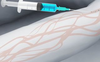
Attention! Contrast agents for MRI are used when the presence of neoplasms is suspected.
Contrast enhancement
Preparing for the examination
If you want to find out in more detail which is better than ultrasound of the spine or MRI, and also consider the features and advantages, you can read an article about it on our portal.
As such, there is no preparation for MRI, except for cases when diagnostics using contrast agents are prescribed. But there are a number of aspects that you need to remember - knowing them, it will be easier to pass the examination.
Step 1. During the tomography you will have to stay inside the equipment for some time. If a person suffers from claustrophobia, this test will be extremely difficult for him to endure. You should tell your doctor if you have claustrophobia. It may well be that the specialist will prescribe sedatives.
Tell your doctor about claustrophobia
Step 2. If there are implants made of metal inside the body, you should definitely tell your doctor about it. Most prostheses or implants can affect the operation of the equipment and also cause pain to the person being examined. In some cases, you will have to refuse an MRI altogether.
Report metal implants in the body
Step 3. It is important to inform the doctor about the presence of pregnancy or serious illnesses, if any. A number of features of the body’s condition may be a contraindication to the study.
Pregnancy is one of the possible contraindications
Step 4. If your doctor previously prescribed any medications, then you do not need to refuse them before the MRI, unless the specialist has said otherwise.
Stop taking medications
Step 5. Before going for an MRI, it is recommended to read about this procedure and watch videos demonstrating how it is performed. This will give you an idea of what to expect in the CT scan room.
Find out more about tomography
Step 6. It is better to go to the MRI accompanied by a friend or relative. The doctor may prescribe sedatives, after which you cannot drive on your own.
Bring a friend or relative with you
Step 7. It is better to arrive for the tomography early than late. The clinics have waiting areas where you can sit before the procedure begins. Moreover, the doctor may ask you to fill out some papers - it is better to do this before the examination, so as not to waste time later.
It's better to arrive earlier
Step 8. It is important to remove all jewelry. You will also need to remove watches, wigs, hair clips and other items from your body. A bra with underwire must also be removed.
Remove all jewelry
Step 9. During the CT scan, it is important to follow the doctor's instructions, do not argue with him and do what he asks. Then the procedure will be as quick and comfortable as possible.
Do whatever the doctor says
If MRI is performed using contrast agents administered intravenously, it is recommended not to eat food approximately 5-6 hours before the examination so that the tomography is done on an empty stomach. If possible, it is worth making an appointment for the examination in the morning, although a regular MRI can be done at any time of the day.
Massage chairs
You should wear comfortable clothing that is easy to take off and put on and does not constrict the body. If data from past examinations is available, it is recommended that you take it with you - the doctor may want to compare the results.
Video – Tomography of the thoracic spine
Tomography is an accurate modern research method widely used to diagnose the condition of the thoracic spinal system. It will help identify virtually any disease that may develop in this area. Unfortunately, in municipal clinics there are always long queues for this examination. It’s easier and faster to get an MRI at a paid medical center.
Source: https://spina-expert.ru/diagnostika/mrt-grudnogo-otdela-pozvonochnika-chto-pokazyvaet/
MRI of the thoracic spine: what it shows
Magnetic resonance imaging of the thoracic spine is one of the most informative diagnostic methods. The technique helps to identify the slightest pathological changes in the local area: damage to the spinal column, infectious foci, tumors, changes in the structure and placement of the vertebrae and a number of other phenomena without harm to the patient’s health.
The essence of the MRI technique of the spine
During MRI diagnostics of the thoracic spine, a diagnostic device is used that is capable of producing a magnetic field, in response to which the tissues independently generate radiation - a dynamic image of the organ being examined is displayed on the screen.
The operation of the equipment is based on the principle of nuclear magnetic resonance: the formation of a powerful magnetic field leads to a subsequent reaction of atomic particles - protons. This response is recorded by the device and translated into a graphical format. The output is three-dimensional sections of the examined anatomical structures.
MRI
Possible symptoms that may be a reason to prescribe an MRI examination of the spine include:
- pain similar to heart;
- discomfort in the area between the shoulder blades;
- intercostal neuralgia (lumbago in the area of the intercostal nerves);
- chest tightness;
- numbness of the local area;
- soreness in the upper, middle part of the abdomen (immediately under the ribs), which becomes stronger after physical activity;
- liver pain;
- dysfunction of the reproductive system.
Magnetic tomography can also be performed to monitor the condition of organs and systems in the case of diagnosis and course of the following pathologies:
- osteochondrosis of the thoracic and cervical spine;
- kyphosis;
- herniated intervertebral discs;
- neoplasms;
- infectious processes.
In some cases, MRI of the spine is performed to prepare the patient for surgical intervention in the local area.
List of contraindications for MRI
Despite the safety of MRI examination of the spine, magnetic tomography has a number of circumstantial contraindications. Objects in the human body can become an obstacle to scanning:
- an implant containing metal;
- vascular clips;
- prostheses;
- insulin pump;
- pacemaker, etc.
Individual limitations to diagnosis include: fear of closed spaces, nervous tics, unexpected seizures. In these cases, the patient is offered sedatives.
MRI examination is contraindicated for pregnant women (in the 1st trimester), as well as for persons whose vitality is supported by special equipment. It is not advisable to scan children under 7 years of age.
If pediatric diagnostics are necessary, anesthesia is used. Magnetic tomography may also be unavailable for people weighing more than 120 kg.
In some cases, it is possible to use specialized equipment.
If an examination with contrast is planned, renal failure, the entire period of pregnancy, and an allergy to the contrast agent are also added to the group of restrictions described.
Preparatory stage
Magnetic tomography is performed on an outpatient basis, rarely in a hospital setting. Preparation for MRI of the thoracic spine includes measures to ensure patient safety:
- before entering the diagnostic room, you must remove objects containing metal from yourself;
- if there are tattoos on the patient’s body (in the examination area), it is necessary to inform the doctor about this, since the pigment of the body pattern may contain metal;
- If you are planning a scan with contrast, you should refrain from eating and drinking 5-8 hours before the MRI. If contrast agent is excluded, there is no need to fast;
- It is important not to forget to warn the diagnostician about existing chronic pathologies or allergic reactions.
The device is an open, closed type, presented in the form of a ring passing through the center of a horizontally placed magnetic capsule. The patient, having changed into a disposable set of clothing, is placed on a movable table, which slowly moves inside the capsule.
During MRI diagnostics of the thoracic spine, you will have to lie on your back. The head, chest, and arms are secured with belts. The patient may be offered a pillow or blanket. The patient must not move or talk (except when it is necessary to contact a specialist through an equipped microphone).
MRI image of the thoracic spine
A patient preparing for an MRI scan of the spine should be prepared to hear a lot of noise from the equipment. If the patient is uncomfortable with the sound, he can ask for earplugs or headphones.
If the subject is nervous, he is offered sedatives. Discomfort or pain during the diagnostic process is not a typical occurrence. There may be a feeling of swelling and stiffness of the body. There will be a feeling of warmth in the area of examination (in the sternum), colic - this is normal and goes away very soon.
If a patient notices signs of nausea, vomiting, pain, dizziness, or difficulty breathing, it is necessary to immediately report this to a specialist.
The scanning time is 20-40 minutes. If a contrast agent is used, the session may last up to an hour.
Osteochondrosis
At the end of the study, the patient can get dressed and leave the office. There are no rest, restrictions on food and drink after MRI of the spine. The results are delivered to the patient after an hour. In severe cases, the conclusion is issued the next day.
Use of contrast agent
The procedure with contrast is not only longer and more complex, but also takes more time to draw up a conclusion.
The reason for performing this type of MRI is the need to obtain detailed information in the presence of a tumor to determine the boundaries of inflammation.
Before placing the patient deep into the magnetic capsule, a contrast agent is injected intravenously, which quickly spreads throughout the body through the bloodstream and accumulates in the pathological focus. This ensures the best visualization quality.
Unlike the iodine-containing drug used in computed tomography, MRI diagnostic contrast is based on gadolinium. This component is more easily tolerated by patients, and in rare cases causes an allergic reaction and side effects.
Before the examination, the patient must undergo an allergy test for the medicine.
Question of price of services
An MRI diagnostic machine is not cheap; large diagnostic centers can afford it. The price for an examination varies from 3,500 to 5,000 rubles and depends on the region of diagnosis, the class of equipment used, the pricing policy of the center and the qualifications of medical personnel.
Additional payment is usually required for the use of a contrast agent, consultation with a doctor, saving study results on a disk or flash card, and other services.
Magnetic tomography of the thoracic spine is carried out in order to identify local pathologies: problems with the structure, placement and joints of intervertebral discs, neoplasms, infections, etc. A common symptom that leads to the need for diagnostics is pain.
In some cases, magnetic tomography is prescribed to monitor the effectiveness of treatment or in the preoperative period. There is no preparation for the study.
The procedure is safe and painless for the patient. If detailed information is needed, a gadolinium-based contrast agent is used.
The list of contraindications in the case of MRI of the spine is not wide and includes both absolute and relative restrictions. The likelihood of adverse reactions to the use of contrast is minimized.
The cost of the examination ranges from 3500-5000 rubles.
Video
Source: https://osnimke.ru/pozvonochnik/mrt-grudnogo-otdela-pozvonochnika.html
MRI of the thoracic spine
Doctors use a variety of hardware techniques to determine health problems and make an accurate diagnosis. One of them is magnetic resonance imaging.
With its help, the condition of the bone and soft tissue of the back is studied in detail, the location of the vascular plexuses and joints is determined, their position and even minor deviations from the norm.
A feature of the technique is its high information content and the absence of discomfort for the patient during the process. Almost all work is automated, and the received data is processed by a computer.
Advantages
Magnetic resonance imaging does not cause pain or discomfort during the process, which is a very big plus.
But the body is exposed to ionizing radiation, which affects health if it is carried out excessively frequently. Therefore, specialists resort to it only in cases of urgent need.
The main advantages of diagnostics using such equipment are:
- The ability to obtain a high-quality image in which all the details of bone tissue are visible.
- High information content and accuracy of data (compared to other methods).
- Visualization of nerve bundles and vascular channels to determine their integrity and general condition.
- The use of a contrast agent that does not cause an allergic reaction.
- The ability to get a complete picture of the condition of the spine in a short time.
- Determining the presence of compression of nerve structures, which is especially important in case of paralysis.
- Detection of changes in tissues at an early stage.
- Ease of tracking the dynamics of changes after operations or medical therapy.
- There is no threat to human life or health, since the procedure is completely safe.
The cost of the technique directly depends on the chosen clinic. Today, even government institutions use modern devices. A comprehensive examination is usually more expensive than just a separate part of the body or area of the spine.
During an MRI, a person needs to relax as much as possible and not move.
When is the study ordered?
An MRI of the thoracic spine is performed only if indicated, since during the process the patient receives a dose of radiation. A referral for the procedure is issued by a neurologist, surgeon, orthopedist, traumatologist or cardiologist, if there is a suspicion of a serious disorder that cannot be determined using other methods.
Usually referred for an MRI in the following cases:
The difference between CT and MRI of the spine
- Violation of the integrity of the bones of the spine or limbs.
- There are developmental disorders or congenital anomalies of the spine.
- Oncology with metastases to bone tissue was diagnosed.
- Osteochondrosis or other vertebral lesions.
- Damage to the myelin sheath of neurons.
- Neoplasm in the vertebrae.
- Intervertebral hernia.
- Vascular pathologies or poor circulation in the back.
MRI is necessary if the patient complains of constant back pain, which is accompanied by other disorders of the general condition (numbness of the arms or legs, difficulty moving, frequent dizziness, muscle spasms). The hardware technique is also used to monitor the progress of treatment of pathology or control relapse in the case of oncology.
An MRI image allows you to obtain complete information regarding the condition of the patient’s spine
What does an MRI of the spine show?
MRI of the thoracic spine is complex, but is most often performed in relation to specific areas.
In the case of the chest area, timely examination is carried out in order to prevent stroke, herniated disc, ruptured disc, cerebral infarction or other problems.
The procedure allows you to fully assess the condition of bones, blood vessels and soft tissues, so it is recommended in case of numbness of the limbs, stiffness of movement, pain between the shoulder blades, or discomfort in the back in the chest area.
What does an MRI show?
- Infectious diseases at any stage.
- Congenital anomalies of the structure of the thoracic region.
- Places of narrowing of the spinal canal.
- Inflammatory processes in soft tissue.
- The onset of deformation or degenerative changes in the vertebrae.
- Development of osteomelitis.
- Areas with insufficient blood supply.
- Damage to nerve fibers.
- Injuries to bone tissue in the area being examined.
- The onset of osteoporosis or osteochondrosis.
- Tumor or metastases in the thoracic region.
- Abnormal neoplasms.
Magnetic resonance imaging allows us to show the exact location of the pathology, the degree of its development and the need to use a radical treatment method.
Usually, concomitant problems with the spine are additionally diagnosed, namely disorders of the intestines, stomach or heart muscle.
Based on the research obtained, the doctor evaluates the anatomical structure of the spine, identifies asymptomatic pathologies, places of injury or inflammation that have formed as a result of surgery.
MRI allows you to identify lesions of the meninges or blood vessels, infectious processes and tumors, arthrosis and the condition of the intervertebral discs in general. Today, the procedure is considered the most informative among all existing ones.
This is what an MRI image looks like using contrast.
Preparation for the procedure
MRI is a modern technique that allows you to obtain highly accurate results. On its basis, decoding and diagnosis are made.
As before any medical procedure, it is necessary to prepare for it in order to eliminate errors or distortion of data.
A special hunger strike or refusal to drink is not required if contrast is not used for the study. If it is intended to be used, the preparation will be as follows:
- The selection of a suitable contrast to exclude allergies is carried out together with the supervising specialist.
- The use of sedatives or tranquilizers if a person is claustrophobic or lacks full control of movement.
- All jewelry and clothing with metal elements are removed.
- Food consumption continues as before.
The use of contrast is prohibited if the patient has a history of bronchial asthma (or other lung problems) or atopic dermatitis. During the procedure, turn off mobile phones, remove coins, keys and other foreign objects from pockets.
Important information! Before an MRI, it is imperative to report tattoos on the body, especially large ones, if a metal-based pigment was used when applying them, as well as if there are plates, braces, metal implants or other elements that cannot be removed. This will allow the specialist to objectively assess all risks and take the necessary measures or prescribe an alternative diagnostic method.
Magnetic resonance imaging does not require much time to perform and is carried out in a separate room. The scanning process starts only when the patient is completely ready. In general, the algorithm of actions when completing the study is as follows:
- The patient takes off his outer clothing and all items containing metal buttons, rivets or inserts.
- You need to sit on the couch as level as possible so that you can spend 15–20 minutes in this position without moving.
- The genitals are protected with special pads.
- The scanning process starts, which may be accompanied by a characteristic sound or noise. During this time you cannot move, talk or sing.
- Receiving and decoding the result.
High-precision images can only be taken if the patient lies absolutely still throughout the entire exposure time.
Any sudden movements can affect the result, so if in doubt, a repeat test is prescribed.
You should also immediately seek help from a specialist if you experience severe nausea, vomiting, dizziness or breathing problems. Most often this is a reaction to the effect of contrast.
Pregnancy is one of the contraindications for MRI.
Contraindications for carrying out
Magnetic resonance imaging is not a universal technique, since it is not prescribed to every patient.
Before signing the referral, the specialist assesses the patient’s general condition, carefully examines his medical history, and asks him about the presence of contraindications to the effects of ionized healing.
This will avoid future complications of existing chronic pathologies.
The main prohibitions for conducting MRI of any part of the spine (be it cervicothoracic or lumbar) are:
- The presence of pacemakers or other electronic devices or implants in the body. During exposure, they may fail or become damaged. Passing is allowed only after consultation with a treating specialist and identification of possible risks.
- High risk of allergic reaction to contrast. In this case, it is possible to prescribe potent antihistamines.
- Pregnancy and breastfeeding. Lactating women are not recommended to use milk for 48 hours after an MRI.
- For claustrophobia. In this case, it is possible to use strong sedatives to ensure immobility for 15–30 minutes.
- Overweight (more than 120 kg). In this case, equipment is looked for that can withstand a weight of up to 180 kg; if the weight is greater, a different technique is used.
- Mental disorders, epilepsy or seizures.
- Acute injuries and the need to promptly obtain research results.
- Confusion or loss of consciousness.
The procedure is absolutely painless and requires minimal preparation, which allows the use of modern diagnostics in the case of any patients. Children's age is not a contraindication for the study, but the doctor decides on the advisability of such an action individually.
Magnetic resonance imaging is a modern way of obtaining complete information regarding the patient’s health status. It is used by doctors in various fields of medicine to determine the most correct treatment tactics for certain disorders. Despite the presence of radiation in the process, the benefits of diagnosis are greater than the harm, so the method is considered relatively safe.
Source: https://spina.guru/diagnostika/mrt-grudnogo-otdela-pozvonochnika
Magnetic resonance imaging (MRI) of the thoracic spine
Magnetic resonance imaging is considered one of the most effective ways to examine the condition of the spine. For the first time, the capabilities of MRI for scanning and diagnosing spinal health were presented to the general public in 1982 at the Paris Exhibition of Radiologists, although the magnetic resonance scanning method itself was developed somewhat earlier - in 1973, when, using the properties of radio waves and a magnetic field, the first MRI images of two tubes filled with water. Since that time, the option of examining the spine using a magnetic resonance imaging scanner has become one of the main ways to identify pathologies and disorders in it at any stage of their development.
The phenomenon of nuclear magnetic resonance was discovered in 1946, and began to be actively studied by scientists at Stanford and Harvard universities. Its essence lies in the ability of hydrogen atoms, when exposed to a magnetic field, to absorb energy and release it in the form of a radio signal.
At that moment, the discoverers probably did not think about the role this principle would play in the development of medical diagnostics. After 1973, the design of magnetic resonance imaging scanners gradually acquired industrial scale.
Today, almost every major medical institution has this device.
The thoracic spine is represented by twelve vertebrae, which together with the ribs and sternum form the rib cage. This part of the spine is susceptible to both injury and all other types of pathologies and lesions of the spine. Problems with the spine in this area can begin due to metabolic disorders, since as a result the nutrition of the intervertebral discs is weakened.
Other reasons are improper distribution of physical activity, poorly selected pillow and mattress, constant stress on the back, intense sports, or vice versa - a complete lack of physical activity in a person’s life.
Painful symptoms in the heart, stomach, liver, kidneys, pancreas in some cases are caused precisely by the presence of disorders in the thoracic spine.
Magnetic resonance imaging images of the thoracic spine can display traumatic, inflammatory, tumor, degenerative disorders and diseases. If it is necessary to study tumors in more detail in the early stages of their development, assess their location and the presence of metastases, the patient may be prescribed an MRI of the thoracic spine with contrast.
The doctor decides to prescribe this scanning procedure in order to:
- determine the presence or absence of diseases and pathologies;
- identify the consequences of injuries to the thoracic spine;
- determine the presence of tumors or metastases;
- clarify the reasons for performing a surgical operation;
- evaluate the effectiveness of previously performed surgery or other types of treatment.
What do MRI scans show? After the procedure, the radiologist interprets the results. In the images obtained during a spinal tomography, he can detect:
- injuries and hernias;
- Scheuermann's kyphosis and adult kyphosis;
- degenerative changes of the vertebrae and discs;
- protrusion, spondylosis;
- neoplasms of any type;
- congenital anomalies of the structure and development of the vertebrae;
- narrowing of the spinal cord canal;
- Bekhterev's disease;
- vascular disorders of the spinal cord;
- violation of the anatomical location of the vertebrae.
Indications for magnetic resonance diagnostics of the spine
Common reasons for sending a patient for an MRI of the thoracic spine:
- pain and burning between the shoulder blades;
- pain syndrome similar to pain in the heart, radiating to the back and sternum;
- chest pain of a girdling nature;
- numbness and stiffness in the chest;
- shooting pain in the area of the intercostal nerves;
- discomfort in the liver and genital areas;
- pain in the epigastric region, manifested after active physical activity;
- the need to clarify the diagnosis;
- monitoring the effectiveness of treatment;
- preparatory measures before surgery.
In cases where the listed symptoms appear systematically, over a certain period of time, magnetic resonance imaging of the thoracic spine is an objective necessity.
Of particular concern among all disorders and diseases of the spine is osteochondrosis, a degenerative process in the intervertebral discs.
Osteochondrosis of the thoracic region is called a “chameleon disease” due to the fact that its symptoms are masked as signs of functional disorders in organs anatomically associated with those groups of nerves that suffer from osteochondrosis. Thus, manifestations of osteochondrosis can be confused with symptoms:
- gastritis;
- colitis;
- peptic ulcer;
- appendicitis;
- hepatic colic;
- angina pectoris;
- heart attack;
- cholecystitis.
In what cases is it impossible to perform an MRI of the thoracic spine?
The scanning procedure with an MRI scanner may not always be beneficial. There are situations when the purpose of this examination method is impractical or even dangerous for the person being studied.
Doctors include contraindications for thoracic MRI:
- the presence of metal implants, prostheses, knitting needles embedded in the human body;
- tattoos, if they were applied using a pigment containing metal compounds;
- pacemaker: the device not only can distort the results of the examination, but there is also a possibility that it will fail when exposed to the strong magnetic field of the tomograph;
- first trimester of pregnancy: at this time the basis of the future organs of the fetus is formed, so doctors try to postpone MRI to a later date, or prescribe alternative diagnostic methods to pregnant women in the early stages.
People with a fear of closed spaces, as well as those who suffer from obesity, are recommended to carry out the procedure on an open tomograph.
For MRI with contrast, in addition to the above limitations, there are others:
- It is not recommended to use a contrast agent on pregnant and lactating women;
- It is prohibited to administer the drug if renal failure is diagnosed;
- Allergy or intolerance to the contrast agent completely excludes the possibility of its use for scanning the spine.
Rules for preparing and conducting the procedure
Preparatory measures for an MRI of the thoracic spine include the fact that the doctor must first establish that there are no contraindications to this type of examination.
The patient is recommended to take the results of all previously performed tests and studies with him to the appointment and give them to the doctor for review.
Women must be informed about their pregnancy.
Before the patient is placed on the CT scanner table, he will be asked to remove all metal objects - jewelry, accessories, gadgets. Items with metal fittings will have to be removed.
Health workers usually offer a change of loose-fitting clothing made from natural fabric and without metal parts.
You can also use earplugs if the loud knocking and crackling of the CT scanner causes discomfort.
Next, the subject is placed on the tomograph table, fastened with belts and fixed with special soft cushions - all this is done in order to ensure complete immobility for the entire period of the procedure, which can last from 10 to 30 minutes.
It takes approximately 1-1.5 minutes for the tomograph cylinder to take 1 picture. About 20 such images are recorded in one diagnostic session.
Their layer-by-layer overlay makes it possible to obtain a three-dimensional image on the tomograph screen with a picture of the state of all deep tissues and processes.
During the entire process, there is the possibility of two-way communication between the patient and the doctor using a special intercom. All atypical, strange and unpleasant sensations that occur during a tomography should be reported to the doctor.
Interpretation and evaluation of the results of examination of the thoracic spine
A radiologist interprets the information received and interprets MRI images. Having received the results of the tomography, the doctor pays attention to a number of indicators of the spinal column in the thoracic region.
It evaluates the size and shape of the spinal cord, as well as the spine, based on the assumption that the spinal cord should be located in the middle, and normally has clear contours. Its width is also determined, and if it is increased, this may indicate the presence of an intramedullary tumor.
The radiologist analyzes the subarachnoid space, looking for the presence of crescent or linear band syndromes, which may indicate hemorrhage in the spinal cord.
- To most accurately determine the localization of pathologies in the image, doctors focus on the level of the second and fifth cervical vertebrae, since images of the thoracic region do not have other, clearer landmarks to which the location of the tumor, source of inflammation or other disorder can be linked.
- Images may also show calcifications (calcium salt deposits) and petrification in soft tissue.
- Non-tumor cysts in the thoracic spinal cord can be identified by MRI results - their presence is indicated by smooth and even contours, the absence of constrictions, low signal intensity in places where cystic cavities are present,
- Ischemic disorders, intramedullary tumor, spinal plaques in multiple sclerosis, tuberculosis, toxoplasmosis, acute encephalitis reflect focal increases in the radio signal along the length of the thoracic region.
- Thickening of the spinal cord can be caused by ischemic changes, post-traumatic deformation, and transverse myelitis.
- The presence of a neuroma can be determined by the posterolateral location of the affected area, the absence of hyperostosis and calcifications, and also by its specific hourglass shape.
Magnetic resonance imaging of the thoracic spine is a procedure that allows for the diagnosis of lesions, injuries and diseases of soft tissues, intervertebral discs, vertebrae, and spinal cord in this area. Based on the results of the procedure, it is possible to identify the presence of neoplasms, circulatory disorders, degenerative changes in the vertebrae, structural anomalies, and other disorders.
Among the advantages of this method of scanning the spine is the absence of radiation exposure to the body, as well as accurate and in-depth examination of tissues, cartilage structures, and the vascular system. Without introducing contrast, this procedure makes it possible to study the spine simultaneously with the spinal cord.
Alternative methods for examining the spine are radiography and computed tomography, but doctors note that they are less informative than MRI.
Author of the article:
Tedeeva Madina Elkanovna
Specialty: therapist, radiologist.
Total experience: 20 years.
Place of work: SL Medical Group LLC, Maykop.
Education: 1990-1996, North Ossetian State Medical Academy.
Training:
1. In 2016, at the Russian Medical Academy of Postgraduate Education, she underwent advanced training in the additional professional program “Therapy” and was admitted to carry out medical or pharmaceutical activities in the specialty of therapy.
2. In 2017, by the decision of the examination commission at the private institution of additional professional education “Institute for Advanced Training of Medical Personnel”, she was admitted to carry out medical or pharmaceutical activities in the specialty of radiology.
Work experience: therapist – 18 years, radiologist – 2 years.
Source: https://FoodandHealth.ru/diagnostika/mrt-grudnogo-otdela-pozvonochnika/