What does a CT scan of the lungs show?
When performing a computed tomography scan, the doctor can evaluate the condition and structure of the tissue of the lung, bronchi, mediastinal lymph nodes, blood vessels and aorta. Thanks to this, CT makes it possible to identify any pathological processes in these structures, both focal and diffuse in nature.
The specialist sees in the resulting images the nature of the pathology, its prevalence, the degree of damage to the lungs and the localization of changes. A CT scan of the chest can evaluate not only the lungs, but also the pleura, muscle structures, subcutaneous fat and mammary glands.
Bone structures are partially visible, since scanning occurs in the longitudinal plane.
The lungs are complex organs. Computed tomography evaluates their density, branching and size of the bronchi, as well as the condition of the blood vessels.
The doctor can identify focal formations in the form of cysts, metastases, primary tumors and extensions of the bronchial tree. Diffuse changes are observed during inflammatory processes.
The pleura may have adhesions, fibrous deposits, areas of calcification, as well as accumulations of fluid, blood or air in the pleural cavity.
Advantages of computed tomography compared to other diagnostic procedures (ultrasound, MRI, etc.):
- the procedure takes from 5 to 30 minutes. Subsequent interpretation of the results is carried out by a doctor within an hour;
- objective results reflecting the condition of the lungs, their normal structure or pathological processes;
- no pain, since the scan is completely non-invasive;
- the total dose of ionizing radiation is low and cannot cause harm to the patient;
- CT allows you to obtain high-quality three-dimensional images of the lungs, which ensures high diagnostic accuracy and a low risk of false results.
Important! Image interpretation should only be carried out by a specialist. Otherwise, an incorrect diagnosis may be made and the underlying pathology may progress.
Results: norms and pathology
A CT scan of the lungs in the absence of pathologies has characteristic features: organs of normal structure without any focal or diffuse changes. The trachea and main bronchi retain their anatomical structure.
There is no free fluid in the pleural cavity. The mediastinum is of normal width and without pathological processes in the lymph nodes. The chest wall is not changed.
The lumen of the aorta, pulmonary arteries and pulmonary trunk is without thrombotic masses.
Pathology in the lungs can take different forms. It depends on the specific disease, the stage of the disease and the individual characteristics of the person.
As a rule, making an accurate diagnosis requires simultaneous assessment of CT changes, clinical symptoms and additional examination methods.
The main pathologies observed in the lungs include: tuberculosis, tumors, bronchitis and pneumonia.
The tumor appears as a lesion measuring 10 mm or more. It has a dense, but not uniform structure, and clearly stands out against the background of the lung tissue. New growths in the lung can grow inside the bronchi. In this case, the doctor sees voluminous structures in the bronchial lumen that partially block it, or can grow into the surrounding tissue.
Tuberculosis in humans occurs in several forms, which have structural differences. With the primary tuberculosis complex, a rounded shadow with indistinct organs appears in the lungs.
The root of the lung expands and it connects with the primary focus with a linear shadow in the form of a thin “path”. If the patient has focal tuberculosis, then rounded shadows appear at the apex of the lung, which can be single or multiple.
The infiltrative form of the pathology appears on CT images as darkening of various shapes with unclear contours and pronounced infiltration.
Pneumonia looks like areas of compaction with a heterogeneous structure and an unclear contour. At the same time, the undisturbed bronchial lumen is clearly visible. Pneumonia is often accompanied by changes in the pleura - it thickens, which is associated with the development of an inflammatory process in it.
Acute bronchitis is a common disease that occurs in patients of all ages. Pathological changes are located in the bronchi, which provide air conduction to the lungs. In the area of the affected tissue, focal infiltrates appear, which are accompanied by an increase in the pulmonary pattern. Tomography allows you to clarify the location and extent of the lesion.
The examination allows us to identify other pathological processes: pleurisy (inflammatory damage to the pleura), abscess (cavity filled with pus), etc.
At the same time, CT has greater accuracy compared to similar methods. Due to this, tomography is used to detect diseases of the respiratory system.
How is a CT scan of the lungs done?
Patients often wonder which doctor orders a CT scan. The main specialist who selects methods for examining the respiratory system is a pulmonologist. He determines a person’s indications and contraindications, selecting the optimal methods of laboratory and instrumental research.
Before performing a CT scan, the patient removes all metal objects, including watches, hairpins, jewelry, piercings, and also removes gadgets.
This avoids the appearance of artifacts on CT images. After this, a medical worker talks with the person.
He explains the rules and procedure for performing a lung scan, and shows how you can contact your doctor if necessary.
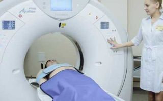
The patient is placed on the tomograph couch in the supine position and hands are placed behind the head. After placing the person, the device turns on. The couch begins to move in a horizontal plane.
At this time, the beam tube and detectors operate according to a given program. The healthcare worker periodically asks the patient to hold his breath. This allows you to better view the lung tissue.
If necessary, contrast agents are administered.
Unlike MRI, CT does not last long - from 5 to 20 minutes. Upon completion of the procedure, the couch returns to its initial position. The nurse helps remove all restraints from the patient. After this, he waits for the results of the examination.
Types of examination
Step-by-step X-ray tomography of the lungs is the first type of CT. During the procedure, the couch with the patient moves inside the device over a certain distance. At each stop, the device takes an x-ray. That is, layer-by-layer images of the lungs are created, which are then processed by a computer. The examination time is 20-30 minutes. The method is highly informative and is used in a large number of clinics.
Spiral computed tomography of the lungs has two differences: the couch moves constantly, and the radiation tube moves around the body in a spiral.
Thanks to this, the doctor receives a more accurate image of the chest organs, and the duration of the examination is reduced. The spiral type of CT allows you to detect minimal changes in the lung tissue.
It has an optimal price/quality ratio.
Multislice CT is characterized by the fact that there are several rows of detectors opposite the X-ray tube. This increases the accuracy of diagnosis, but increases its cost. Thanks to several detectors, the method is comparable to MRI in imaging the chest organs. Multispiral examination is considered the most modern and informative in medicine.
Any type of CT scan can be performed with contrast. Thanks to its introduction, the doctor can evaluate the bloodstream, bronchi, etc. In medicine, they use contrasts that are safe for patients, which are soluble in water and are excreted from the body along with urine.
Indications - contraindications
CT scan of the lungs is performed for certain indications. Thanks to this, the harm of X-ray exposure is reduced and overdiagnosis of diseases of the chest organs does not occur. The main indications for performing computed tomography include:
- symptoms of tumor or metastatic lesions of the trachea, bronchi, mediastinum or pulmonary parenchyma;
- differential diagnosis between benign and malignant neoplasms;
- determination of the stage and extent of tumor tissue spread on CT sections;
- diagnosis of acute and chronic lung diseases: pneumonia, tuberculosis, sarcoidosis, pneumosclerosis, emphysema, COPD, alveolitis, bronchiolitis, etc.;
- symptoms of pleurisy or pleural empyema;
- long-term symptoms of lung diseases in the form of shortness of breath, cough, fever and chest pain with negative results of other examination methods;
- search for structural anomalies of the bronchi, lungs, arterial and lymphatic vessels, as well as other organs of the chest;
- chest injuries. CT can exclude pneumothorax, hydrothorax, hemothorax and lung rupture.
Computed tomography, like MRI, is suitable for identifying any pathological processes in the organs of the chest cavity - from malignant neoplasms to disorders of blood flow in the vessels of the lung.
Contraindications for CT scanning are divided into two groups: in the case of using contrast and without it. If contrast is not administered, scanning is not performed if:
- any stage of pregnancy;
- the patient's age is less than 14 years;
- inappropriate behavior of the patient.
If a person has prostheses, implants or metal structures, the study is carried out, however, but the images will be of low quality.
When using contrast, the list of contraindications is longer:
- allergic diseases with severe course, including bronchial asthma;
- individual intolerance and allergy to iodine;
- severe renal or liver failure;
- age under 14 years and over 70 years;
- tumor diseases of the thyroid gland and hyperthyroidism;
- pheochromocytoma;
- long-term use of beta-blockers, non-steroidal anti-inflammatory drugs and diuretics;
- taking iodine-containing products the day before the procedure.
Patients are interested in how often a CT scan of the lungs can be done. Limitations relate to the harm and radiation dose the patient receives during each scan. The optimal interval between two examinations is 6-12 months. In patients with lung tumors, CT may be performed more frequently for medical reasons.
Preparation
You should stop eating 6-8 hours before the scan. This avoids nausea and vomiting during the examination. Also, 2-3 days before the CT scan, it is recommended to exclude from the diet foods and dishes that contribute to the development of flatulence. A large amount of free air in the intestine can lead to elevation of the diaphragm and the appearance of artifacts in the images.
Before the procedure itself, the person takes off his watches, accessories (watches, hairpins, jewelry), dentures, etc. After this, he puts on a hospital gown. The doctor talks with him and tells him about the upcoming procedure.
CT, unlike MRI, is performed quickly, which reduces discomfort for the patient, however, understanding the examination process reduces stress. After the patient is positioned on the couch, the nurse places a cushion under the neck and fixes the arms.
This allows you to avoid body movements and “blurring” of pictures.
During the scan, the doctor is in the next room and monitors the progress of the procedure. This allows you to avoid errors during CT scanning and monitor the patient’s condition.
Photo CT scan of the lungs
Infiltrative tuberculosis
Single cylindrical bronchiectasis (bronchitis)
Price
The price for a lung examination depends on the specific city and diagnostic center. The cost of scanning without contrast is from 1,500 to 3,000 rubles.
For the procedure with the introduction of a contrast agent you will have to pay more than 3 thousand rubles. For medical reasons (suspicion of a lung tumor, developmental defects, etc.)
) the study can be performed free of charge at the expense of the Compulsory Medical Insurance Fund.
Source: https://mrtu.ru/organy/kt-legkih-chto-eto-takoe-chto-pokazyvaet-kak-delayut.html
Which method of examining the lungs will give the best result and why?
21.11.2017
To examine the lungs, specialists can prescribe any of the specified diagnostic methods (CT, X-ray or MRI).
When prescribing any of these research methods, the doctor takes into account such nuances as: duration of the procedure, indications, contraindications, which is better visualized by each of the diagnostics. It is difficult to say unequivocally which is better than CT or MRI.
To do this, you should familiarize yourself with the differences in the principle of their operation, advantages/disadvantages, indications/contraindications.
The fundamental difference between computed tomography and magnetic resonance imaging
The main difference between computer diagnostics and magnetic resonance diagnostics is the operating principle:
- MRI provides informative images of the lungs thanks to nuclear magnetic resonance of hydrogen atoms. There is no radiation exposure to the body.
- CT scans of the lungs use X-rays. X-ray radiation passes through tissue, providing doctors with images in specific sections.
CT and MRI are used for different purposes:
- CT scans are more often used to examine bone structures.
- MRI ideally visualizes soft tissues and blood vessels.
There is a difference between the diagnostic methods under consideration and in the pricing policy:
- MRI is more expensive due to the high cost of the tomograph itself. It is chosen by patients who do not want to receive radiation exposure to the body.
- CT is cheaper, so CT scanners are used in most public health institutions.
For many patients, the timing of the procedure also plays an important role in the study of body systems. The difference in the duration of MRI and CT of the lungs is significant:
- A computed tomography scan of the lungs takes only 15 to 20 minutes.
- Magnetic tomography of lung tissue is performed over 1 – 1.5 hours.
Advantages and disadvantages of MRI and CT for examining the lungs
The specialist decides which diagnostic to choose, MRI or CT of the lungs. Computed tomography of the lungs is performed using x-rays. To obtain a complete picture of the developing pathology, doctors study a series of images of given sections. A diagnostic procedure such as CT makes it possible to visualize in detail all segments, sections of the lung tissue and bronchi.
The advantages of examining the lungs using a computed tomograph are:
- Obtaining images of spiral sections. The doctor can change the viewing angle at his discretion. CT images of the lungs are digitally presented as three-dimensional images.
- Speed of the procedure. This point is considered decisive if the patient has internal bleeding.
- Ability to detect internal bleeding and hematomas.
Computed tomography is used to easily, quickly detect damage that a patient has suffered as a result of trauma (broken ribs, abnormal lung structure, bleeding).
The disadvantages of CT are:
- Harm to health caused by X-rays.
- Possibility to carry out the procedure a limited number of times.
Magnetic resonance imaging of the respiratory system is considered an absolutely safe diagnostic method. Instead of X-rays, a strong electromagnetic field is used to take pictures of the lungs. MRI provides doctors with high-quality 3D images.
Magnetic resonance imaging has its advantages over CT:
Disadvantages include:
- Poor quality of images of hollow organs. They come out blurry.
- Duration of the procedure. It takes approximately 1.5 hours.
- Poor quality of images of an organ that is in constant motion.
- A large list of contraindications for the procedure.
The attending physician decides which diagnostic method to choose. Thanks to CT, chronic diseases and respiratory injuries can be seen, but more complex pathologies are examined using a magnetic resonance imaging scanner.
Indications and contraindications
Regular fluorography can sometimes give results that raise suspicion among the doctor. In this case, he may refer the patient for a more detailed examination of the lungs (CT, MRI).
Diagnostic methods such as CT and MRI are equally considered effective in diagnosing the following pathologies:
- atelectasis;
- pneumonia;
- sarcoidosis;
- pneumofibrosis;
- lung sequestration;
- pulmonary vascular abnormalities;
- pulmonary nodes;
- tuberculosis;
- pulmonary artery aneurysm;
- pulmonary failure (acute, chronic);
- pulmonary vascular abnormalities;
- mesothelioma;
- vasculitis;
- clarification of the stage of cancer;
- arteriovenous malformations;
- pleural effusion of unspecified etiology.
CT and MRI are almost equally effective in diagnosing inflammatory processes and tuberculosis. But if internal bleeding is suspected, preference should be given to CT, and to diagnose neoplasms it is better to use MRI. Below we will consider cases when it is better to perform MRI or CT.
In what cases is MRI of the lungs and bronchi prescribed?
A doctor may prescribe an MRI of the lungs and bronchi in the following cases:
- differential diagnosis of mediastinal oncology;
- cystic fibrosis;
- assessment of respiratory function;
- differential diagnosis of atelectasis, tumors;
- pulmonary circulation disorders (hypoxic pulmonary vasoconstriction, embolism);
- suspicion of oncology, likelihood of metastasis to the mediastinal organs;
- chronic pleurisy;
- enlargement of intrathoracic nodes;
- monitoring the effectiveness of the operation performed;
- suspected tuberculosis;
- inflammatory processes that provoke lung damage;
- preparation for surgery.
MRI also clearly shows bronchial wall thickening and dilatation of the central bronchi.
When to choose computed tomography
CT of the lungs and bronchi is more effective than MRI in diagnosing the following pathologies:
- interstitial lung disease;
- pulmonary nodes;
- emphysema.
This diagnosis is considered the main one in the study of cystic fibrosis and bronchiectasis in adults. CT has better spatial resolution and is able to show morphology in more detail than MRI.
Due to limitations in the resolution of the magnetic resonance imaging scanner, specialists cannot clearly examine the small vessels of the respiratory tract (3rd, 4th generation). Therefore, CT is used in the diagnosis of the smallest vessels.
A CT scan of the lungs is done when other diagnostic methods do not provide specialists with a clear picture of the pathology. You should check your lungs using CT if you suspect:
- metastases;
- inflammation of the pleura;
- internal bleeding;
- pneumonia;
- accumulation of fluid inside the pleural cavity;
- pulmonary embolism;
- bronchiectasis;
- vascular pathologies;
- emphysema;
- tuberculosis;
- injuries, soft tissue ruptures;
- arterial aneurysm;
- neoplasms;
- foreign body inside the cavity of the lungs, bronchi, small bronchioles.
Contraindications for diagnostics
MRI and CT have a number of general and individual contraindications. General contraindications for diagnosis are:
- pregnancy (this condition is considered a relative contraindication for MRI);
- claustrophobia;
- childhood (this item is considered a relative contraindication. If necessary, the child is given a sedative so that he lies motionless, then a suitable diagnosis of the lungs is carried out);
- mental disorders;
- individual restrictions.
CT scan of the lungs is also contraindicated in the following cases:
- The patient is taking medications that are incompatible with x-ray exposure.
- Weight more than 150 kg.
- A pathological condition manifested in impaired hematopoietic function.
Contraindications for magnetic resonance imaging are significantly different:
- Presence of metal implants. Performing the procedure on patients with metal pins, plates on the skeleton, or hearing aids may affect the diagnostic results (MRI data will be false). In addition, performing magnetic tomography in the presence of an electrical pacemaker or heart pacemaker is dangerous for human life. These nuances do not have a significant impact on the CT results.
- Attacks of uncontrollable coughing.
- Weight more than 130 kg.
- Tattoos on the skin with metallic inclusions.
- Thyroid diseases.
- Diseases of the epidermis.
Comparison of examination costs
MRI of the lungs is not a cheap procedure. Its cost is much higher than CT, radiography, and fluorography. In Moscow, the average price for lung diagnostics using a magnetic resonance imaging scanner is 6,000 rubles.
The cost of the procedure is influenced by many factors (the rating of the clinic where the diagnosis is carried out, the equipment used, the qualifications of the specialist, the scope of the task, the use of a contrast agent).
The cost of CT is much lower. In Moscow, computer diagnostics of the lungs can be done for 3,500 rubles and more. The cost of the procedure depends on the above factors, as well as the need for recording to disk and building a three-dimensional model.
Summarizing
It is difficult to say which of the modern diagnostic methods is better for examining the lungs. Everyone has their own strengths and weaknesses.
MRI does not show the condition of tissues that are in constant motion well enough. But when studying functional changes in hemodynamics, perfusion, assessing pulmonary ventilation, and their work, it is better to use magnetic resonance imaging.
Computed tomography is considered more informative in examining the hollow structures of the lungs and assessing the condition of small vessels of the respiratory tract, but its results in the study of neoplasms are inferior to MRI data.
The choice of lung diagnostic method should be left to specialists.
Which method of examining the lungs will give the best result and why Link to main publication
Source: https://diagme.ru/mrt/bryushnaya-i-grudnaya-polost/chto-luchshe-dlya-obsledovaniya-legkih
What does a CT scan of the lungs show?
Computed tomography of the bronchi and lungs is a modern diagnostic method that allows you to assess the condition of these organs and identify diseases of the respiratory system.
The procedure, due to the minimal level of radiation, can be prescribed to assess the condition of the organ and identify lung diseases even in newborn children.
When performing a CT scan, the layers of the lungs are studied and information is obtained about all pathological processes, including those that are at an early stage of development.
The essence of the technique
Lung CT is a modern diagnostic method related to x-ray examinations. The procedure is intended to study the condition and functions of the respiratory system and identify pathological processes extending to the specified area.
Using CT, all segments of the lungs are examined and any changes occurring in the tissue structures of this organ are identified. In addition, this diagnostic method allows:
- identify the location of pulmonary tumors and their size;
- determine the stage of development of the ongoing pathological process;
- assess the condition of the bronchial vessels, as well as lymph nodes;
- detect focal changes in the structure of the organ.
The most advanced type of examination is spiral CT. Thanks to the latest equipment, detailed images of the organ being examined are created and displayed on the monitor screen.
Computed tomography can be performed either with or without the introduction of a contrast agent into the patient’s blood. In the second case, before the procedure, the doctor injects a solution based on iodine into the patient’s vein. Contrast is only required in certain cases to differentiate pathology and better visualize target structures.
Tomography is characterized by high sensitivity to diseases of the respiratory system.
Indications and contraindications
A CT scan of the lungs is indicated for:
- chest injuries;
- suspicion of the presence of malignant or benign neoplasms in the lungs, bronchi, and pleura;
- penetration of a foreign body into the respiratory organs;
- enlarged thoracic lymph nodes;
- suspected tuberculosis or sarcoidosis;
- congenital heart pathologies;
- the need to detect the exact source of inflammation;
- parasitic lesions of soft tissues;
- the need to identify the location of metastases;
- pneumonia and pleurisy.
There are the following indications for performing a CT scan of the lungs with contrast:
- the need to identify in detail the boundaries of the existing tumor;
- determining the true dimensions of the node;
- violation of the integrity of blood vessels in the chest area;
- assessment of the patient’s condition after surgery and his readiness for radiation therapy;
- suspected pulmonary embolism.
Contraindications to CT scanning are:
- severe dysfunction of the heart and kidneys;
- severe diabetes mellitus;
- thyroid diseases;
- pregnancy period;
- mental disorders;
- allergy to iodine (relevant for CT scanning with contrast);
- overweight (more than 130 kg). Under such conditions, the subject objectively does not have the opportunity to fit into the scanning device;
- claustrophobia (the study takes place in a confined space);
- general serious condition of the patient.
If the patient has no categorical contraindications, the specialist sets a date for the study.
Preparation
Tomography of the lungs does not require long preliminary preparation. To prevent serious complications, tests may be ordered to identify contraindications to CT scanning.
Immediately before the procedure, the patient is required to:
- remove all metal parts and jewelry from the body;
- wear loose clothing that does not restrict movement;
- refuse food 6-7 hours before the scan (if a CT scan with contrast is prescribed);
- inform the specialist about existing chronic diseases and the characteristics of the body’s reaction to certain irritants and substances.
Carrying out the procedure
A CT scan without contrast is performed as follows:
- the patient is placed on the table of the device in the form of a cylindrical chamber;
- the table slides into the tomographic tunnel;
- X-ray radiation is activated. The rays are directed directly to the chest of the subject;
- The organ segments are recorded and the resulting images are immediately transferred to the monitor.
This study takes a few minutes. During a CT scan, the subject cannot move, otherwise the images may turn out blurry and, accordingly, insufficiently informative.
A CT scan of the lungs with contrast is performed in the same way. The only difference is the preliminary intravenous administration of a substance containing iodine. Thanks to this connection, individual objects in the image that are of particular interest for study become extremely clear.
In terms of time, a CT scan using a contrast agent takes up to 35 minutes.
Using Contrast
CT scan of the lungs with contrast makes it possible to easily detect pathological processes in the pleural area. This procedure is necessary for diagnosing the vessels of the chest area, as well as for identifying pathological changes in tissues that have symptoms similar to tumors.
Computed tomography of the lungs allows you to visualize signs of the following diseases:
- tumors and neoplasms in the chest area;
- pleural effusion (accumulated fluid);
- deviations in the structure and functionality of the aorta;
- large tuberculosis lesions in the lungs, which can also be differentiated from tumors on CT;
- bronchopleural fistula;
- bronchitis.
Computed tomography of the bronchi allows you to assess whether the condition of the lungs and bronchi is within normal limits. In the photographs, inflammation and other changes look clear, which makes it possible to make an accurate diagnosis.
How often can a CT scan of the lungs be done?
Examination of the lungs and bronchi is prescribed as needed, so it is carried out as many times as necessary to make an accurate diagnosis. The radiation exposure rate is not exceeded if you undergo a CT scan of the lungs and bronchi no more than twice a year.
From what age is it used in pediatrics?
A CT scan of a child’s lungs can be performed from the first days of life, but only if it is really required. Despite the fact that this diagnostic procedure is safe, during its implementation the body is still exposed to x-ray radiation. That is why children undergo a CT scan of the lungs only when other research methods do not allow an accurate diagnosis to be made.
Advantages of the method
Computed tomography of the lungs has the following advantages:
- the ability to obtain detailed images that provide comprehensive information about the condition of the lungs and bronchi;
- short duration of the procedure;
- ease of implementation;
- quick results;
- high accuracy;
- absence of unpleasant sensations during the study.
A CT scan of the lungs is not harmful if it occurs no more than twice a year.
Decoding the results
Lung tomography is a valuable informative method. It does not take much time to decipher the results of a lung CT scan. After the examination, the specialist examines all the obtained images.
Each image is assessed for the density of organ segments and the presence of sarcoid granulomas in the tissues.
The procedure makes it possible to determine the boundaries of existing cancer tumors and the presence of single or multiple pathological foci.
The patient receives the results of a CT scan of the lungs and bronchi 2 hours after the procedure.
Price
The cost of the study depends on the city, as well as the status of the clinic where the patient undergoes a CT scan.
In Moscow, the cost of this study varies from 2,700 to 12,500 rubles.
Alternatives
Computed tomography of the lungs, if necessary, can be supplemented or replaced by other diagnostic methods. This:
- MSCT of the lungs (multispiral computed tomography). The examination is carried out using modern equipment, which allows one area of the body to be examined using several complexes at once;
- PET CT examination of the lungs (positron emission tomography). The study is carried out using a special drug that reduces the radiation dose to the subject. The method is suitable for detecting tumors in various locations, including the esophagus or fistula;
- coronary angiography (coronary angiography). This is an x-ray examination of the coronary vessels using contrast.
RCT (radiographic computed tomography) is a valuable research method that can be used to identify pathological processes occurring in the lungs and bronchi. If necessary, the study is carried out with the introduction of a contrast agent, which makes it possible to reliably determine the localization of pathological foci.
Source: https://iDiagnost.ru/kt/chto-pokazyvaet-kt-legkih
When is a lung tomography prescribed?
In the area of the chest cavity there are organs that play an important role in ensuring human life - the heart, pulmonary arteries and aorta, lungs, bronchi, lymph nodes.
The primary way to visualize disorders of these organs is often x-rays, fluorography, and ultrasound. Computed tomography is a clarifying technique that allows a more detailed study of pathologies detected using radiography.
Moreover, CT has the highest sensitivity and specificity for detecting chest pathologies.
In what cases is a CT scan of the lungs prescribed?
For CT scans of the lungs, the indications are as follows: prolonged cough, including with atypical contents, blood, elevated body temperature, general weakness, headaches and chest pain, previous chest injuries, suspicion of the presence of foreign bodies in the lungs, cyanosis of the lips and etc.
Lung tomography is prescribed to assess the extent of surgical intervention, when assessing the effectiveness of radiation and chemotherapy, and also in cases where alternative methods do not reliably and unambiguously identify the pathological focus.
CT scan of the chest - what the diagnosis shows
Primary diagnosis using radiography usually reveals large lesions ranging in size from 1 cm, while the sensitivity of X-rays for recognizing single lumps is very low.
CT plays a special role in the early diagnosis of respiratory disorders - by obtaining a series of thin layer-by-layer images of the chest, doctors can assess the general condition of the structures in this area, verify the smallest inflammatory, infectious, oncological and other processes.
A CT scan of the chest cavity shows the presence of the following pathologies:
- focal formations in the lungs, bronchi;
- enlarged mediastinal lymph nodes, their sizes;
- fungal infections;
- acute and chronic bronchitis;
- lymphoma and other disorders of the lymphatic system;
- fibrosis;
- calcifications;
- presence of free fluid in the chest;
- lung contusion, hematoma, other consequences of traumatic injury;
- acute and chronic forms of pneumonia, tuberculosis;
- obstructive pulmonary diseases;
- emphysema;
- atelectasis;
- pulmonary edema, CT shows the localization of edema, severity, etc.;
- bronchiectasis;
- malignant and benign space-occupying formations of the lungs, bronchi and other structures;
- hamartoma, other congenital anomalies of organ development;
- inflammation of the pleura;
- thromboembolism of pulmonary and great vessels;
- inflammation leading to damage to lung tissue and capillaries;
- systemic lung diseases.
Computed tomography of the lungs detects the smallest nodules, which are characteristic of the initial stage of lung cancer.
Chest CT without contrast is often used in clinical examinations as a screening method.
CT scans of the heart, bronchi, and CT scans of the lungs are normally characterized by the presence in the images of organs and tissues of normal density, homogeneous structure, without the presence of foreign bodies, focal changes, without vasodilatation, infiltrates and free fluid in the cavity. In this case, the pulmonary fields, roots, bronchi, trachea, heart structures are not changed, the mediastinal lymph nodes are not enlarged, bone-traumatic disorders are not detected, the great vessels are unchanged, and the heart is of normal configuration.
Lung cancer on CT photo
Can a CT scan be used to diagnose lung cancer? Computed tomography for malignant tumors of the lung is the main informative type of diagnosis after obtaining primary information using chest radiography.
In case of peripheral lung cancer, the conclusion may contain information about the disintegration of the node and the active nature of the growth of the pathology.
Adenocarcinoma appears as a round or irregular nodule on imaging, with a maximum thickness of less than 3 cm. Invasive adenocarcinomas may have a variable appearance, ranging from consolidation to multifocal nodules.
Squamous cell carcinoma, when centrally located on CT images, is characterized by intraluminal obstruction, which causes obstructive pneumonitis or lung collapse. If the formation is located on the periphery, a solid nodule with or without a clear border is visualized. Characterized by infiltrative growth, cavitation is common.
The small cell form of lung cancer is most often located centrally, growing from the lobar bronchi. On images it is therefore visualized as an intrathoracic disorder, which is accompanied by widening of the mediastinum. The mediastinal lymph nodes are often involved in the process. This form of cancer is a common cause of obstruction, thrombosis or direct infiltration.
Rarely, bronchioloalveolar cancer is a type of adenocarcinoma. In most cases, such a formation is peripheral; on photographs it appears as a diffuse decrease in the density of the lung parenchyma, like ground glass.
A CT scan of the mediastinum shows the involvement of other structures of the thoracic region and lymph nodes in the tumor process, and to assess the spread of the disease, the radiologist makes fine frontal and sagittal reformatting to visualize the pulmonary roots and the affected part of the chest.
CT diagnosis of lung cancer also verifies tumor growth into the lumen of the bronchus, which causes narrowing of the latter. In the photographs, the tumor node itself is visualized as white in color in the lumen of the bronchus. You can also notice an increase in the density of the corresponding lobe or segment that is ventilated by this bronchus.
MSCT of the lungs is also done to assess the spread of tumor cells to the lymph nodes. For example, with lymphangitis, you can notice the appearance of specific “paths to the root” in the photographs.
The lungs can act not only as the site of primary tumors, but also as the location of metastases for cancer of the stomach, testicle, esophagus and other organs, since cancer cells easily penetrate into the mediastinum through the lymphatic system. Thanks to CT with contrast, it is possible to recognize secondary tumors whose size does not exceed a fraction of a centimeter.
Note that to diagnose primary tumors and metastases in the lungs, CT with contrast is performed; thanks to this enhancement, the exact sizes of the foci, their blood supply and connection with adjacent organs and tissues are assessed, it is possible to accurately verify not only single foci of the tumor process, but also numerous volumetric formations of minimal sizes.
Will a CT scan show sarcoidosis?
The main signs of this disorder are areas of compaction in the lungs on computed tomography, symmetrical bilateral tissue compaction in the form of multiple small foci of the lungs on CT images, which are located along the vessels, pleural layers, bronchi of the perilymphatic type. Also, sarcoidosis is characterized by pleural effusion in the active stage of the disease; in advanced forms, fibrous changes, traction bronchiectasis, etc. are visualized in the images.
Areas of compaction in sarcoidosis are characterized by a ground glass type, the nodes often have calcifications along the periphery. Each node is usually quite distinct, which distinguishes sarcoidosis from neoplastic lymphadenopathies.
What does pulmonary emphysema look like on CT images?
The main signs of the disease include the presence on tomograms of a rarefaction of the pulmonary pattern, dilatation of the branches of the pulmonary arteries, which abruptly break off along the periphery, flattening of the domes of the diaphragm, etc.
CT scan of the lungs for pneumonia
Viral lesions of lung tissue appear on images as diffusely located acinar foci, ground-glass compactions in the lung, as well as the presence of pulmonary consolidations, weakening or absence of the vascular pattern of lung tissue.
Mycoplasma pneumonia is characterized by a ground glass symptom - a hazy decrease in the transparency of the lung tissue with a pattern of blood vessels and bronchi clearly visualized against this background.
CT scan for pulmonary tuberculosis
Diagnosis of pulmonary tuberculosis should be performed not only according to CT data, but also a set of patient tests and an assessment of clinical symptoms.
With tuberculosis of the intrathoracic lymph nodes, the main symptom is an increase in their size. In the miliary form of the disease, multiple small foci of the lungs are visible on the images, located diffusely in all areas of the lung on CT images. In this case, differentiation from small focal metastases is required.
In the case of a focal form of tuberculosis, a few compactions are visualized on the images, which are located in one lobe of the lung, along the bronchi and in the interlobular septa. The size of the lesions does not exceed 1 cm, they can be homogeneous in structure or look like areas of increased density in the case of calcium deposition.
Source: https://msk-mrt-kt.ru/kt-grudnoj-kletki-kogda-nuzhno
CT scan of the lungs: what are the indications and contraindications
Environmental deterioration, poor nutrition and smoking have provoked a sharp increase in respiratory diseases. For high-quality diagnostics, lung tomography is increasingly being used - a modern study that helps to quickly identify any pathologies and inflammations. It successfully replaces standard x-rays, reducing the risk of misdiagnosis to 0.5–1%.
Description of lung tomography
X-ray examination is actively used in examining the human respiratory system. Tomography of the lungs and bronchial tree is a more advanced diagnostic method that allows one to assess the condition of organs, blood vessels or bone tissue in one session.
Unlike conventional X-rays, the doctor receives a unique image of a longitudinal section, in which it is easy to see pathologies and lesions ranging in size from 1–3 mm, and evaluate the anatomy of the lungs for neoplastic processes.
The essence of the bronchial CT technique is the ability of rays, when passing through the patient’s body, to linger in tissues of different densities. Depending on the degree of absorption, a certain signal is generated, which the tomograph processes into a clear image. To ensure a highly accurate image, the device scans the object from several sides.
X-rays are produced by a special tube, which synchronously sends a signal to movable sensors located on the back of the body.
During operation, the scanner passes over the table several times, removing information in thin layers of 1 mm. Depending on the number of sensors, the examination takes from 10 to 60 minutes.
SCT of the lungs, which takes images in a spiral, is considered a more advanced technique.
CT scan of the lungs using contrast
Many diseases of the respiratory system are accompanied by changes in blood vessels and the formation of tumors. If a detailed examination of the processes is necessary, a CT scan of the lung area with contrast, better known as angiography, is used.
The procedure involves the introduction of a special substance that passes through the circulatory and lymphatic systems, ideally coloring the tumors in the pictures.
Additionally, the doctor receives information about the nature of the tumor, its size and position, and the presence of blood supply in it.
When using contrast, the CT procedure of the lungs is divided into two parts. In the first, the specialist conducts a standard examination of the lungs and bronchial tree. In the second, a contrast agent is injected, after which the scan is repeated. This allows you to get maximum information, compare the picture in dynamics and get high clarity.
Multislice CT
If it is necessary to search for small metastases and inflammatory foci in the lungs, multislice computed tomography is recommended. Unlike a standard CT scan, the tomograph scans in a spiral, turning around a stationary patient at high speed.
Such devices use an increased number of high-precision sensors that capture the slightest signals and responses of X-rays. It requires less time, so the spiral SCT procedure is recommended even for critically ill patients with simultaneous artificial ventilation.
Benefits of CT
More and more medical institutions are abandoning standard X-rays in favor of CT scans of the lungs due to its high information content and comfort for the patient. Obvious advantages over classical diagnostics of the respiratory system:
- All sensors are highly sensitive, so the boundaries of organs are visible in the images without distortion. A tomogram of the lungs makes it possible to examine the smallest details, blood vessels in different planes, and even determine the type of tumor by its outline.
- Preparation for computed tomography of the lungs does not contain any special requirements: it can be done on the day of treatment.
- Does not cause pain or discomfort when carried out.
- A CT scan of the lungs can be interpreted directly during scanning.
- Replaces painful bronchoscopy and endoscopy.
- Allows you to detect tumors no larger than 0.1–0.3 cm in size.
- There is a lot of open information about how CT scans of the lungs are done. Therefore, you can first familiarize yourself with the course of the examination in order to feel confident and calm.
- The images can be used for three-dimensional modeling during prosthetics or before a complex operation.
- CT diagnostics with contrast for the lungs combines the properties of radiography and ultrasound examination, which significantly saves time in establishing an accurate diagnosis of the disease.
What does a CT scan show?
If a computed tomography scan of the lungs is prescribed, patients are interested in what this procedure shows during the study:
- forms and consequences of tuberculosis;
- advanced form of pneumonia with lung damage;
- pathologies of the chest organs;
- location and size of malignant tumors;
- ulcers and fistulas;
- tumors in the bronchi;
- damage to large vessels;
- aortic aneurysm;
- lesions of the diaphragm;
- disorders of the blood supply to the mediastinal organs.
Using a regular X-ray, it is difficult for doctors to correctly diagnose the nature of the tumor: its contours are blurred and do not provide information about the nature of the tumor. This causes misdiagnosis.
When describing the CT scan procedure of the lungs, it should be noted that the picture shows in different planes: high accuracy allows you to examine blood vessels, cysts or fatty deposits that negatively affect the breathing process and provoke shortness of breath.
Indications for tomography
CT examination of the lungs and bronchi is not used to diagnose ordinary pneumonia or bronchitis. This type of complex examination is used when a patient shows signs of developing oncology or other painful conditions that cause concern to the specialist. Indications for computed tomography:
- accumulation of fluid in the lungs;
- aortic rupture or aneurysm;
- the appearance of neoplasms in the esophagus or mediastinum;
- lung puncture;
- inflammatory foci in the bronchi;
- pain of unknown origin in the chest;
- X-ray detection of changes in the structure of lung tissue;
- suppuration;
- prolonged cough with bloody sputum;
- the appearance of severe shortness of breath;
- suspected thromboembolism;
- inflammation of the pericardium;
- rib fractures, destruction or change in bone structure;
- atypical form of tuberculosis;
- inhalation of a foreign object;
- intercostal neuralgia;
- enlargement of one or a group of lymph nodes.
Indications for performing lung therapy may be a patient presenting with symptoms characteristic of severe lung or heart diseases:
- prolonged cough;
- aching chest pain;
- constant increase in temperature to low-grade levels;
- feverish state without signs of viral infection;
- blue discoloration of the nasolabial triangle;
- complaints of constant weakness and drowsiness;
- severe weight loss while maintaining a normal diet;
- deterioration of the patient's condition after pneumonia.
A CT examination helps pulmonologists assess the condition of tissues and bronchi in patients who have presented with an advanced form of tuberculosis. It is indispensable if pleurisy is suspected; it is performed before an operation to pump out fluid or transplantation.
Of particular importance is the diagnosis of CT scans of the lungs with contrast in the treatment of oncology. It is this type of research that helps to notice the appearance of a tumor at an early stage, when treatment is not difficult.
Thanks to high-precision images, the number of abdominal diagnostic operations has significantly decreased: contrast CT shows the affected area in detail, allowing a removal plan to be drawn up in advance with minimal complications for the patient.
With the help of tomography, it is easier for the radiologist to prepare the patient for radiation therapy and calculate the area for irradiation.
Thanks to CT, you can find out what healthy lungs look like without pathologies and developmental anomalies. The use of a vascular angiogram of the pulmonary veins with contrast is the only way to examine the blood supply to the tumor, which is important for assessing the possibility of its removal.
Contraindications for tomography
Computed tomography of the bronchi and lungs has virtually no absolute contraindications. If you do not plan to use a contrast agent, then the following may be prohibited for the study:
- Mental disorders that prevent the patient from being in a static position: when the scanner is operating, a person may be disturbed by buzzing, blinking of sensors and other features. In addition, the retractable table places the patient in a closed space of the capsule, which is categorically unacceptable for claustrophobia.
- During pregnancy at any stage: it should be remembered that X-rays have a negative effect on the formation of the fetus and cause severe anomalies.
- The patient's weight is over 120 kg: this is a relative contraindication. Increasingly, medical centers are purchasing powerful equipment that can withstand body weight up to 180 kg.
- Lactation period: if SCT of the lungs is indicated for a woman for urgent diagnosis of cancer, pneumonia or pleurisy, the procedure should be performed despite contraindications. For several days, the patient is advised to express milk and not put the baby to the breast.
More serious contraindications arise if CT or MSCT with contrast agents is to be performed. Fabric dyes contain chemicals that are neutralized by the liver and excreted by the human kidneys. Therefore, their use is prohibited in the following cases:
- kidney dysfunction;
- cirrhosis of the liver;
- blockage of the liver ducts;
- diabetes;
- hemorrhagic stroke;
- acute heart attack.
Everyone understands how CT scans are performed on adults, but there are a number of peculiarities when performing the procedure on children. The technique is used only in situations where all other diagnostic methods have proven ineffective. Children are frightened by the scanner and become capricious, interfering with the operation of the device. Therefore, general anesthesia is recommended for them. The procedure is contraindicated for infants under 3 years of age.
Preparation for the procedure
There is no preliminary preparation for a CT scan of the lungs. All the patient needs to do is arrive at the appointed time and show a referral from a specialized doctor. Experts recommend preparing light cotton clothing that does not irritate the skin. Metal jewelry is best left at home.
Is it possible to eat before a CT scan of the lungs? If the doctor plans to administer a contrast agent, you must refrain from eating for at least 6 hours and do not drink water for 4 hours. This helps reduce the risk of nausea or vomiting during X-ray exposure.
CT technique
An X-ray scan of the lungs begins with changing into loose clothing. The patient lies down on a special table that slides into the scanner cavity. The arms should be placed behind the head to free the chest for examination. The body can be secured using straps to help maintain position.
While a CT scan of the lungs is being performed, doctors leave the room so as not to be exposed to X-rays.
They control the process from a specially equipped room, communicating via intercom with the patient.
The computer monitor receives information and the first images, allowing one to draw conclusions about the nature of the disease. How long a CT scan takes depends on the need to use contrast.
When choosing where to undergo a CT scan of the lungs, it is better to give preference to trusted medical centers and clinics. They regularly conduct advanced training courses for their specialists, participate in the latest research, so the results are distinguished by accuracy and quality.
How often can CT scans be done?
X-rays are not completely safe for health. Unless there is an urgent need, one lung scan per year is sufficient. In such situations, experts recommend using spiral tomography, the load of which is more gentle on the patient’s body.
Decoding the results
When interpreting the results, doctors evaluate CT scans of the lungs without contrast, comparing the images with contrast ones if necessary.
This helps to accurately identify benign and malignant tumors, problems with blood supply, and assess the condition of tissues.
What matters is the density of the pulmonary pattern, inclusions in the presence of metastases. On average, it takes 1–2 hours to fully describe all areas.
Comparison of CT and MRI
For some patients, there is no difference between a lung MRI and a CT scan. In fact, research methods differ radically in terms of technique, research methodology and impact on the patient’s body:
- CT scans better determine the structure of bone tissue, hollow organs, and calcifications.
- MRI is ideal for viewing soft tissue.
Despite the absence of X-rays, MRI is not used to examine the respiratory system. But the procedures can be combined when diagnosing one disease, if there is a need to search for metastases in the abdominal cavity or intestines.
Source: https://mrtdom.ru/diagnostika-kt/kt-grudnoj-kletki/kt-legkih-kakie-pokazaniya-i-protivopokazaniya