MRI of the lumbosacral spine is an effective diagnostic procedure prescribed for various back diseases.
With its help, you can identify almost all types of pathologies, malignant/benign formations, or even assess an injury in the lumbosacral region.
With all this, the procedure is absolutely painless and does not have a negative effect on the body, as happens during an x-ray examination.
MRI of the lumbosacral spine
What it is
Magnetic resonance imaging (MRI) of the lower back is a safe and, perhaps, the most accurate diagnostic method that allows you to identify any disease.
Its essence lies in the reaction of the human body to the influence of a strong magnetic field. As you know, all tissues consist of hydrogen and water, which are responsible for this effect.
A special device records the response, showing the smallest changes in the boundaries of the internal organs.
Lumbosacral spine
During the procedure, the tomograph takes a large number of images in the examined area, and this is done without any implantation. As a result, doctors receive a three-dimensional image with which they can study the condition of the lumbosacral region. The tomograph takes pictures in three projections at once, one of which takes pictures of the intervertebral discs, and the rest go along the ridge.
If you want to learn in more detail how the MRI procedure of the spine is performed, and also consider alternative treatment methods, you can read an article about it on our portal.
How MRI works
Note! The lumbar region is less mobile than the cervical region, and therefore is constantly exposed to heavy loads. This creates conditions for the development of intervertebral hernia or displacements in the vertebrae.
And if in most cases pathologies are accompanied by pain and other pronounced symptoms, which makes it possible to quickly and easily make an accurate diagnosis, sometimes diseases do not manifest themselves in the form of any symptoms.
An MRI is required for accurate diagnosis.
Magnetic resonance imaging
Purpose of MRI of the spine
People often seek help from doctors complaining of back pain.
The spine is an important structure of the human body, therefore, when characteristic pain appears in any of its parts, timely and accurate diagnosis plays an important role.
MRI is certainly an effective diagnostic procedure with a wide range of indications. But there are also contraindications to this study that must be taken into account.
Indications for spine examination
Indications
As a rule, doctors prescribe MRI of the lumbosacral spine for the following diseases:
- lumbar osteochondrosis;
- mechanical damage to the vertebrae resulting from trauma;
- intervertebral hernia or protrusion;
- development of tumors of a malignant or benign nature;
- abnormal development of the spine, for example, its curvature;
- demyelinating diseases of the central nervous system (multiple sclerosis, Devika's disease, etc.);
- disruption of metabolic processes in the body, due to which a sufficient amount of blood does not flow to the spinal cord;
- inflammatory diseases of the spine in this region, for example, ankylosing spondylitis;
- development of tuberculosis or osteomyelitis;
- pathological changes in the structure of blood vessels.
Ankylosing spondyloarthritis
MRI is also necessary to assess the patient's health after recent surgery. This allows you to monitor the slightest deviations in tissue development and healing.
Contraindications
Despite the many advantages, MRI has some contraindications. They can be divided into absolute and conditional. The first ones include:
- the presence of temporary atraumatic vascular clips;
- various electronic devices in the body, such as ferromagnetic implants;
- Patient use of pacemakers.
Absolute contraindications to MRI
If there are conditional contraindications, magnetic resonance imaging can be performed, but only with caution (using specially designed tomographs). Such contraindications include:
- heart failure;
- a pathological condition in which the patient cannot remain motionless for a long time;
- the presence of dentures made of metal ceramics;
- claustrophobia (fear of closed or small spaces);
- presence of metal fragments in the body;
- patient weight over 130 kg.
Relative contraindications
On a note! During pregnancy, an MRI can be performed, but this is recommended only in the 2nd and 3rd trimester. In rare cases, doctors may prescribe this procedure at the initial stage of pregnancy.
What does it show
Thanks to MRI of the lumbosacral region, doctors can diagnose the following diseases and pathological conditions:
- purulent tissue inflammation (abscess);
What does an MRI show? - inflammatory processes in the lumbar region;
- multiple sclerosis;
- infectious bone diseases;
- infantile (baby) hemangioma;
- myelitis (inflammation of the spinal cord);
- various metastases and neoplasms;
- cauda equina syndrome;
- spinal osteoporosis;
- narrowing of free space in the spinal canal (spinal stenosis);
- intervertebral hernias and protrusions;
- mechanical damage to different parts of the spine;
- lumbar osteochondrosis.
Prices for lumbosacral corset
The MRI is interpreted by a qualified radiologist.
All of the above diseases affect not only the structure of bones and cartilage tissue, but also the human nervous system, which has a direct connection with soft tissues and various organs. Using MRI, specialists will be able to study intervertebral discs, spinal cord and cartilage tissue without any harm to the patient’s health.
Preparatory stage
Since MRI of the spine is a simple procedure, no significant or specific preparation is required before it is performed.
There is no need to take special medications or follow a diet, and the event itself can be held absolutely at any time of the day.
But there are still certain instructions that must be followed when performing magnetic resonance imaging.
Table. Preparation for MRI of the lumbosacral region.
Steps, photo Description of actions| Step one | If you suffer from claustrophobia, be sure to inform your doctor. During the procedure, you will have to spend some time in a special apparatus, which is shaped like a tunnel. If you have claustrophobia, you will feel very restless. |
| Step two | Some types of metal implants may negatively affect your test results, so be sure to tell your doctor about them before starting the procedure. |
| Step three | Before the procedure, tell your doctor if you have any health problems, as some of them may be contraindications. First of all, we are talking about conditions such as diabetes, allergy to gadolinium or kidney failure. |
| Step four | Do not stop taking your medications before the scan. It is necessary to maintain the usual routine. |
| Step five | Follow all doctor's recommendations. Typically, once the MRI is completed, the patient can return to his or her normal life, but certain medical problems may require changes in sleep patterns, diet, or medications. |
Note! Patients with claustrophobia are often prescribed a sedative, so you should arrange for someone to pick you up from the hospital in advance. Even if you remain conscious throughout the procedure, the presence of a relative or friend will not be superfluous.
Preparing for an MRI
How does the procedure work?
To begin with, the patient is given special clothes (hospital clothes) to change into. Then he lies on the table, which, during the diagnostic examination, is placed in a cylindrical tube - a tomograph.
During the examination, the inside of the tomograph is quite noisy, as it creates loud knocking sounds. Therefore, some clinics offer their patients to use earplugs.
To avoid panic, there will be two-way audio communication between the doctor and the patient throughout the session.
Conducting research
To keep the patient's back stationary, doctors can use special belts to fix the body position. Depending on the part of the spine being examined, the duration of the procedure may vary.
As a rule, without the use of contrast, the duration of an MRI is 30-40 minutes, and with the use of contrast - 60-90 minutes. Once the procedure is complete, you can remove your shoe covers and change into your regular clothes.
Decoding
Upon completion of the study, the doctor draws up a detailed description and makes a conclusion based on the images obtained. Specialists write all the information onto a disk on which a special program for viewing these files is already recorded. The patient is also given printed MRI images, with which he can then go to the attending physician to prescribe a therapeutic course.
How are MRI results interpreted?
On a note! Only a qualified specialist will be able to correctly decipher the results of an MRI, so it is not recommended to independently study the images for the purpose of self-medication. Your attending physician will provide you with all the necessary information about the diagnosis and course of therapy.
Depending on the specific clinic, the waiting time for MRI results may vary, but often the patient can receive all the necessary information within a few hours after completion of the procedure. In rare cases, you may have to wait several days to get results. As soon as the results of the study are ready, the patient will receive a corresponding message on his mobile phone.
Carrying out MRI
Approximate cost of diagnostics
Despite the erroneous opinion of many, this type of diagnosis is not the most expensive. The cost of the study may depend on various factors.
For example, the price of an MRI depends on the region in which you live, because in big cities and capitals all medical services cost an order of magnitude higher than in small towns. This rule also applies to magnetic resonance imaging.
Also, the cost of the service may vary depending on the type of tomograph, the qualifications of the specialist or the level of the clinic itself where the procedure will take place.
Approximate cost of MRI
The average cost of an MRI of the lumbar region is 3000-5000 rubles. Accordingly, without using contrast, the price decreases (3000-4000), and with use, on the contrary, it increases (4500-5000).
How is magnetic resonance imaging performed?
Corrective corset for the spine
Source: https://spina-expert.ru/diagnostika/mrt-poyasnichno-krestcovogo-otdela-pozvonochnika/
Indications for MRI of the lumbosacral spine: preparation
When the tissues and organs of the human body are exposed to a powerful magnetic field, the object being examined responds with radio frequency resonance, which is then recorded and subjected to computer processing.
In this way, a three-dimensional representation of the anatomical structure under study is provided. We are talking about magnetic resonance imaging.
This technique has become widespread in the diagnosis of back diseases and is applicable for the assessment of lumbosacral injuries.
MRI of the lower back makes it possible to visualize a clear picture of the corresponding part of the spine, demonstrating it in detail.
The image clearly shows the visualized vertebral bodies that were in the study area, in addition to the intervertebral discs, spinal cord and nearby tissue.
MRI demonstrates visualized symptoms of the following pathological processes in the local area:
- osteoarthritis, osteoporosis and osteochondrosis of the lower back;
- injuries and lesions of the lumbosacral region;
- intervertebral hernia;
- spinal stenosis;
- pathological vertebral fusion;
- cauda equina syndrome;
- tumors (benign and malignant);
- metastases in the local area of the spine;
- myelitis;
- bone infections;
- multiple sclerosis;
- hemangioma;
- abscesses;
- areas of inflammation of bone, soft tissue, etc.
Diagnostics helps to identify pathologies that cannot be determined by other research methods.
Osteochondrosis
What is needed to pass the examination
To undergo magnetic tomography of the lower back, the patient will have to take a referral for the procedure from the attending physician. If you plan to undergo another MRI, the patient must have the results and a transcript of previously taken images with him - specialists will be able to assess the dynamics of the disease and monitor therapy.
If a person is indicated for MRI diagnostics of the lumbosacral spine with contrast, the interval between procedures should be at least three days (with internal contrast) and seven days (with the introduction of contrast into the spinal cord canal).
List of contraindications
All existing restrictions on diagnostics can be divided into two groups: absolute and conditional. The first includes:
- presence of vascular clips;
- the presence of implantable electronic devices in the patient’s body;
- use of a built-in pacemaker.
When it comes to conditional contraindications, you can undergo an MRI of the lower back, but with caution (using devices of a specific design). Conditional restrictions include:
- heart failure;
- the patient’s condition in which he is unable to remain in a stationary position for a long period;
- the presence of metal-ceramic dentures;
- intolerance to confined spaces;
- detection of metal fragments in the patient’s body;
- the body weight of the subject is more than 120 kg.
MRI of the lower back in women shows lesions of the vertebrae and nearby tissues. Tomography diagnoses oncology of the internal organs of the reproductive system. The same method reveals pathological processes in the bladder and intestines, as well as metastases in the lymph nodes and subcutaneous fat.
In gynecology, MRI is used to determine the cause of a patient’s sterility, inability to bear a child, abdominal pain, and the presence of blood clots in urine and feces. The result of tomography makes it possible to diagnose congenital or acquired diseases of the reproductive organ, ovaries and fallopian tubes.
MRI of the lower back during menstruation
The presence of menstrual bleeding when diagnosing pathologies of the local area can distort the tomography data. Therefore, MRI is performed after the end of menstruation. The seventh day of the cycle is considered preferable for scanning.
MRI of the lower back during pregnancy
At the moment, there is no evidence of the negative impact of MRI on the fetus. however, magnetic tomography is contraindicated during the formation of the child’s internal organs and tissues - this is the first trimester. during the rest of the pregnancy, MRI can be performed in cases where the risk to the child can be neglected due to a possible danger to the health of the mother.
how to prepare for diagnosis
Due to the simplicity of the procedure, there is no need for serious or specific preparation for MRI of the lumbosacral spine. Tomography is carried out at any time of the day without prior use of any means or diet. You can eat and drink before MRI diagnostics of the lower back.
Magnetic resonance imaging has simple step-by-step instructions that must be followed on the eve of the procedure:
- Warn the specialist about the fear of confined spaces. The internal shape of the machine where the patient is located is similar to a tunnel - people suffering from claustrophobia may experience anxiety. As an alternative, you can resort to tomography in open-type equipment.
- Tell your doctor if you have metallic implants or electronic devices in your body that regulate vital organs (for example, a pacemaker), as they may have an adverse effect on the operation of the equipment and distort the results of the examination.
- The patient should inform the doctor about the general state of health, as well as the presence of chronic diseases, since some of them may be contraindications to magnetic tomography. For example, if you have diabetes or kidney failure, an MRI scan should not be performed.
- It is worth maintaining your usual lifestyle without stopping taking medications. Open type MRI
During the procedure, specialists try to relieve phobia of confined spaces by prescribing sedatives, so for this group of people it is advisable to constantly have a relative or friend nearby.
How is an MRI of the spine performed?
MRI of the local spine is performed according to the following scheme:
- the patient assumes a horizontal position;
- to reduce the likelihood of data distortion, the patient’s head, arms, and legs are fixed in a stationary position using special fasteners;
- the couch with the person moves inside the tomograph;
- during diagnostics, a person can hear faint noises that accompany the rotational movements of the wheels of the device;
- in addition to maintaining a motionless state, it is required to follow the instructions of the diagnostician, who from the next room gives the patient the necessary instructions through a special microphone;
- During the tomography process, the patient usually does not experience pain;
- At the end of the MRI, you should leave the office and wait for the doctor’s report.
When performing an MRI with contrast, before moving the patient into the magnetic tunnel, the patient is injected with a contrast agent and waits for some time.
Duration of MRI diagnostics
Typically, magnetic tomography of this part of the back lasts from 15 to 30 minutes. If a contrast agent is used to improve the effectiveness of the examination, the MRI scanning time is slightly extended.
Research results
After the tomography is completed, the data obtained is processed - after some time, the MRI diagnostic conclusion and a series of attached images of the local area are handed over to the patient. Tomography results are saved to a memory card or external storage device.
This approach helps specialists in other fields quickly make a diagnosis, assess the dynamics of the disease, and plan treatment.
How to interpret the results of an MRI study
Interpretation of MRI is the prerogative of a specialist. Attempting to analyze data on your own will not lead to success. Usually the doctor completes the task in an hour. In rare cases (for cancer, metastases), decoding may take a day.
Based on the MRI results, the specialist refers the patient to a doctor of a certain profile. Among them:
- neurologist (for diseases of the spine, spinal cord);
- neurosurgeon (if surgery is prescribed);
- oncologist (when diagnosing an oncological neoplasm);
- traumatologist (in case of injury in a local area).
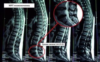
Intervertebral hernia of the lumbar spine on an MRI image
Intervertebral hernia is a pathology in which displacement of discs between vertebrae is diagnosed as a result of a violation of the integrity of the fibrous ring.
What does the pathology look like on a successful MRI image? The radiologist notices a small protrusion from the intervertebral space. The more advanced the disease, the larger the protrusion.
There are cases when the hernia is so small that it is practically not visualized on the image. Under such circumstances, the source of pathology is detected by indirect symptoms.
How often can you get an MRI?
MRI diagnostics of the lumbosacral spine is a safe research method.
For this reason, there are no restrictions on magnetic tomography. The exception is scanning with contrast.
MRI with contrast is indicated when it is necessary to improve the accuracy and efficiency of the upcoming procedure. Often, scanning with the introduction of a contrast agent is resorted to in cases of suspected tumor development.
Before starting MRI diagnostics, the subject is injected with a contrast component intravenously or into the spinal cord canal. Before using the drug, it is necessary to conduct an allergy test.
X-ray, CT or MRI - what to choose
It is impossible to answer the question posed unambiguously, since each of the given methods is strong in its own field.
The decision to prescribe a specific study is made by the doctor, who analyzes a number of factors, including the location of the suspected pathology, the patient’s condition, the degree of progression of the disease, and characteristics of the symptoms.
To conduct a comparative analysis of the procedures, we provide a table of key selection criteria.
First let's compare MRI and X-ray:
MRIX-ray| Special medical centers are equipped with the device | All healthcare institutions are equipped with the device |
| No radiation exposure | Based on X-ray radiation |
| Duration - 15-30 minutes | Duration: a few seconds |
| Expensive procedure | Low cost |
| Can be performed on pregnant women (2nd, 3rd trimester) | The study is contraindicated for pregnant women |
| The examination is applicable in the diagnosis of soft tissues, ligaments, discs between vertebrae, etc. | The procedure is informative in diagnosing bone tissue |
CT is comparable in information content to magnetic tomography, however, it is based on X-ray radiation. This option is cheaper (compared to MRI), but is not suitable for examining pregnant women and young children.
The most harmful diagnostic method
Where and at what price can you get an MRI of the lower back?
Magnetic tomography of the lumbosacral spine can be performed in specialized medical centers for a fee or free of charge.
In the first case, an MRI will cost the patient an average of 3,000 rubles (without contrast) and 4,500 (with contrast). The price depends on the region in which the center is located, the qualifications of the staff and the level of the clinic.
You can be examined free of charge if this tomography is included in the list of services under the policy.
Magnetic tomography of the lower back is performed to diagnose local pathologies, injuries and monitor treatment. MRI of the local area is short in time, painless and safe for human health.
The risk of adverse reactions is minimized, especially in the absence of contrast.
MRI of the local area has a number of advantages over computed tomography and traditional x-rays, however, it is applicable if it is impossible to diagnose anomalies using alternative methods.
Video
Source: https://osnimke.ru/pozvonochnik/mrt-poyasnichno-kresttsovogo-otdela.html
MRI of the lumbosacral spine
MRI of the lumbosacral spine is a promising and reliable method of radiological diagnostics.
It allows you to make an accurate diagnosis and prescribe the correct treatment. The examination is carried out in two projections: transverse and sagittal.
An MRI of the lumbosacral spine is performed as prescribed by a doctor or when pain occurs in the lumbar region.
Content
Description of the diagnostic method:
- Time: from 30 to 60 minutes (time increases if MRI is performed with contrast).
- The need to administer a contrast agent: depends on the indications, prescribed by a doctor.
- Necessity of preliminary preparation: not required
- Presence of contraindications: yes
- Restrictions: the weight of the subject should not be more than 180 kg, waist height - less than 32 cm, waist circumference - up to 140 cm.
- Conclusion preparation time: from 30 to 60 minutes
- Children: MRI is performed for patients aged 7 years and older.
Indications for tomography
MRI of the lumbosacral spine is prescribed in the following cases:
- Determining the cause of acute or chronic back pain, making an accurate diagnosis.
- Confirmation of osteochondrosis or other degenerative diseases.
- Confirmation or absence of musculoskeletal diseases that can cause pain in the lumbosacral spine.
- Examination of the department for the presence or absence of hemangiomas, which put pressure on the spinal bodies, epidural space and blood vessels of the spinal cord.
- Diagnosis of diseases associated with damage to intervertebral discs: protrusion, hernia, dorsopathy.
- Detection of primary and metastatic tumors, infectious diseases affecting the functioning of the musculoskeletal system.
- Study for the presence or absence of congenital anomalies of the lumbosacral spine, surrounding tissues and bones of the pelvic column.
- Examination for the presence of transitional vertebrae, spinal column injuries, osteoporosis.
- Diagnosis of the patient's condition with ankylosing spondylosis, spondylosis, sacroiliitis.
- Evaluation of spinal cord tissue and blood circulation in this area.
Contraindications
Not in all cases, patients with problems in the lumbosacral spine are prescribed an MRI.
This procedure has contraindications:
- MRI cannot be performed if there are metal objects : pacemakers, vascular clips, etc. This is dangerous to health, since the magnetic fields of tomography lead to disruptions in the operation of these devices and heat up the metal. An exception is structures in the human body that are not attracted by a magnet, for example, braces, titanium dentures, dental implants, etc.
- Claustraphobia may be a limitation for MRI . In this case, the doctor suggests performing the procedure in a state of medicated sleep or on an open-circuit device.
- MRI with contrast should not be performed during pregnancy and lactation . The drug has a negative effect on the fetus and milk production.
- Allergic reaction to contrast agent.
- Chronic renal failure , due to which the body will not be able to remove the contrast agent for a long time.
| If you have one of the contraindications to the procedure, then it is better to refuse MRI and choose alternative research methods. In any case, you should first consult a specialist. |
Advantages of the method
The main advantages of MRI of the lumbosacral spine are time savings and no need to use other diagnostic methods.
Previously, during the examination, surgical manipulations (puncture with examination of the cerebrospinal fluid), radiographic techniques (discography, pneumomyelography), and radiographic examination (images of the spinal column in different projections with a static load and at rest) were used. Now MRI can replace them all.
The MRI result is known within a few minutes after the examination. The specialist performing the MRI provides the images and a description of them; in many medical centers, a recording of the examination can be obtained on digital media.
How to do an MRI of the lumbosacral spine
MRI is the most accurate examination of the body at the moment.
- It is carried out by experienced specialists using modern equipment.
- The procedure is absolutely harmless, but you still need to know about some safety measures.
- First, the patient must fill out a questionnaire that will help identify contraindications to the procedure.
- Before an MRI, you need to remove clothing with metal elements, metal jewelry, and remove a USB flash drive and mobile phone from your pockets.
The procedure is performed horizontally. First, the subject lies down on the table, his head and arms are fixed, then the table is moved into the annular part of the apparatus. The patient's legs remain exposed only.
During an MRI, you need to lie still to get high-quality images. If the noise from the operating device is unpleasant, you can insert earplugs.
The contrast agent is injected into the vein a few seconds before the MRI; there is no discomfort.
MRI can be performed in several modes:
- Tomography with contrast . First, a special substance is injected, then pictures are taken. Contrast helps improve image quality and is required to determine the development of tumor and vascular malformations.
- Native tomography . It allows you to take a series of images without prior preparation and administration of a contrast agent.
Preparation for the procedure
No special preparation is required to perform an MRI.
A person can lead his usual lifestyle, not change his diet or stop taking medications. To undergo an examination, you must take a referral from a doctor, who also decides whether to carry out the procedure with or without contrast.
For the MRI, you need to bring pictures from previous studies, an extract from the outpatient card and medical documents that contain information about the disease.
Contrast enhancement
Gadolinium-based drugs are used for contrast enhancement in MRI.
These drugs increase the information content of the study. The dye is non-toxic, created on the basis of gadolinium chelate complexes , and has no side effects. TO
- Contrast enhancement cannot be used when conducting examinations in the first trimester of pregnancy and in case of individual intolerance to the drug.
- After entering the body, the contrast agent is retained in the tissues and allows them to be better visualized during MRI.
- The drug is eliminated from the body within 24 hours.
Doctor's report
After the pictures are taken, the doctor conducting the examination writes a report.
In most cases, this does not take more than an hour, but sometimes the specialist needs more time. Most clinics in large cities issue examination results on electronic media and send them by email.
With the obtained MRI results, you must contact the doctor who sent you for examination to establish a diagnosis and treatment. If the MRI was performed on the patient’s initiative, then after the examination, if pathologies are detected, you should contact the following specialists:
- traumatologist , if problems in the lumbosacral spine are associated with injuries or their consequences;
- to a neurosurgeon if surgical intervention is necessary;
- neurologist for diseases of the spinal cord and spine;
- to an oncologist if a tumor is discovered during research.
Exploration opportunity for children
Children can have an MRI of the lumbosacral spine.
- Doctors usually prescribe examinations for patients over 7 years of age, but these are only conditional indicators.
- The most important thing is that the child should lie quietly and not move during the examination.
Prices in Moscow and other major cities
MRI is one of the most expensive and effective procedures. The cost of the examination depends on the location, and the price difference between different medical centers can be significant.
To save on a complex examination, you need to do an MRI and choose a specialist for consultation in one clinic. But you shouldn’t skimp on MRI.
The average cost of an examination in Moscow is 4,500 rubles , the time is from 15 to 30 minutes. Some clinics offer discounts and examinations at night.
In other large cities of Russia, prices for MRI may vary significantly.
In St. Petersburg, the average cost of an examination complex is 3000 - 3500 rubles, in Ufa 2000 - 2300 rubles, in Tula about 3000 rubles, in Yaroslavl - 2500 rubles, in Yekaterinburg - 2800 rubles.
Description of some clinics
- In Moscow, MRI can be done in many specialized centers and clinics.
- At the Central Committee of the Russian Academy of Sciences in Moscow, an examination of the lumbosacral region will cost 4,700 rubles, with the introduction of a contrast agent 12,200 rubles.
- At the MRI center you will have to pay at least 3,380 rubles, and an MRI with contrast costs 6,200 rubles.
In the centers of V.I. Dikul prices for examination range from 3200 to 4700 rubles.
At the Medic-City clinic, an MRI costs 4,000 rubles, and to save money, you can come for an examination at night.
At the Stolitsa Medical Center, the cost of an MRI of the lumbosacral spine is 4,300 rubles, with the introduction of a contrast agent – 9,200 rubles.
In St. Petersburg, you can sign up for an MRI at the specialized center “Health Workshop”; open equipment is used here.
The cost of examination of the lumbosacral region is 3,300 rubles. The center offers discounts that allow you to save from 290 to 900 rubles.
At the National Diagnostic Center of St. Petersburg, an MRI of the lumbosacral spine will cost 3,000 rubles.
The cost of MRI in other large cities differs from prices in Moscow and St. Petersburg. For example, in Ufa, at the Outpatient MRI Center, an examination will cost no more than 2,100 rubles, and at the Tesla LLC MRI Center the cost does not exceed 2,300 rubles. In regional centers, the cost of an MRI without contrast injection is from 2,000 to 3,000 rubles.
Reviews
Among all methods for diagnosing diseases of the lumbosacral spine, MRI is the most effective and reliable .
With its help, you can see even minor deviations in the condition of the spine. Many who have already undergone this examination highlight its advantages and disadvantages.
- The main advantages of MRI : accuracy of results and time saving (20-40 minutes is enough).
- But there are also disadvantages that most people doing MRI note - the discomfort that occurs during the procedure and the high cost.
| MRI is an effective examination that will help identify diseases and pathologies of the lumbosacral spine. Tomography allows not only to make a correct diagnosis, but also to monitor the process of treatment and restoration of the spine after injury. This method has many advantages, but there are contraindications and disadvantages. |
Source: http://vashaspina.com/diagnostika_zabolevaniy/mrt/poyasnichno_kresttsovogo_otdela_pozvonochnika.html
MRI of the lumbosacral spine: indications, contraindications
MRI is a modern diagnostic method, recognized by all specialists as the most informative. It is used in a variety of areas of medicine, as it helps to establish the presence of pathology at an early stage, when other methods are not yet effective.
Orthopedists and vertebrologists also actively prescribe this study to diagnose the cause of pain.
Magnetic resonance imaging of the lumbosacral spine is a popular diagnostic method that allows you to prescribe the most effective therapy.
Collapse
An MRI of the lumbosacral spine is performed if an accurate diagnosis cannot be made in any other way. This is explained by the fact that diagnostics are complex and expensive. MRI is mandatory in the following cases:
- Uncertain diagnosis;
- Back pain of unknown etiology;
- Suspicion of birth trauma in the newborn;
- After a significant injury - a traffic accident, a fall from a height;
- Systemic developed disorders, when MRI makes it possible to diagnose other pathologies (when internal organs are compressed due to severely impaired posture);
- Lack of effect from the treatment;
- Suspicions of active involvement of soft tissues in the process;
- There are restrictions on the use of other diagnostic methods (x-rays).
Since diagnosis is carried out as a last resort, it cannot be abandoned, since there are no alternatives.
Contraindications
There are few contraindications to MRI of the lumbosacral spine. The technique is one of the safest and has no restrictions in use. But sometimes MRI is carried out with a number of precautions. These are the following features:
- The patient’s weight is more than 150 kg (in recent years, devices have appeared that can informatively examine a patient weighing up to 170 kg, but no more, and they are not yet very widespread);
- The patient is pregnant (there are no direct contraindications, but the exact effect of the magnetic field on fetal development has not been studied, however, the study is carried out using special protective aprons if there are vital indications);
- The presence of tattoos made with metal-containing ink (the metal will interact with the magnet);
- The presence of metal-containing microdermals, permanent metal piercings, for the same reasons;
- Intolerance to a contrast agent is a contraindication for diagnostics with contrast;
- The presence in tissues of fragments, implants made of materials capable of interacting with a magnetic field (now almost never encountered);
- Fear of closed spaces - during the process the patient spends a long time in a cramped space (the problem is solved by choosing open-type MRI machines).
Contraindications are conditional; most patients can perform the procedure.
What does it show?
The MRI machine in which the patient is placed generates a magnetic field that interacts with the fluid in the body's cells. Depending on the difference in the reaction of cells to the field, a diagnostic picture is constructed.
When deciphering this picture, it is possible to determine in which tissues the disturbances occurred.
The advantage of the method is that it is informative for all types of tissues - soft, nervous, vascular, bone, articular (unlike x-rays, which can correctly display only the condition of bone tissue).
If correctly interpreted, MRI results can be used to diagnose:
- Presence, nature of the neoplasm;
- Injuries of soft, bone, joint tissues;
- Inflammatory processes;
- Accumulations of liquids;
- Formation/overgrowth of cavities;
- Displacements, dislocations;
- Areas of tissue degradation/destruction;
- Changes in the cellular composition of tissues.
Thanks to these features, MRI can diagnose almost any disease that manifests itself in changes in organs and tissues.
How to prepare?
Preparation for an MRI of the lumbosacral spine does not take long. However, before making a diagnosis, it is better to follow the recommendations:
- Choose thin, not too tight clothes;
- Check the absence of metal elements on clothing - rivets, zippers, buttons;
- Remove jewelry, piercings;
- On the eve of the procedure, it is advisable not to eat foods that can cause bloating, but they will not have a significant effect on the results.
No discontinuation of medications is required. The only condition is that if an MRI with a contrast agent is indicated, it is necessary to do an allergy test for the drug 24 hours before the procedure (it is applied cutaneously). Intolerance to it is extremely rare; this is a mandatory procedure.
How do they do it?
The scanning procedure itself lasts approximately 20 minutes (depending on the device). But along with the preparation time for it, the subject will stay in the medical facility for about 40-60 minutes. Then you will need to wait 20-90 minutes for the study to be decrypted.
With contrast
The subject puts on shoe covers and enters the office. There he is given a contrast agent intravenously. Then he goes into the second room with a tomograph located in it. Here he removes outer/metal-containing clothing (there is no need to completely undress for diagnosis), jewelry, piercings, and is placed on the extended couch of the MRI machine.
The device turns on, the couch slides into the tube of the device. The device extends a special arch that generates a magnetic field. A characteristic hum is heard, indicating the operation of the apparatus and the generation of a field. Then a tapping sound appears indicating the start of the scan. The patient must remain relatively motionless throughout the procedure.
After scanning is completed, the noise disappears and the device turns off. The table moves out of the tube, the patient gets up, gets dressed and can leave the office.
No contrast
A study without contrast is carried out in completely the same way, but the patient is not first injected into a vein with a contrast agent. You also do not need to do allergy tests for the drug first. The procedure itself, due to the absence of an injection, takes 5-10 minutes less. It also usually costs much less.
Decoding the results
Interpretation of the MRI results of the lumbosacral region is carried out by the doctor who performed the diagnosis. The patient is given a printed transcript, confirmed by a personal seal and signature of a specialist.
But the most authoritative opinion will be the conclusion of the doctor who prescribed the procedure, since he is able to assess the dynamics, the nature of the changes, and correlate them with the ongoing pathology and the general characteristics of the patient’s health.
Therefore, even if, according to the doctor’s opinion, there are no MRI pathologies, it is necessary to consult an orthopedist about the indicators.
Pictures
- An MRI of the lumbosacral region produces the images shown in the photo below.
Are there any side effects?
The procedure is completely safe, has no side effects or complications. However, people prone to claustrophobia may experience increased nervousness, excitability, and anxiety for some time after it.
Actions after the procedure
After the study, a person receives his images within 20 minutes to an hour and a half. The time varies depending on the specialist’s workload.
The subject receives a set of images printed on a special film and a description of the results, printed and signed, with the doctor’s stamp.
In some medical institutions, lists are recorded on disk, but the transcript must still be issued in the form of a document.
After receiving the MRI materials, the patient’s task is to get to a consultation with their therapist who prescribed the MRI as soon as possible with these images. Only he is able to make a diagnosis and prescribe therapy.
What is the price?
The cost of an MRI of the lumbosacral spine varies, depending on the popularity of the medical center, the type of machine used, and whether the study is performed with or without contrast. The average cost of the procedure without an intravenous injection of a contrast agent ranges from 3,000 to 7,000 rubles. MRI of the lumbosacral spine with contrast – 6,000 – 9,000 rubles.
It is also possible to carry out research for free. Some insurance companies include this service in the list of free services for patients under compulsory medical insurance. But in this case, you must show up strictly on the specified date (usually there is not much choice) and wait in line. Urgent free diagnostics are carried out only for vital indications, in a hospital setting.
Where to do it?
Conclusion
Most specialists try to avoid prescribing an MRI in order to save the patient money. Magnetic resonance imaging is as informative as possible, and therefore can replace all other diagnostic procedures when there are contraindications to them.
Source: https://columna-vertebralis.ru/diagnostika/mrt/mrt-poyasnichno-kresttsovogo-otdela-pozvonochnika.html