IT IS IMPORTANT TO KNOW! An effective remedy for restoring vision without surgery or doctors, recommended by our readers! Read more…
The inner layer of the eye is called the retina. It regulates the interaction between the central nervous system and the organs of vision. It is she who is responsible for translating light signals perceived by the organs of vision into corresponding nerve impulses sent to the brain.
A number of visual impairments, which are characterized by changes in the retina and are irreversibly destructive, are called dystrophy. Pathological changes in retinal tissue always lead to a stable deterioration of vision. To prevent its loss and progression of the disease, it is necessary to treat retinal dystrophy.
Symptoms
Symptoms depend on the type of disorder, but they also have general deviations from the norm. The most common symptoms of the disease:
- loss of vision;
- image distortion;
- the patient feels like there are dark spots before the eyes;
- objects without clarity;
- twilight lighting sharply worsens perception;
- decrease in peripheral vision (and even its complete loss).
Causes of the disease
The impetus for the development of retinal dystrophy (acquired), a dangerous disease for eye health, can be many reasons of a different nature:
- any type of eye injury;
- toxic effects on the body of any origin;
- other diseases of the visual organs, for example, myopia, inflammation, etc.;
- complication after surgery;
- infectious diseases;
- systemic health problems (high blood pressure, kidney disease, diabetes, etc.).
All of these listed reasons can contribute to the development of the disease, but they are just risk factors. With a hereditary predisposition, the risk of the disease can be considered extremely high. For your information! Even stress, pregnancy, excess weight, and direct solar radiation can play a role as a trigger for the onset of the disease.
Diagnostics
Effective treatment of the disease requires competent, timely diagnosis and examination by an ophthalmologist. Among the studies that can be recommended are:
- retina;
- light sensations;
- Ultrasound;
- fundus;
- eye structures;
- determination of the boundaries of peripheral vision;
- visual acuity test;
- electrophysiological examination, which gives a picture of the state of the retina itself and nerve cells;
- fluorescein angiography to study the vessels of the eye.
Types of retinal dystrophy
The disease is divided into types, which is associated with its origin and the location of the inconsistent pathological process of the retina. There are hereditary and acquired forms of the disease.
Hereditary. This is an inherited dystrophy; it includes several types. But the most common are: dotted white and pigmented.
- Spot white. This pathology is congenital. Development occurs from early childhood, which leads to a deterioration in visual perception even before school.
- Pigmented. This is a genetic eye disease. Dystrophy of this type is characterized by disruption of the functioning of photoreceptors, which are important for human twilight vision.
The disease has a slow course with stable deterioration, despite alternating periods of remission after exacerbations. Most often the disease manifests itself while studying at school. By the age of 20, the disease clearly manifests itself and a diagnosis is made. With age, the condition worsens so much that it can lead to complete loss of vision.
Acquired dystrophy
This type of disease is typical for older people. It may occur in combination with other diseases of the visual organs associated with age-related changes. It is impossible to completely cure it conservatively. Depending on the affected area, there are:
- Generalized (with this type of dystrophy, damage to the retina affects all its areas).
- Central (macular).
- Peripheral.
Central dystrophy. Macular degeneration of the retina is named for its location in the area of the retina (macula) that is responsible for the area of clearest vision. Types of macular degeneration:
Depending on the pathology and damage to the retina, the following types are distinguished:
- serous choriopathy;
- age (wet or dry);
- colloidal;
- cone (congenital);
- Best's disease;
- Franceschetti's disease;
- Stargardt's disease.
Important! In the central form of dystrophy without damage to peripheral areas, the development of the disease does not lead to blindness.
Patients experience discomfort and complain to the ophthalmologist of the following manifestations:
- doubling of objects;
- the image of objects is distorted.
Age-related dystrophy. Treatment for macular degeneration of the retina depends on the clinical form (dry or wet) and the degree of pathology. Both forms of the disease are characteristic of the age group over 60 years.
The central part of the retina is damaged due to age-related changes. It is the macula that is responsible for the eye's ability to distinguish small objects.
But even in cases of severe disease, the peripheral parts of the retina continue to perform their functions and blindness occurs in very rare cases.
The peculiarity of the wet form is the penetration of fluid and blood into the retina. The loss of vision occurs extremely quickly, up to several days. Treatment of this condition is complex and surgical.
The most common is the dry form, in which the deterioration occurs gradually. The disease is characterized by the accumulation of cellular breakdown products between the retina and the lining of blood vessels.
Patients with age-related macular degeneration are recommended to undergo inpatient treatment once every six months, give up bad habits, and be required to wear sunglasses.
Peripheral. This type of retinal lesion is characterized by damage only to the peripheral area without affecting the macular area. Of the manifestations of the disease, a person can only note the appearance of “flies” before the eyes.
A feature of peripheral dystrophy is its difficult diagnosis. When an ophthalmologist examines the patient's fundus, the peripheral areas are practically invisible. Pathology can only be diagnosed using special equipment. Classification of peripheral dystrophy:
- pigmented;
- fine-grained;
- frost-like;
- lattice.
Often, against the background of myopia, retinal detachment may occur. In this case, the patient complains of a feeling of a veil before the eyes, but without surgery, vision can no longer be restored.
Treatment methods
Dystrophy is a serious disease that can lead to complete blindness. Already lost vision at the onset of the disease and exacerbations cannot be restored. For the most part, treatment is supposed to be symptomatic, since, except for secondary ones, any types of degeneration have a hereditary predisposition. Treatment is mainly aimed at the following actions:
- stabilization of the condition;
- prolongation of periods of remission;
- strengthening the muscles of the eyes and blood vessels;
- improvement of metabolic processes in the organs of vision.
Methods of treating the disease:
We recommend!
To treat eyes without surgery, our readers successfully use a proven method. Having carefully studied it, we decided to offer it to your attention. Read more…
- medicinal;
- physiotherapy;
- surgery;
- laser coagulation.
In some cases, eyes are treated with folk remedies, which can be used in combination with other treatment methods, but always under the supervision of the attending physician.
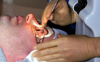
Laser coagulation
This treatment method is designed to prevent a serious complication of dystrophy - retinal detachment and prevent vision loss. The laser allows you to provide a targeted effect without damaging healthy tissue. During the manipulation, the damaged areas are cauterized to the desired areas of the eye to the specified depth.
Surgery
Whether it is possible to do without surgical intervention is determined by the doctor after a comprehensive examination of the patient. Retinal dystrophy is treated surgically most often in cases where the disease was diagnosed late and when it no longer makes sense to hope that eye injections will help.
To improve metabolic processes and normalize blood supply, patients undergo vasoreconstructive surgery. When the wet form is diagnosed, treatment of macular degeneration of the retina is aimed at preventing the accumulation of fluid in the retinal tissue. To prevent retinal destruction, the following surgical methods are used:
- Vasoreconstruction, which is based on the use of transplants;
- Revascularization results in an increase in the lumen of functioning vessels.
Physiotherapy
For retinal dystrophy, physiotherapy is prescribed in the initial stages of the disease to strengthen the eye muscles and the retina itself. Several methods of physiotherapy exist and are used:
- ultrasound therapy;
- phonophoresis;
- electrophoresis;
- microwave therapy;
- laser irradiation of blood (intravenously).
Drug treatment
Retinal dystrophy can be treated with medications only at the earliest stages of the disease. In other situations, the positive effect of such conservative treatment alone is impossible. The following medications are indicated for patients:
- vitamins E and A;
- angioprotectors;
- corticosteroids;
- products with lutein;
- agents that strengthen the walls of blood vessels;
- local acting vasodilators;
- antioxidants;
- general action vasodilators.
Prevention
As preventive measures, those who are at risk for eye diseases are recommended:
- spend less time under sunlight;
- perform eye exercises;
- lead a healthy lifestyle;
- take vitamin complexes;
- give your eyes a chance to rest;
- good workplace lighting;
- lack of heavy physical activity;
- periodic medical examination by an ophthalmologist.
Treatment with traditional recipes
As an additional therapy and an integrated approach to the treatment of dystrophy, treatment with folk remedies can be used.
Pine decoction. To prepare you will need:
- 1 l. water;
- 4 tsp each crushed rose hips and onion peels;
- 10 tsp. pine needles.
Preparation:
- Connect all components.
- Fill with warm water.
- Cook for 10 minutes over low heat.
- Cool.
- Strain.
- Take throughout the day, distributing evenly.
- The course is 30 days.
- Garlic drops
- To prepare you will need:
- 1 l. vodka;
- 1 kg garlic.
Preparation:
- Pour vodka over garlic in a jar.
- Close the container tightly with a lid.
- Place in a warm place.
- Leave for 2 weeks, stirring the contents periodically.
- Strain.
- Take 13 K before meals three times a day.
The course of treatment is 60 days, then a break for 1.5 weeks.
Serum drops
For preparation you will need: 2 tsp. water and the same amount of fresh goat whey.
Preparation: mix the ingredients.
Application: Drop 1 drop into the eyes. Blindfold your eyes with a cloth. Lie down for 30 minutes. without moving your eyes.
Course - 7 days.
Treatment at home should be carried out after consultation with an ophthalmologist.
Conclusion
At the moment, there is no remedy that would provide a painless cure for retinal dystrophy. Therefore, it is so important to undergo medical examinations by an ophthalmologist on time, monitor your health, and, if necessary, undergo treatment prescribed by an ophthalmologist.
By secret
- Incredible... You can cure your eyes without surgery!
- This time.
- No trips to the doctors!
- That's two.
- In less than a month!
- That's three.
Follow the link and find out how our subscribers do it!
Source: https://aokulist.ru/anatomiya/setchatka/kak-lechit-distrofiyu-setchatki-glaza.html
Modern methods of treatment of central and peripheral retinal dystrophies
To date, treatment for retinal dystrophy does not completely eliminate the disease. Using various techniques, doctors can only stop the development of dystrophic processes. In some cases, ophthalmologists are able to achieve a temporary increase in visual acuity.
However, in the absence of supportive therapy, the disease soon begins to worsen again.
Any dystrophy causes irreversible organic damage to the retina of the eye, due to which it ceases to function normally. Since the retina has a complex structure, it is almost impossible to restore its structure and functions. This means that a person with dystrophy is unlikely to be able to see well again.
Which doctor treats retinal dystrophy?
The diagnosis and treatment of pathology is carried out by a retinologist - an ophthalmologist who specializes in diseases of the retina. If surgical intervention is necessary, a vitreoretinal surgeon comes to his aid. This specialist performs complex operations on the posterior segment of the eyeball.
Methods
Using what methods and how to treat retinal dystrophy? Medical tactics depend on the location of dystrophic foci, their size and type. The choice of treatment method is also influenced by the presence (or absence) of concomitant diseases and complications.
Uncomplicated peripheral dystrophies are usually treated with medications. If a patient is diagnosed with excessive thinning or ruptures of the retina, he undergoes laser treatment. But for macular degeneration, laser is not used, since this is fraught with serious complications.
In the initial stages, medications are used to combat central degenerations. If the wet form of macular degeneration develops or macular edema appears, the patient is administered anti-VEGF drugs.
Due to the abundance of forms and types of retinal dystrophies, the question of treatment tactics for each of them is decided on an individual basis. First, the doctor carefully examines the patient and makes a diagnosis. After this, he tells the patient in detail about his illness and possible methods of combating it.
Based on the information received, the person himself makes the final decision. No one has the right to force a patient to buy expensive medicines or agree to an expensive operation.
Medication
Drug treatment is used for age-related, pigmentary and peripheral retinal dystrophies. Patients are prescribed drugs that improve blood supply to the tissues of the eyeball and saturate the retina with nutrients. Medicines can be taken orally or administered parabulbarly.
Drugs used to combat retinal degeneration
A drugAction
Features of application
| Eufillin | It has an antispasmodic effect, that is, it relieves spasm from the vessels supplying the retina of the eye. Improves blood circulation in the tissues of the eyeball, thereby slowing down the course of the disease | The drug is taken orally, in the form of tablets or capsules |
| Emoxipin | The medicine strengthens retinal vessels, has an antiplatelet and antihypoxic effect. Thanks to this, it improves blood circulation in the retina, which slows down its destruction | For the treatment of retinal dystrophy, the drug is used in the form of parabulbar or subconjunctival injections |
| Cavinton | Antispasmodic agent with antihypoxic effect. Normalizes microcirculation, thins the blood, slows down platelet aggregation, protects tissues from the effects of free radicals and oxygen starvation |
The drug can be prescribed in tablets or administered intravenously. |
| Retinalamine | The manufacturer positions Retinolamine as a drug that improves metabolism and normalizes energy processes in the retina. Restores some damaged cells and prevents the death of others | The medicine is administered intramuscularly or parabulbarly |
| Adruzen Tsinko | It is a dietary supplement containing a lot of active ingredients. Improves metabolism, preventing retinal destruction | Available in capsule form to be taken orally |
| Visiomax | The medicine contains a complex of vitamins, minerals and pigments that actively nourish the retina | Taken orally in tablet form |
Physiotherapy
Along with medications, physiotherapeutic methods are used to treat central and peripheral chorioretinal dystrophies of the retina. They are prescribed in courses lasting 10–14 days. A person must undergo at least two courses of physiotherapy per year.
Methods that may include in treatment:
- magnetic therapy;
- photo- or electrical stimulation of the retina;
- stimulation of the retina with low-energy laser radiation;
- electrophoresis with nicotinic acid, No-Spa or heparin.
If there is no therapeutic effect from conservative therapy, the patient is recommended to use more effective methods.
Intravitreal administration of anti-VEGF factors
In modern ophthalmology, anti-VEGF drugs are used to treat the wet form of macular degeneration and macular edema of the retina. The drugs are administered intravitreally, that is, directly into the eyeball. The procedure is performed under local anesthesia.
The administration of Lucentis or Aylia helps to remove macular edema and even improve the patient’s vision. To get a good, lasting result, you need to perform several injections. Due to the high cost of drugs, not every person can afford this procedure.
Description of anti-VEGF factors
Lucentis
Ailia
| Active substance | Ranibizumab | Aflibercept |
| Mechanism of action | Medicines suppress angiogenesis - the process of growth of new blood vessels. Thus, they help to avoid neovascularization, macular edema, and hemorrhage. They also reduce the permeability of the vascular walls, thereby preventing hemorrhagic leakage of the retina | |
| Indications for use |
|
|
| Possible side effects and complications |
|
|
| Average cost per package | 42,500 rubles | 38,000 rubles |
| How many injections are needed? | Three injections at 1 month intervals |
Avastin is also an anti-VEGF factor. However, the drug is intended exclusively for the treatment of cancer. It should absolutely not be used in ophthalmology. Intravitreal administration of the drug can lead to unpredictable consequences and serious complications.
Surgical intervention
Central retinal dystrophy, complicated by a macular hole, is treated surgically. The essence of the surgical intervention is to remove the vitreous. After this, an air bubble or a special liquid is introduced into the cavity of the eyeball, which puts pressure on the retina and prevents its detachment.
Peripheral chorioretinal retinal dystrophy can also be treated surgically. The indication for vitrectomy is the formation of adhesions between the retina and the hyaloid membrane of the vitreous body. Removal of the strands in this case is necessary to prevent retinal detachments and ruptures.
Stages of surgical treatment:
- Exploratory survey . Before the operation, the patient is sent for consultations with specialists and for tests. This is necessary to identify concomitant diseases that can cause intraoperative complications.
- Preoperative preparation . A few days before surgery, the patient begins to put drops prescribed by the doctor (antibiotics, non-steroidal anti-inflammatory drugs) into the eye. Such a preventive measure helps reduce the risk of complications in the postoperative period.
- Pain relief . The operation is performed under local retrobulbar anesthesia. The anesthetic is injected into the postorbital space.
- Operation . During surgery, the patient lies on his back. His head is carefully fixed. The operation usually takes about 30-40 minutes. After this, the patient is transferred to the recovery room.
- Rehabilitation . After the operation, the patient must use the medications prescribed by the doctor for some time. In the future, he will need to be monitored by an ophthalmologist.
Laser
Laser coagulation is used to protect areas of degeneration and suppress pathological vascular growth. This allows you to slow down the development of the disease and reduce the risk of complications. Timely laser coagulation of newly formed vessels makes it possible to avoid hemorrhages, which are fraught with severe deterioration or even loss of vision.
The laser is used to treat lattice degeneration, retinal pigmentary degeneration, cochlear track and cobblestone dystrophies. Laser coagulation is not performed in the macular area due to the high risk of complications. Because the mesh in this area is very thin, it can tear or peel off.
Since it is impossible in principle to cure retinal dystrophy, laser coagulation has only a temporary effect. Don’t think that it will help you forget about the disease forever. Unfortunately, over time, the patient may develop new degenerative lesions. Therefore, after the procedure, it is necessary to continue to be observed by an ophthalmologist.
Traditional methods
How to treat retinal dystrophy with folk remedies and is it worth doing it at all? Unfortunately, traditional medicine is in many ways inferior to traditional medicine. It is powerless against macular edema, ruptures and detachments. The use of various tinctures and decoctions in this case will only harm the patient.
Treatment at home using folk remedies is possible only for slowly progressive peripheral and age-related retinal dystrophies. It should be noted that traditional medicine methods can only be used after consultation with your doctor.
Folk remedies that are used for dystrophies:
- crushed sprouted wheat;
- infusion of nettle leaves;
- a decoction of blueberries, rowan and sea buckthorn;
- a collection of chamomile, lingonberries, calendula, dandelion and dried cucumbers;
- blueberry infusion.
The above remedies are taken orally. But to wash the eyes, use a decoction of nettle and lily of the valley leaves, aloe juice, a mixture of water and fresh goat milk. It is believed that these remedies nourish the eyes and help cope with the disease.
Is eye dystrophy curable? Yes, there are many methods that can successfully combat the disease. However, the second question is, is it possible to completely cure retinal dystrophy and forget about the problem forever? Unfortunately, this is impossible to do these days. However, despite this, sick people should not despair.
Thanks to the achievements of modern medicine, it is possible to slow down the progression of the disease and preserve vision. To combat the disease, medications, physiotherapy, surgery and laser methods are used to treat retinal dystrophy. They can be used separately or combined.
Alina Lopushnyak, ophthalmologist,
specially for Okulist.pro
Source: https://okulist.pro/bolezni-glaz/setchatka/lechenie-distrofii-setchatki-glaza.html
Retinal dystrophy: types and methods of treatment
The retina of the eye is the thinnest membrane consisting of nerve cells. It covers the entire eyeball from the inside. Light-sensitive cells in the retinal tissue convert light impulses into electrical impulses. Then, along the optic nerve and optic tract, electrical signals enter the human brain, where they are deciphered and transformed into visual images that we see before our eyes.
Causes of retinal dystrophy
The development of pathology can be triggered by many factors. Basically, dystrophy occurs:
- in old age, as a consequence of aging processes in the body
- by hereditary predisposition
- for hypertension, atherosclerosis and other vascular diseases
- as a complication of diabetes
- for vitamin deficiencies and poor nutrition
- if you are overweight
- from the harmful effects of smoking
- as a result of stress and nervous shock
- after a viral illness
- as a result of harmful effects of ultraviolet radiation
Up to 40% of different types of this retinal pathology are observed in myopic people. With farsightedness, dystrophy is observed only in 8% of cases, from 2 to 5% occur in people with normal vision. All causes of the disease can be classified into local and general.
The first include:
- genetic predisposition;
- eye injury;
- myopia;
- inflammatory and infectious eye pathologies
Common reasons include:
- diabetes
- atherosclerosis, hypertension
- various types of intoxication
Symptoms of dystrophy
Dystrophy may have different symptoms, depending on the form of the disorder, but basically, several general signs of the disease can be named. So, you should be concerned if you have:
- Visual acuity decreases
- Decreased or lost peripheral vision
- Disorientation at dusk
- Dark spots appear before the eyes
- The clarity of the outlines of objects is lost
- Visible images are distorted
- Straight lines appear crooked
- Letters fall out when reading
Retinal dystrophy is a general concept that includes various forms of the disease that have different characteristics, symptoms and effects on vision.
Types of retinal dystrophy
All types of retinal dystrophy have common symptoms, which include the progression of visual dysfunction and degenerative changes in the retinal fiber. It is worth highlighting several main types of retinal dystrophy. First of all, retinal dystrophy is divided into:
Congenital dystrophy is a genetically determined, inherited disease. The most common form of congenital dystrophy is retinal pigmentary dystrophy. All congenital dystrophies are incurable, steadily progress and lead to significant irreversible vision loss.
Acquired dystrophies can be divided into:
- central
- peripheral
Central retinal dystrophy
(macular degeneration
)
It affects the central fovea of the retina, that is, the area that is responsible for the most accurate vision and discrimination of small details. It is characterized by impaired central vision, while peripheral vision remains normal. It often affects the eyes of those who suffer from myopia.
With central retinal dystrophy, serious problems arise with driving, writing, reading and drawing. Age-related macular degeneration is the most common cause of vision loss in older people in developed countries. The progression of the disease can be stopped if treatment is started in time.
Age-related macular degeneration can cause significant vision loss, but it never leads to complete blindness.
Peripheral dystrophy
It affects the periphery of the retina, that is, the area that does not participate in vision. It is not accompanied by deterioration of vision, but is dangerous because it can lead to the development of such a serious complication as retinal detachment. This form mainly occurs in nearsighted people.
Sometimes the presence of peripheral dystrophy may be indicated by the appearance of floaters in front of the eye. In this case, you need to urgently consult a doctor for a thorough examination of the periphery of the retina with mandatory pupil dilation.
If peripheral retinal dystrophy or tear is detected, then urgent laser treatment will be necessary to prevent retinal detachment.
Video - Professor M.E. Konovalov talks about macular degeneration of the retina
Watch from the 30th minute of the program “Live Healthy” with Elena Malysheva (release dated 10/08/2010).
Treatment of retinal dystrophy
Modern medicine has a sufficient number of methods aimed at treating retinal dystrophy. With their help, you can improve vision and stop the progression of the disease. The goal of treatment is to reduce the likelihood of complications that could lead to permanent vision loss.
The prognosis and course of the disease depend on how it is carried out. Timely consultation with a doctor increases the patient’s chances of restoring vision. But, nevertheless, only a few manage to regain their former sharpness, since in most cases, retinal dystrophy is caused by age-related changes.
If the patient comes to the clinic at the initial stage of the disease, he is prescribed drugs containing lutein, which is necessary for the normal functioning of the retina. It is also recommended to eliminate bad habits, if any, and protect your eyes from ultraviolet radiation. Vitamins will support vision, preventing the eyes from becoming overly tired due to visual stress.
In the wet stages of dystrophy, it is recommended to introduce special drugs into the vitreous body of the eye to relieve swelling of the central zone of the retina.
Timely and correct treatment will help you maintain good vision for many years!
Code: A03.26.019.002
Included in the price:
- OST diagnostics
- consultation with a laser surgeon
Code: A03.26.019.002
Included in the price:
- OST diagnostics
- consultation with a laser surgeon
Code: A03.26.003
Zenina M.L., Gorensky A.A., Trikhacheva E.A. 2,000 rub.
Code: A22.26.029
The price for the operation is per eye
Code: A22.26.029
The price for the operation is per eye
Code: A22.26.029
The price for the operation is per eye
Code: A22.26.029
The price for the operation is per eye
Code: A22.26.010
The price for the operation is per eye
Code: B04.029.001
Included in the price:
- visual acuity control – 3
- laser surgeon consultation – 3
- OST of the macula (as prescribed by a doctor)
Code: A16.26.082
Ophthalmologist RUB 60,000. The price includes:
- surgeon's work
- Consumables
- postoperative observation for 1 month from the date of surgery
The price for the operation is per eye
Code: A16.26.081
Ophthalmologist RUB 24,000. The price includes:
- surgeon's work
- Consumables
- postoperative observation for 1 month from the date of surgery
The price for the operation is per eye
Code: A16.26.142
Ophthalmologist RUB 40,000. The price includes:
- surgeon's work
- Consumables
- postoperative observation for 1 month from the date of surgery
The price for the operation is per eye
Code: A16.26.089.001
Professor Konovalov M.E. 36,000 rubles. Ophthalmologist 22,000 rubles. The price includes:
- surgeon's work
- Consumables
- postoperative observation for 1 month from the date of surgery
The price for the operation is per eye
Code: A16.26.089
Ophthalmologist RUB 67,000. The price includes:
- surgeon's work
- Consumables
- postoperative observation for 1 month from the date of surgery
The price for the operation is per eye
Code: A16.26.089
Ophthalmologist RUB 106,000. The price includes:
- surgeon's work
- Consumables
- postoperative observation for 1 month from the date of surgery
The price for the operation is per eye
Code: A16.26.089
Ophthalmologist RUB 145,000. The price includes:
- Consumables
- surgeon's work
- postoperative observation for 1 month from the date of surgery
The price for the operation is per eye
Code: A16.26.089
Ophthalmologist RUB 189,000. The price includes:
- surgeon's work
- Consumables
- postoperative observation for 1 month from the date of surgery
The price for the operation is per eye
Code: A16.26.089
Ophthalmologist RUB 20,000. The price for the operation is for one eye.
Code: A16.26.089
Ophthalmologist RUB 9,000. The price for the operation is for one eye.
Source: https://www.konovalov-eye-center.ru/info-for-patient/articles/dystrophia_setchatki/
Surgical treatment of macular degeneration - effective restoration of vision - Medical Center
Due to age-related sclerotic changes in the macula, the central part of the retina, macular degeneration may occur. The cells located in this sector ensure visual acuity, allow reading, and performing precise work.
With this disease, due to disturbances in capillary flow, the cones of the retina are destroyed, soon the patient develops a dark spot and the surrounding vision becomes blurred. Light-sensitive cells do not function normally due to the growth and appearance of new blood vessels, defective vessels with impenetrable walls. Lack of treatment leads to vision loss.
Causes and main symptoms
In this disease, central vision impairment occurs due to macular degeneration. The specific causes of this condition have not been determined by medicine. But among all the factors, age is the main one. Also, factors that provoke macular degeneration include:
- age over 55 years;
- gender, occurs twice as often in women;
- hereditary predisposition;
- overweight;
- smoking;
- injuries to eye tissue.
Factors such as poor ecology, lack of vitamins, and prolonged direct exposure to UV radiation have a bad effect on the condition of the macula of the eye.
As the disease progresses, it goes through several stages. There are dry and wet forms of macular degeneration. The dry type is observed in 90% of cases and is considered the initial stage. If treatment is not followed, the condition progresses to the more severe wet stage. New pathogenic, very fragile blood vessels are formed in the body of the macula.
This further leads to the formation of yellow accumulations in the retina, drusen. The walls of blood vessels and neoplasms do not allow blood and fluid accumulated under the retina to pass through. As a result, blind spots are created in the central vision area.
Vision is lost, difficulty reading occurs, coordination is impaired, hallucinations and a false lack of lighting occur.
Diagnosis and treatment
Macular dystrophy is diagnosed in different ways. After an initial examination by an ophthalmologist, fundus photography, examination of the retinal vessels, as well as the Amsler test and electrophysiological study, an accurate diagnosis is made and treatment is prescribed.
None of the methods guarantees a complete cure for the disease. Treatment of the wet form of AMD is carried out using laser surgery, therapy, and internal injections.
A surgical intervention such as submacular surgery is prescribed to remove newly formed pathogenic vessels.
Surgery destroys unnecessary tumors, thereby preventing further vision loss. Drusen are also removed and bleeding is prevented.
Application of innovative technologies for surgical treatment using modern ophthalmological equipment at the ophthalm Eye Diagnostics and Eye Surgery Center.
com gives high results in vision restoration, reducing the risk of relapses and disease progression.
You have no rights to post comments
Source: http://medic-dok.ru/article/eyes/1594-makulodistrofii.html
Treatment of retinal dystrophy
The retina is the most important structure of the eye, which has a complex structure that allows it to perceive light impulses. The retina is responsible for the interaction between the optical system of the eye and the visual parts of the brain: it receives and transmits information. Retinal dystrophy is usually caused by disorders in the vascular system of the eye. It mainly affects older people, whose vision gradually deteriorates. With retinal dystrophy, the photoreceptor cells responsible for distance vision and color perception are affected. At first, retinal dystrophy can be asymptomatic and often a person does not even suspect that he has such an insidious disease.
Retinal dystrophies can be divided into:
- Central and peripheral. Peripheral retinal dystrophy is most often present in myopic people. Reduced blood circulation in the eye with myopia leads to a deterioration in the delivery of oxygen and nutrients to the retina, which is the cause of various peripheral retinal dystrophies.
- Congenital (genetically determined) and acquired.
- “Senile” dystrophy develops most often after 60 years. This type of retinal dystrophy can be combined with the development of senile cataracts caused by the aging of the body.
- Retinal pigmentary dystrophy is associated with disruption of the photoreceptors responsible for twilight vision. This type of retinal dystrophy is quite rare and is a hereditary disease.
- White dot retinal dystrophy - usually occurs in childhood and progresses with age. This type of dystrophy is hereditary.
Causes of retinal dystrophy
The causes of retinal dystrophy are different, but most of them are general diseases (diabetes mellitus, atherosclerosis, hypertension, kidney disease, adrenal glands) and local (myopia, uveitis), as well as genetic predisposition.
Symptoms of retinal dystrophy:
- decreased visual acuity;
- loss of peripheral vision and the ability to navigate in poorly lit spaces.
How to diagnose retinal dystrophy?
In order to confirm or refute the diagnosis of retinal dystrophy, it is necessary to undergo a thorough examination of the visual system. At the Excimer ophthalmology clinic, diagnostics are performed using a complex of modern computerized equipment and allow you to get a complete picture of the patient’s vision.
Examination of patients with suspected retinal dystrophy includes:
- determination of visual acuity;
- study of visual fields (perimetry) in order to assess the condition of the retina in its periphery;
- optical coherence tomography;
- electrophysiological study - determination of the viability of nerve cells of the retina and optic nerve;
- ultrasound examination of the internal structures of the eye - A-scan, B-scan;
- measurement of intraocular pressure (tonometry);
- fundus examination (ophthalmoscopy).
Treatment of peripheral retinal dystrophy with laser (peripheral preventive laser coagulation)
Very often, degenerative changes in the retina accompany moderate and high degrees of myopia. The fact is that usually in this case the size of the eyeball increases, and the retina lining its inner surface stretches, which leads to dystrophy. Modern treatment of this condition, as well as other types of dystrophies (many inflammatory and vascular diseases of the retina lead to dystrophies), occurs using an argon laser. The main goal of this treatment is to strengthen the retina.
The laser is a unique surgical instrument that has given ophthalmic surgeons completely new opportunities. The principle of treatment is based on the fact that laser exposure leads to a sharp increase in temperature, which causes coagulation (clotting) of the tissue.
Thanks to this, the operation is bloodless. The laser is very precise and is used to create fusions between the retina and uvea of the eye (i.e., strengthening the retina) to prevent retinal detachment.
To perform the operation, a special lens with an anti-reflective coating is placed on the patient's eye. It allows radiation to completely penetrate the eye.
Laser radiation is supplied through special light guides, and the surgeon has the ability to control the progress of the operation through a stereomicroscope, direct and focus the laser beam.
The main goal of PPLC (peripheral preventive laser coagulation) is precisely prevention - reducing the risk of retinal detachment, and not improving vision.
What kind of vision will be after surgery largely depends on whether there are any concomitant eye diseases that affect the ability to see well.
The main thing is not to delay solving the problem.
Article rating: 4.5/5 (65 ratings)
Source: https://excimerclinic.ru/retina/distrofiaretinal/
Retinal dystrophies - causes and treatment in Moscow, prices and reviews
Retinal dystrophy is a degenerative change in the tissue of the retina. Depending on the location, central and peripheral forms of the disease are distinguished. In this article we will look at central (macular) degeneration.
There are two types of macular degeneration of the retina - a wet and a dry form, let's look at them.
Macular degeneration of the retina - “dry form”
Age-related macular degeneration (AMD) or involutional central dystrophy, senile macular degeneration. This is the name of a degenerative disease of our retina, which leads to decreased central vision.
The main cause of the disease is the irreversible aging process of the entire human body, including its eye organs. Also, retinal dystrophy can be a consequence of trauma, previous inflammatory or infectious diseases, developed myopia, and sometimes this is due to the negative influence of heredity.
Smoking, diseases of the cardiovascular system, and radiation can provoke the development of the disease.
The first symptoms that indicate retinal dystrophy are:
- Curvature of contours and blurring of objects.
- Difficulty reading due to noticeable “breaking” of letters.
- The brightness of images decreases.
- At later stages, a kind of transparent spot appears in the central part of the visual field.
If such changes are detected, it is extremely important not to hesitate to contact an ophthalmologist in order to diagnose the disease promptly and correctly. The initial stages of the disease can be treated much more effectively than advanced retinal dystrophy.
In the early stages, in cases of the “dry” form of this disease, doctors usually use drug treatment to improve the functional state and nutrition of the retina.
Depending on the type of disease, retinal dystrophy undergoes laser correction or surgery.
Laser procedures are painless and only special eye drops are used as anesthesia. As a result of this operation, accumulated waste is removed from the affected area of the retina.
Usually one procedure is enough, but in special cases the doctor may prescribe it again.
Surgical treatment is often aimed at preserving visual functions in patients with dystrophic processes of the fundus.
modern method of surgical correction of hemodynamics (separately or together with a conservative course of treatment) involves the use of special drugs injected into the vitreous body of the eye and thereby reducing swelling in the central part of the retina. This operation is also performed using local anesthesia.
Retinal dystrophy treatment and course of surgery
A collagen implant (width – 6 mm, length – 20 mm) is impregnated with an antioxidant or vasodilator drug and inserted through an incision in the conjunctiva into the sub-Tenon’s space (inferonasal or inferotemporal quadrant, 8 mm from the limbus) without sutures. During 10 postoperative days, instillations are carried out against the occurrence of inflammation.
Results:
The collagen sponge “Xenoplast” is introduced into the sub-Tenon’s space to dilate blood vessels due to the developing aseptic inflammation in the surrounding tissues of the microvasculature. This stimulates the growth of connective tissue along with newly formed vessels.
After 1-2 months from the day of the operation, granulation tissue forms at the site of sponge insertion. After 2-3 months, the sponge completely resolves, and the degree of vascularization of the newly formed episcleral tissue remains quite high.
Improving blood flow in the choroid, which is involved in the blood supply to the retina and optic nerve head, is a factor that leads to an increase in acuity by 61.4%, as well as a 75.3% expansion of the visual field.
- The operation, during which retinal dystrophy is corrected, can be performed repeatedly, but not more often than after 2 months from the date of the previous one.
- Indications:
- Correction of visual acuity up to 0.4 D:
- In the presence of retinal pigmentary abiotrophy
- In the presence of a dry form of chorioretinal central retinal dystrophy
Not all patients diagnosed with retinal dystrophy can be recommended for surgical intervention. Therefore, it is necessary to know in advance about the available contraindications:
- Age over 75 years
- Vision with acuity below 0.02 D
- Severe uncompensated somatic diseases (collagenosis, stage III hypertension, cancer, etc.)
- Diabetes
Also, retinal dystrophy cannot be treated during a period when any inflammatory processes occur in the body, including those related to eye diseases.
Macular degeneration "wet form"
Nowadays, one of the most effective methods for treating “wet” forms of macular degeneration is considered to be the injection of Lucentis (an inhibitor of the growth of newly formed vessels) into the vitreous body. This leads to a slowdown in the progression of central vision decline, as well as to the restoration (partial) of visual acuity in approximately 25-40% of patients who applied, and its stabilization in 95%. 0.05ml (0.5 mg).
Retinal dystrophy is corrected in several stages: the first three injections with a frequency of 1 r/month are performed sequentially for three months.
After this, treatment with Lucentis is stopped, a stabilization phase begins, and visual acuity is checked at least once a month. An interval of 1 month is required between two doses of the administered drug.
Treatment of the disease “Retinal Dystrophy” with Lucentis should only be carried out by an ophthalmologist, observing aseptic conditions.
Photodynamic therapy
Another most effective treatment technique used to treat central retinal dystrophy of the “wet” form during the formation of a neovascular subretinal membrane. Treatment is carried out as follows: the photosensitizer Visudin (a special substance that accumulates under the retina in the neovascular pathological membrane) is injected into the patient’s vein.
After this, the central zone of the retina, which is absorbed by this photosensitizer, is irradiated by a laser (by a specialist with a specified wavelength). The neovascular pathological membrane is destroyed under the retina, as a result of which the disease retinal dystrophy is partially cured.
For a sustainable effect, 3 sessions of photodynamic therapy are required, the interval between which is 2-3 months.
Barrage of the macular region
This operation is performed to treat certain forms of central “wet” retinal dystrophy. Laser coagulants are applied in a circle near the central zone of the retina. Typically, after this procedure, retinal swelling goes away, with partial or complete restoration of visual functions.
Like any other disease, retinal dystrophy is easier to prevent than to treat. Therefore, following simple preventive measures helps reduce the likelihood of developing the disease.
Namely: a healthy lifestyle, a balanced diet with essential microelements and vitamins, mandatory moderate physical activity, systematic visits to an ophthalmologist (once or twice a year is enough, if there are no complaints) and mandatory treatment of chronic diseases.
Source: https://doctor-shilova.ru/distrofiya-setchatki-glaza/