R'SЂRѕРЅС…РѕСЃРєРѕРїРѕЏ – это SЃРѕРІСЂРµРјРµРЅРЅРѕРµ RґРёР°РіРЅРѕСЃС‚ическое исследование SЃР»РёР·РёСЃС‚ых оболочек трахеи Рё Р± SЂРѕРЅС…РѕРІ СЃ RїРѕРјРѕС‰СЊСЋ SЃРїРµС†Реального RѕRїS‚ического RїСЂРёР±РѕS ЂР° – Р±СЂРѕРЅС…РѕСЃРєРѕРїР°. RS‚Рѕ единственный метод, RїРѕР·РІРѕР»СЏСЋС‰РёР№ непосредственно оценить внутреннюю RїРѕРІРµСЂС…ность Р±СЂРѕРЅС…РѕРІ, РёР·С ѓС‡РёС‚СЊ RyoS... RєRѕРЅС„Регурацию, С΀ельеф SЃР»РёР·РёСЃС‚РѕР№ оболочки Рё РµС' СЃРѕСЃСѓR ґРёСЃС‚ый СЂРёСЃСѓРЅРѕРє, Р° РїСЂРё RѕР±РЅР°СЂСѓР¶РµРЅРёРё патологически пзмененного SѓS‡Р°СЃС‚РєР° слизистой — РїСЂРѕРё R·РІРµСЃС‚Рё Р±РѕРїСЃРёСЋ для RїРѕСЃР»РµРґСѓСЋС‰РµРіРѕ RјРѕСЂС„РѕР»РѕРіРёС ‡РµСЃРєРѕРіРѕ анализа. Р'СЂРѕРЅС…РѕСЃРєРѕРїРёСЏ СѲвляется S‚акже важнейшим Рё эффективным СЃРїРѕСЃРѕР±РѕРј лечения больны S… SЃ S…SЂРѕРЅРёС‡РµСЃРєРёРјРё воспалительными гнойными R·Р°Р±РѕР»РµРІР° РЅРёСЏРјРё легких.
R›Р°СЂРёРЅРіРѕСЃРєРѕРїРїРѕЏ – это метод ввзуального исследованРеСЏ гортани. СпецРеалистом осматриваются RїРµСЂРµРґРЅСЏСЏ оаддняя С‡Р°СЃС ‚Рё гортани, складки преддверни Рё гстинные голосовые SЃРєР»Р°РґРєРё. Rсследование S‡Р°С‰Рµ RІСЃРµРіРѕ выполняется R›РћР -всачом R їСЂРё помощгортанного зеркала (непрямая ларРеРЅРіРѕСЃРєРѕРїРїРѕЏ) либо СЂРеРіРёРґРЅРѕРіРѕ ларингоскопа (прямая R»Р°СЂРёРЅРіРѕСЃРєРѕРїРёСЏ). RћРґРЅР°РєРѕ РїСЂРё RѕРїСѓS…олевых RїРѕСЂР°Р¶РµРЅРЅСЏS… RіРѕСЂС‚ани RѕСЃРјРѕС‚СЂ СЃ Р їРѕРјРѕС‰СЊСЋ SЂРѕРіРёРґРЅС‹С… Ренструментов бывает Р·Р °С‚СЂСГРґРСЅС'РЅ ограннченных условий РѕР±Р·РѕСЂР°, СѓРіСЂРѕР·С‹ РїРѕ вреждения РѕРїСѓS…оли Рё кровотечения , Р° также РеР·-Р·Р° болевых ощущений пацРеента, которыми РЅР µРјРёРЅСѓРµРјРѕ SЃРѕРїСЂРѕРІРѕР¶РґР° ется это исследование. R' RѕS‚делении ССРЅРґРѕСЃРєРѕРїРёРё RќРњРР¦ RѕРЅРєРѕР»РѕРіРёРё РёРј. Rќ.Рќ.Петрова для выполнення R»Р°СЂРеРхРіРѕСЃРєРѕРїРїРё Рё Р±СЂРѕРЅС…РѕСЃРє РѕРїРёРё применяется цифровая РІРІрґРµРѕСЃРјСЃС‚ема Olympus EVIS Exera III СЃ РіРїР±РєРё Rј RІРІРґРµРѕСЌРЅРґРѕСЃРєРѕРїРѕРј , диаметр RєРѕС‚РѕСЂРѕРіРѕ SЃРѕСЃС‚авляет РЅРµ более 5 RјРј. RS‚Рѕ позволяет безопасно выполно‚СЊ RїРѕР»РЅРѕС†РµРЅРЅС‹Р№ РѕСЃ RјРѕС‚СЂ даже наиболее S‚СЂСѓРґРЅРѕРґРѕСЃС‚ SѓРїРЅС‹С… участков гортани RїСЂРё RјРёРЅРёРјР°Р»СЊРЅС‹С… негативны S… ощущениях Сѓ пацРеента, R ° также произвести прицельную Р±РёРѕРїСЃРёСЋ RѕRїSѓS…оли, необхРѕРґРёРјСѓСЋ для постановки диагноза.
Показанводля проведения плановой бронхоскоппии:
- Подозрение РЅР° опухоль S‚рахеи Рё Р±СЂРѕРЅС…РѕРІ
- Кровохарканье
- Подозрение РЅР° налпросвете РґС ‹С…ательных путей
- Ожоги РЅРежних дыхательных путей
- RџРЅРµРІРјРѕРЅРёСЏ СЃ затяжным S‚ечением, рецидивирующие RїР Sевмонии
- ДеструктРевная/аспирационная RїРЅРµРІРјРѕРЅРёСЏ, абсцесс легкого
- Хронические заболеванния бронхов и легких невыясненРSой причины
- RџСЂРёР·РЅР°РєРё RґРёСЃСЃРµРјРёРЅРёСЂРѕРІР°РЅРЅС‹С… патологических РїСЂРѕС†РµС ЃСЃРѕРІ РЅР° SЂРµРЅС‚гене (мелких очагов, RєРёСЃС‚, полостей)
- RR»РёС‚ельная одышка (РїСЂРё псключении бронхиальной астмS ‹ Рё сердечной РСРµРґРѕСЃС‚Р°С ‚очности)
- RќРµРјРѕС‚ивванный кашель, продолжающийся SЃРІС‹С€Рµ 1 RјРµ SЃSЏS†Р°
Противопоказаннк к проведению Ёследованния:
Р' настоящее время врачи SЃРЅРёР¶Р°СЋС‚ RєРѕР»РёС‡РµСЃС‚РІРѕ проти RІРѕРїРѕРєР°Р·Р°РЅРёР№ Рє Р±СЂРѕРЅС…РѕСЃРєРѕРїРёРё. RќРѕ RїSЂРё некоторых патологоЏС… RѕР±СЃР»РµРґРѕРІР°РЅРёРµ RјРѕР¶РµС‚ РїСЂР ёРЅРµСЃС‚Рё больше вреда, чем RїРѕР»СЊР·С‹.
- Обостренке S…ронической обструктивной болезни легРєРёС… (РҐРћР'Р›) Рё Р±СЂРѕРЅС…Реальной Р°СЃС RјS ѓСЃРёР»РёС‚СЊ SЃРїР°Р·Рј Р±СЂРѕРЅС…РѕРІ Рё SѓСЃСѓРіСѓР±РёС‚СЊ SЃРѕСЃС‚РѕСЏРЅРёРµ больногР*) .
- мнфаркт РјРеокарда Рё меренесенда менее 4 РЅ едель назад (стресс Рё спазм SЃРѕСЃСѓРґРѕРІ Рё некоторая нехватка ккклорода РІРѕ время процедуры RјРѕРіСѓС‚ вызв ать повторный случай нарушения РєСЂРѕРІРѕРѕР± ращения).
- Неконтролируемая аритмия
- Р'ыраженные нарушенвисвертываемости РєСЂРѕРІРё (тромбоцитопения менее 20000)
- RђРЅРµРІСЂРёР·РјР° аорты
- РџСЃРёС…Реческие R·Р°Р±РѕР»РµРІР°РЅРёСЏ, такие RєР°Рє S€РєР·РѕС„рения Рё SЌРїРёР»РµР їСЃРёСЏ (стресс Рё снижение RєРѕРЅС†РµРЅС‚СЂР° S†РёРё ковлорода РІ РєСЂРѕРІРё RјРѕРіСѓS‚ SЃРїСЂРѕРІРѕС†РІРєРѕРІР°С‚СЊ РїСЂРѕЃС‚С ѓРї СЃСѓРґРѕСЂРѕРі)
RџРѕРґРіРѕС‚РѕРІРєР° Рє Рє исследованию
- псследование РїСЂРѕРІРѕРґСЏС‚ строго натощак, полность СЋ исключается прием пищи Р·Р° 8-10 часов Рё жидкости Р·Р° 4–6 часов РґРѕ начала процедуры. Р'ечером накануне Ѓсследованнм (РґРѕ 18:00) — Р»С'РіРєРёР№ ужин. Р' день ксследования РѕС‚ курения SЃР»РµРґСѓРµС‚ РІРѕР·РґРµСЂР¶Р°С‚СЊС ЃСЏ.
- Отменить пероральные антикоагулянты (РїСЂРµРїР°СЂР°С‚С ‹ для разжижения РєСЂРѕРІРё) накануне вЁследования, пауза Рї/Рє введения пепарина Р·Р° 4-6 С‡Р°С ЃРѕРІ РґРѕ процедуры.
- РќР° обследованРеРµ РїСЂРё SЃРµР±Рµ необходимо ометь R°РјР±СѓР»Р°С‚РѕСЂР ЅСѓСЋ карту, результаты РљРў РіСЂСѓРґРЅРѕР№ клетки или опмсанне SЂРµРЅС‚генограммы легк полотенце (так как после процедуры РІРѕР· можно непродолжительное кровохарканье). Если Р'С‹ страдаете бронхиальной астмой, то РЅРµ забудьтРµ РеРСРіР° R»SЏS‚РѕСЂ.
- Р'Рѕ время предварительной беседы СЃРѕРѕ±С‰РёС‚Рµ врачу РѕР± аллергии РЅР° медикаменты (особенно если есть аллергия РЅР° обезболивающие препараты) Рё имеющРеС…СЃСЏ Сѓ Р'ас С… SЂРѕРЅРёС‡РµСЃРєРёС… заболеваниях (бронхиальная астма, сердечРSая недостаточРСРѕСЃС‚ SЊ).
псследование РїСЂРѕРІРѕРґСЏС‚ РІ положении SЃРёРґСЏ. РџСЂРё этом нельзя вытягивать вперед голову РџСЂРё РІС‹РіРёР±Р°С ‚СЊ РіСЂСѓРґРЅСѓСЋ клетку, чтобы аппарат РЅРµ травмировал слизистую дыхательных путей.
РЎ целью местной анестезии непосредственно RїРµСЂРµРґ осследованвем проводится RѕР±СЂР°Р±РѕС‚РєР° РЅРѕСЃРѕРІРѕР№ Рё ротоРІРѕР№ полост Ryo 10% R›РёРґРѕРєР°РеРЅ-спреем. РћРЅ вызывает онемение неба, чувство RєРѕРјРєР° РІ горле, легкую заложеРСность РЅРѕСЃР°.
RђРЅРµСЃС‚езия RїРѕРјРѕРіР°РµС‚ RїРѕРґР°РІРІРёС‚СЊ RєР°С€Р»РµРІРѕР№ Рё SЂРІРѕС‚ный СЂРµС „Декс. Р' процессе псследованвия анестетРеРєРѕРј RїРѕСЌС‚апно РѕСЂР ѕС€Р°РµС‚СЃСЏ SЃР»РёР·РёСЃС‚ая гортани, голосовых SЃРІСЏР·РѕРє, S‚рахеи Рё Р±СЂРѕРЅС…РѕРІ.
R'опреки S‚ревожным RѕР¶РёРґР°РЅРёСЏРј R±РѕР»СЊС€РІРЅСЃС‚РІР° RїР°С†РёРµРЅС‚Р ѕРІ, РІРѕ время Р±СЂРѕРЅС…РѕСЃРєРѕRїРёРё РѕРЅРё SЃРѕРІРµСЂС€РµРЅРЅРѕ РЅРµ S‡СѓРІСЃС‚РІСѓS ЋС ‚ боли.
РўСЂСѓР±РєР° Р±СЂРѕРЅС…РѕСЃРєРѕРїР° имеет очень RјР°Р»РµРЅСЊРєРёР№ дмаметр, Рї оэтому дыханию обследуемого РѕРЅр° РЅРµ РјРµС€Р°РµС ‚.
R'Рѕ время RїСЂРѕРґРІРІР¶РµРЅРёСЏ S‚СЂСѓР±РєРё RїРѕ дыхательным RїSѓS‚СЏРј R І РЅРёС… может ощущаться легкое РґР° RІР»РµРЅРёРµ, РЅРѕ выраженного RґРёСЃРєРѕРјС„орта R'S‹ RїSЂРё этом РЅРµ Ryo SЃРїС‹С‚ываете.
Чтобы снизить рвотный SЂРµС„лекс РІ момент введения R ±СЂРѕРЅС…РѕСЃРєРѕРїР°, рекомендуем R'ам дышать RїРѕРІРµСЂС…ностно ккак RјРѕР¶РЅРѕ S‡Р°С‰Рµ.
После процедуры чувство онемения остается РЅР° РїС ЂРѕС‚яжении получаса. RќRµ SЂRµRєRѕRјRµРЅРґСѓРµС‚СЃСЏ RєSѓSЂРёС‚СЊ Ryo RїSЂРёРЅРёРјР°С‚СЊ RїРёС‰Сѓ RЅР° протя жении 2-С… часов после завершения процедуры.
Процедура Р±СЂРѕРЅС…РѕСЃРєРѕРїРёРё, выполненная РЅР° современн RѕRј S†РёС„СЂРѕРІРѕРј RѕR±РѕСЂСѓРґРѕРІР°РЅРёРё, SЃРѕРїСЂРѕРІРѕР¶РґР°РµС‚СЃСЏ S„иксацией получ енного материала РІ РІРІРёРґРµ S„ото– или видеозаписи, что РїР ѕР·РІРѕР»СЏРµС‚ проследить изменения SЃРѕСЃС‚РѕСЏРЅРёСЏ SЃР»РёР·РёСЃС‚РѕР№ органов РІ динамике.
Рћ результатаС... Осследования R'ам SЃРѕРѕР±С‰РёС‚ врач–эндос РєРѕРїРёСЃС‚ сразу же после обследованния, результаты S†РёС‚ологического РѕЃСЃР»РµРґ ования Р±СѓРґСѓС‚ готовы через 3-4 РґРЅСЏ . 8–12 РґРСей
R»RѕRїRѕR»РЅРёС‚ельные RґРёР°РіРЅРѕСЃС‚ические Rе R»РµС‡РµР±РЅС‹Рµ RјР°РЅРёРїS ѓР»СЏС†РеРё РІРѕ время Р±СЂРѕРЅС…РѕСЃРєРѕРїРёРё:
- R'РёРѕРїСЃРёСЏ SЃР»РёР·РѕСЃС‚РѕР№ оболочки/новообразования
- RЎРјС‹РІ СЃРѕ стенок Р±СЂРѕРЅС…РѕРІ
- Р'ронхоальвеолярный лаваж
Неясные мененнмогут быть инфекционной, РЅРµРёРЅС „екцРеРѕРЅРЅРѕР№, злокачественной СЌС‚Реологии. важе РІ тех случаях, РєРѕРіРґР° лаваж РЅРµ SЏРІР»СЏРµС‚СЃСЏ РґРёР°РіРЅРѕСЃС‚РёС ‡РµСЃРєРёРј, РїРѕ резул ьтатам его можно предположить диагноз, Рё тогда РІРЅРёРј ание врача будет сфокусировано РЅР° нужных дальнейших исследованияS…. Например, даже РІ нормальной лаважной жидкости высокР° вероятность обнаружения различных нарушений. Р' дальнейшем бронхоальвеолярный лаваж потенцРеально используется РІ установлении степени R°РєС‚ивностзаболевания, для РѕР їСЂРµРґРµР»РµРЅРёСЏ РїСЂРѕРіРЅРѕР·Р° Рё необходимой тера RyRyoRyo.
- Санация трахеобронхРеального дерева
Санация трахеобронхиального дерева — это лечебная РјР µСЂР°, позволяющая СѓСЃС ‚ранить скопление слизи РЅР° пораженных бронхах.
Основными задачами санационной Р±СЂРѕРЅС…РѕСЃРєРѕРїРёРё являюS ‚СЃСЏ воздействРеРµ РЅР° характер секрета слизистых желез, улучшение дренажной С„СѓРЅРєС †РеРё Р±СЂРѕРЅС…РѕРІ Р·Р° СЃС‡ ет удаления секрета, проведение противовоспалительной терапии. ОднократРые РєСѓСЂСЃС‹ лечебной санационной Р±СЂРѕРЅС…РѕСЃРє RѕRїРёРё эффективны RїСЂРё RїРЅРµРІРјРѕРЅРёРё, нагноившейся RєРёСЃС‚ Рµ легкого, абсцессе легкого, Р° РїСЂРё S…ронической обстр SѓRєS‚РёРІРЅРѕР№ болезни легккв…, хроническом обструктРеРІРЅРѕРј бронхите, бронхоэктазах, муковисциРґРѕР·Рµ необходимо RјРЅРѕРіРѕРєСЂР°С‚РЅРѕРµ RєСѓСЂСЃРѕРІРѕРµ леч ение.
RљР°РєРёРµ РјРѕРіСѓС‚ быть осложнения?
Как правило, данное Ксследованве S…орошо RїРµСЂРµРЅРѕСЃРёС‚СЃS Џ пацРеентами, РЅРѕ РёРЅРѕРіРґР° возннкают потеря или РѕС…СЂРеплость голоса, боль РІ горле, Р° РІ случае Р±РеРѕРїСЃРёРё RјРѕР¶Рµ С‚ наблюдаться кровохарканье. RS‚Рё явления РЅРѕСЃСЏС‚ временный характер. Р'ас должны насторожить длительное кровохарканье, R eРSтенсивная неослабевающ ая боль РІ РіСЂСѓРґРё, появление РѕС‚С'РєР° РЅР° лице Рё РІРѕРєСЂСѓРі S€РµРё, С‚РѕС €РЅРѕС‚Р° Рё рвота, Р° С ‚акже повышение температуры тела Рё РѕР·РЅРѕР±. РџСЂРё появленви указанных симптомов немедленно РѕР±СЂР°С ‚итесь Рє врачу.
Прохождение процедур Р±СЂРѕРЅС…РѕСЃРєРѕРїРёРё ларингоскопии РІ нашем центре возможно S‚олько RїРѕСЃР»Рµ предваритель PSPsP No. записи РІ реггстрацРеРѕРЅРЅРѕРј журнале (СЃРј. раздел РљРѕРЅС‚Р°РєС ‚С‹), РїСЂРё наличии РСР° СЂСѓРєР°С... результатов РљРў РіСЂСѓРґРЅРѕР№ клетки оли RѕRїРёСЃР°РЅРёСЏ рен S‚генограммы легких.
R'SЂRѕРЅС…РѕСЃРєРѕРїРёСЏ варингоскопнвыполняются RСКЛЮЧРТЕЛЬНО РїРѕРґ местной анестезией.
РќР° амбулаторном SЌS‚апе процедура выполняется РЅР° РїР» атной РѕСЃРЅРѕРІРµ. Оплатить всследование RјРѕР¶РЅРѕ РІ SЂРµРіРёСЃС‚ратуре RїРѕР»РёРєР» РёРЅРёРєРё центра РЅР° 1-Рј этаже.
Направленве РѕС‚ РґСЂСѓРіРѕ… специалистов для РїСЂРѕС…РѕР¶РґРµРЅРёС Џ исследования РЅРµ требуется.
Source: https://www.niioncologii.ru/patients/screening-and-diagnosis/research-types/ei/bronchoscopy
Bronchoscopy of the lungs
One of the most important research methods in pulmonology is bronchoscopy.
In some cases, it is used not only as a diagnostic method, but also as a therapeutic method that allows one to effectively eliminate certain pathological changes.
We will talk about what lung bronchoscopy is, what are the indications and contraindications for this study, what is the methodology for conducting it, in this article.
What is bronchoscopy
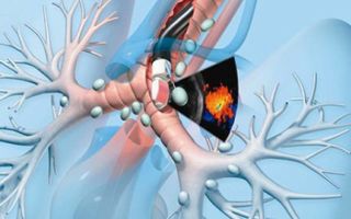
Bronchoscopy is a method of examining the bronchi using a long flexible tube with an optical system at the end - a bronchoscope.
Bronchoscopy, or tracheobronchoscopy, is a method of examining the lumen and mucous membrane of the trachea and bronchi using a special device - a bronchoscope. The latter is a system of tubes - flexible or rigid - with a total length of up to 60 cm. At the end, this device is equipped with a video camera, the image from which, magnified many times, is displayed on the monitor, i.e. the specialist conducting the study personally observes the condition of the respiratory tract in the mode real time. In addition, the resulting image can be saved in the form of photographs or video recordings, so that in the future, comparing the results of the current study with the previous one, it is possible to assess the dynamics of the pathological process. (Read about bronchography in our other article.)
A little history
Bronchoscopy was first performed back in 1897 by the doctor G. Killian. The purpose of the procedure was to remove a foreign body from the respiratory tract, and since it was very traumatic and painful, cocaine was recommended to the patient as an anesthetic.
Despite the large number of complications after bronchoscopy, it was used in this form for more than 50 years, and already in 1956, the scientist H. Fidel invented a safe diagnostic device - a rigid bronchoscope. Another 12 years later, in 1968, a fiberoptic bronchoscope, a flexible bronchoscope made from fiber optics, appeared.
An electronic endoscope, which allows you to repeatedly enlarge the resulting image and save it to a computer, was invented not so long ago - in the late 1980s.
Types of bronchoscopes
Currently, there are 2 types of bronchoscopes - rigid and flexible, and both models have their advantages and are indicated in certain clinical situations.
Flexible bronchoscope or fiberoptic bronchoscope
- This device uses fiber optics.
- It is primarily a diagnostic device.
- It easily penetrates even into the lower sections of the bronchi, minimally damaging their mucous membrane.
- The examination procedure is performed under local anesthesia.
- Used in pediatrics.
Consists of a smooth flexible tube with an optical cable and light guide inside, a video camera at the inner end and a control handle at the outer end.
There is also a catheter for removing fluid from the respiratory tract or supplying a drug into it, and, if necessary, additional equipment for diagnostic and surgical procedures.
Rigid or rigid bronchoscope
- It is often used for the purpose of resuscitation of patients, for example, in case of drowning, to remove fluid from the lungs.
- Widely used for medical procedures: removing foreign bodies from the respiratory tract, expanding the lumen of the trachea and bronchi.
- Allows for diagnostic and therapeutic manipulations in the area of the trachea and main bronchi.
- If necessary, in order to examine thinner bronchi, a flexible bronchoscope can be inserted through a rigid bronchoscope.
- If certain pathological changes are detected during the study by this device, they can be eliminated immediately.
- When examining with a rigid bronchoscope, the patient is under general anesthesia - he is asleep, which means he does not experience fear of the examination or the expected unpleasant sensations.
A rigid bronchoscope includes a system of rigid hollow tubes with a light source, video or photographic equipment at one end, and a manipulator for controlling the device at the other. The kit also includes various mechanisms for therapeutic and diagnostic procedures.
Indications for bronchoscopy
Bronchoscopy can be used for both diagnostic and therapeutic purposes.
Indications for fibrobronchoscopy are:
- suspicion of the presence of a tumor in the lungs;
- the patient has symptoms that are inadequate to the diagnosed disease, such as a prolonged unexplained cough, a prolonged intense cough when its severity does not correspond to other symptoms, severe shortness of breath;
- bleeding from the respiratory tract - in order to determine the source and directly stop the bleeding;
- atelectasis (collapse of part of the lung);
- pneumonia, characterized by a protracted course, difficult to treat;
- isolated cases of pleurisy;
- pulmonary tuberculosis;
- the presence of a shadow (or shadows) on the chest x-ray, the nature of which needs to be clarified;
- upcoming lung surgery;
- blockage of the bronchi with a foreign body or blood, mucus, purulent masses - in order to restore the lumen;
- purulent bronchitis, lung abscesses - for washing the respiratory tract with medicinal solutions;
- stenoses (pathological narrowings) of the respiratory tract - in order to eliminate them;
- bronchial fistulas - in order to restore the integrity of the bronchial wall.
Examination using a rigid bronchoscope is the method of choice in the following cases:
- with large foreign bodies present in the trachea or proximal (closest to the trachea) bronchi;
- with intense pulmonary bleeding;
- if a large amount of stomach contents mixed with food enters the respiratory tract;
- when examining the respiratory tract of a child under 10 years of age;
- for the purpose of treating bronchial fistulas, stenotic (narrowing the lumen) scar or tumor processes in the trachea and main bronchi;
- for washing the trachea and bronchi with medicinal solutions.
In some cases, bronchoscopy is necessary not as a planned, but as an emergency medical intervention necessary to quickly make a correct diagnosis and eliminate the problem. The main indications for this procedure are:
- intense bleeding from the respiratory tract;
- foreign body of the trachea or bronchi;
- ingestion (aspiration) of stomach contents by the patient;
- thermal or chemical burn of the respiratory tract;
- status asthmaticus with blockage of the bronchial lumen by mucus;
- airway damage due to trauma.
For most of the above pathologies, emergency bronchoscopy is performed in intensive care conditions through an endotracheal tube.
Contraindications to bronchoscopy
In some cases, bronchoscopy is dangerous for the patient. Absolute contraindications are:
- allergy to painkillers administered to the patient before the study;
- acute cerebrovascular accident;
- myocardial infarction suffered in the last 6 months;
- severe arrhythmias;
- severe heart or pulmonary failure;
- severe essential arterial hypertension;
- stenosis of the trachea and/or larynx of the 2nd–3rd degree;
- exacerbation of bronchial asthma;
- acute stomach;
- some diseases of the neuropsychic sphere - consequences of a traumatic brain injury, epilepsy, schizophrenia, etc.;
- oral diseases;
- pathological process in the cervical spine;
- ankylosis (lack of mobility) of the temporomandibular joint;
- aortic aneurysm.
The last 4 pathologies are contraindications only for rigid bronchoscopy, and fiber-optic bronchoscopy is acceptable in these cases.
In some conditions, bronchoscopy is not contraindicated, but it should be temporarily postponed until the pathological process resolves or clinical and laboratory parameters stabilize. So, relative contraindications are:
- 2nd and 3rd (especially 3rd) trimesters of pregnancy;
- period of menstruation in women;
- diabetes mellitus with high blood sugar levels;
- IHD;
- alcoholism;
- 3rd degree enlargement of the thyroid gland.
Preparing for the study
Before the study, the doctor tells the patient in detail the essence of the upcoming procedure, warns about possible complications, and the patient, in turn, signs consent to the study.
Before bronchoscopy, the patient must undergo a series of examinations prescribed by the doctor. As a rule, this is a general blood test, biochemical blood test, pulmonary function tests, chest x-ray or others, depending on the disease of the individual patient.
Immediately before the study, the patient will be asked to sign a consent for this procedure.
It is important not to forget to inform the doctor about any allergies to medications, especially to anesthesia drugs, if any, pregnancy, medications taken, acute or chronic diseases, since in some cases (see above) bronchoscopy is absolutely contraindicated.
As a rule, a planned study is carried out in the morning. In this case, the patient has dinner the night before, and is prohibited from eating in the morning. At the time of the study, the stomach should be empty to reduce the risk of reflux of its contents into the trachea and bronchi.
If the patient is very worried about the upcoming bronchoscopy, he may be prescribed mild sedatives a few days before the examination.
How is bronchoscopy performed?
Bronchoscopy is a serious procedure that is performed in a room specially equipped for this purpose in compliance with all sterile conditions. Bronchoscopy is performed by an endoscopist or pulmonologist who has been trained in this type of examination. An endoscopist assistant and an anesthesiologist also take part in the study.
Before the examination, the patient must remove glasses, contact lenses, dentures, hearing aids, jewelry, unbutton the top button of his shirt if the collar is tight enough, and empty his bladder.
During bronchoscopy, the patient is in a sitting or supine position. When the patient is sitting, his torso should be slightly tilted forward, his head slightly back, and his hands should be lowered between his legs.
When performing fibrobronchoscopy, local anesthesia is used, for which a lidocaine solution is used. When using a rigid bronchoscope, general anesthesia, or anesthesia, is required - the patient is put into a state of medicated sleep.
In order to dilate the bronchi for easy advancement of the bronchoscope, a solution of atropine, aminophylline or salbutamol is administered to the patient subcutaneously or by inhalation.
Source: https://otolaryngologist.ru/1529
Bronchoscopy
Tracheobronchoscopy (full name of the procedure) is a modern diagnostic and treatment method for visualizing the internal surfaces of the trachea and bronchi.
The examination is performed with a special optical device – a fiberoptic bronchoscope. Essentially, this is a multifunctional endoscope, which consists of a flexible cable with a light source and a video/photo camera at the end and a control handle with an additional manipulator.
Indications for bronchoscopy
The decision to perform bronchoscopy is made by a pulmonologist. He also determines the scope and frequency of the examination, taking into account the preliminary diagnosis and age of the patient.
Bronchoscopy is prescribed in the following cases:
- Darkening (disseminated lesions) on x-rays;
- Suspicion of cancer;
- Suspicion of the presence of a foreign body;
- Chronic shortness of breath not associated with diseases of the cardiovascular system or bronchial asthma;
- Hemoptysis;
- Abscesses or cysts in the lungs;
- Long-term recurrent pneumonia;
- Protracted inflammatory processes in the bronchi;
- Bronchial asthma (to determine the cause);
- Abnormal expansion or narrowing of the lumen of the bronchi;
- Monitoring the condition of the upper and lower respiratory tract before and after surgical treatment.
Manipulations that can be additionally performed during the procedure:
- selection of pathological contents to determine sensitivity to antibiotics;
- biopsy – collection of biomaterial for histological analysis;
- administration of a contrast agent necessary for other diagnostic procedures;
- removal of foreign bodies;
- washing the bronchi from pathological contents (sputum, blood);
- targeted administration of drugs (directly to the area of inflammation);
- elimination of abscesses (foci with purulent contents) by drainage (suction of fluid) and subsequent introduction of antibacterial drugs into the inflamed cavity;
- endoprosthetics - installation of special medical devices to expand the lumen of abnormally narrowed airways;
- determining the source of bleeding and stopping it.
Bronchoscopy is performed even on newborn children, but in this case it is performed to examine only the upper respiratory tract and only under general anesthesia.
Contraindications
There are also a number of contraindications to this procedure, the absolute ones of which are:
- stenosis of the larynx and trachea 2 and 3 degrees;
- respiratory failure 3rd degree;
- exacerbation of bronchial asthma.
These three conditions carry a risk of bronchial injury when the endoscope is inserted.
- Aortic aneurysm - nervous overstrain of the patient and manipulation with the endoscope can provoke rupture of the aneurysm.
- Heart attack and stroke less than 6 months old;
- Bleeding disorders;
- Mental illnesses (schizophrenia, psychosis, etc.). Stress and acute lack of oxygen during the procedure can significantly worsen the patient’s condition, causing another attack of the disease.
- Individual intolerance to painkillers. A reaction to them can provoke an allergy in any degree of its manifestation, up to the most severe – anaphylactic shock and suffocation.
Among the relative contraindications - conditions in which it is desirable to postpone the procedure to a later date, are:
- acute course of infectious diseases;
- menstrual bleeding (due to decreased blood clotting during this period);
- asthmatic attack;
- 2-3 trimester of pregnancy.
However, in cases of resuscitation (emergency), bronchoscopy is performed regardless of the presence of contraindications.
Preparation for bronchoscopy
Before bronchoscopy, it is necessary to undergo a number of diagnostic tests:
- radiography of the lungs,
- ECG (electrocardiogram),
- blood tests (general, for HIV, hepatitis, syphilis),
- coagulogram (blood for clotting)
- and others according to indications.
- You can take mild sedatives the night before;
- Dinner should be no less than 8 hours before the procedure;
- On the day of the study, smoking is prohibited (a factor that increases the risk of complications);
- Bronchoscopy is performed strictly on an empty stomach;
- In the morning, do a cleansing enema (prevention of involuntary bowel movements due to increased intra-abdominal pressure);
- It is recommended to empty your bladder immediately before the procedure.
If necessary, the doctor will prescribe mild sedatives on the day of the procedure. Patients with bronchial asthma must have an inhaler with them.
For people suffering from cardiovascular pathology, preparation for bronchoscopy is carried out according to an individually developed program.
Methodology
- The duration of bronchoscopy is 30-40 minutes.
- Bronchodilators and painkillers are administered to the patient subcutaneously or by spraying, facilitating the advancement of the tube and eliminating discomfort.
- The patient's body position is sitting or lying on his back.
It is not recommended to move your head or move. To suppress the urge to vomit, you need to breathe often and not deeply.
- The bronchoscope is inserted through the oral cavity or nasal passage.
Source: https://www.diagnos.ru/procedures/manipulation/bronhoskopia
Bronchoscopy
Bronchoscopy is a diagnostic method that allows the doctor to examine the airways. This procedure is performed by inserting a special endoscopic instrument, a bronchoscope, through the nose or mouth and down the throat to reach the lungs. There are many different methods for diagnosing the respiratory system, such as bronchography, chest radiography, chest CT, spirography - all of them are widely used, including bronchoscopy, which, in some cases, is vital.
Bronchoscopy first came into clinical use in 1897, when Killian removed a pig bone from the right main bronchus of a German farmer. Early clinical applications of bronchoscopy were limited to the removal of foreign bodies.
As lighting and optical technology, particularly the Hopkins rod and lens system, improved, bronchoscopy became more widely used. Wood and Flink first described the use of a flexible bronchoscope in children in 1978. In 1981, fiberoptic bronchoscopes thin enough for use in children became widely available.
Since then, there has been a rapid increase in the use of flexible bronchoscopy, as well as its improvement.
Types of bronchoscopy
Flexible bronchoscopy is done using a long, thin, lighted tube that is designed to look at the airway.
A flexible bronchoscope is used more often than a rigid bronchoscope because it usually does not require general anesthesia, is more comfortable for the person, and offers a better view of the smaller airways.
It also allows the doctor to take small tissue samples (biopsy).
Solid bronchoscopy is usually performed under general anesthesia and a straight metal tube is used during the procedure. It is used when there is bleeding that may block the view of a flexible bronchoscope, or when large tissue samples need to be taken for biopsy, or to remove foreign bodies in the airway that a flexible bronchoscope cannot handle.
Indications for bronchoscopy
Bronchoscopy is most often a diagnostic procedure that is done to diagnose lung diseases, tumors, chronic cough, and infections. Depending on the condition and disease of the patient, during bronchoscopy you can find: blood, mucus, signs of an infectious process, swelling, puffiness, the presence of a foreign body, tumor.
Indications for bronchoscopy:
- to detect the cause of the problem (eg, bleeding, chronic cough, difficulty breathing);
- to take tissue samples when other tests, such as a chest X-ray or CT scan, show problems with the lung or lymph nodes in the chest;
- to diagnose lung disease by collecting samples of tissue or mucus (sputum);
- to determine the degree of lung cancer;
- to remove foreign bodies that block the airways;
- for brachytherapy;
- for the diagnosis of bronchial tuberculosis (bronchoscopy is performed for differential diagnosis with other diseases).
Preparation for the procedure
Before starting the procedure, the patient needs to remove dentures, glasses, contact lenses, hearing aids, if any of the above are available. During bronchoscopy, a local anesthetic spray is used, which is applied to the throat and nasal cavity. The patient may also be given a sedative to help them relax.
A patient scheduled for bronchoscopy should not eat or drink 6-12 hours before the procedure, so it is worth undergoing bronchoscopy in the first half of the day. It is worth talking to your doctor about which medications you should stop taking before the procedure.
Before the procedure, you should empty your bladder. You need to remove all or most of your clothing. The procedure is performed by a pulmonologist and an assistant. During the procedure, heart rate, blood pressure and blood saturation levels will be checked. A chest x-ray must be performed before the procedure.
Before performing a bronchoscopy, the doctor may prescribe other tests, such as a complete blood count, coagulogram, and pulmonary function tests.
Algorithm for bronchoscopy
Flexible bronchoscopy algorithm
Source: https://FoodandHealth.ru/diagnostika/bronhoskopiya/
Indications and contraindications for bronchoscopy and bronchofibroscopy
Bronchological studies occupy one of the leading places in the diagnosis of respiratory diseases; in many cases they are of decisive importance both in establishing a diagnosis and determining the extent of the pathological process.
In the existing fundamental works devoted to bronchological studies, due to the diversity of the presentation of the material and its significant volume, it is difficult for a practicing physician to identify the basic information necessary to solve specific diagnostic problems.
In addition, the literature does not reflect indications for bronchoscopy and bronchography based on clinical and radiological signs of respiratory diseases, which makes it difficult to select patients for bronchological examination and select the most informative diagnostic method.
Extensive clinical experience in the use of bronchoscopy with both flexible and rigid endoscopes has allowed us to develop a differentiated approach to each method.
Based on our own experience and literature data, indications and contraindications for bronchological research methods - bronchoscopy and bronchography, as well as an assessment of the corresponding symptoms for various lung diseases are presented in a concentrated form.
- Indications for diagnostic bronchofibroscopy
- It is advisable to formulate indications for bronchoscopy based on clinical and radiological symptoms that indicate the likelihood of bronchial damage, but do not allow a diagnosis to be made without a bronchological examination.
- I. Indications for bronchoscopy based on clinical symptoms:
- In all cases when the doctor is forced to note a protracted chronic inflammatory process in the lungs. In this situation, it is always secondary, and the disease that caused the inflammation is localized, as a rule, in the bronchi.
- Unmotivated cough (prolonged cough as the only symptom of the disease).
- Inappropriate symptomatic cough (severe, prolonged cough that cannot be explained by the nature of the diagnosed pathological process).
- Shortness of breath not adequate to the volume of the lesion.
- Hemoptysis and pulmonary hemorrhage.
- Sudden changes in the amount of sputum in a short period of time (possibly an obstruction in the bronchi).
- Bacillary and oligobacillarity in the absence of obvious tuberculosis of the lungs (possible bronchial tuberculosis, bronchonodular fistulas).
- The need for bacteriological, cytohistological examination of pathological material from the bronchi.
II. Indications based on radiological symptoms:
- Presence of signs of bronchial obstruction: reduction of the lung or part of it in volume; presence of hypoventilation; atelectasis; swelling of the lung or parts of it.
- Prolonged and chronic pneumonia (protracted or chronic inflammation most often occurs against the background of another disease).
- The presence of shadows of unknown etiology in the hilar, middle sections, as well as in the root of the lung and in the mediastinum.
- Rapid change in the size of the intrapulmonary cavity (with cavernous tuberculosis or abscesses).
- Disseminated lung diseases.
- Pulmonary tuberculosis.
- Pleurisy of unknown etiology.
III. Bronchoscopy is necessary in all cases before surgical treatment.
Indications for therapeutic bronchoscopy
- The need to eliminate bronchial obstruction with mucus, pus, blood, and foreign bodies.
- Stopping pulmonary hemorrhage with lobar bronchus tamponade.
- Treatment of purulent bronchitis.
- Removal of pus from intrapulmonary cavities.
- Treatment of bronchopleural and bronchonodular fistulas.
- Treatment of post-inflammatory stenosis of the trachea and bronchi.
In acute and progressive chronic respiratory failure due to bronchial obstruction, there is a need for urgent bronchoscopy:
- Massive pulmonary hemorrhage.
- A large foreign body lodged in the trachea or bronchi.
- Postoperative atelectasis and pulmonary hypoventilation.
- Aspiration of gastric contents.
- Asthmatic status with obstruction of the bronchi by viscous mucus.
- Chest injury with damage to the trachea and bronchi.
- Thermochemical damage to the respiratory tract.
The purpose of emergency bronchoscopy is urgent diagnosis and elimination of the main cause of bronchial obstruction, improvement of pulmonary gas exchange.
In case of emergency conditions indicated in paragraphs. 1-2, rigid bronchoscopy is performed under general anesthesia in an operating room; in paragraphs 3-7 - emergency bronchofibroscopy through an endotracheal tube against the background of artificial ventilation in the operating room or in the intensive care unit.
Indications for rigid bronchoscopy
Despite the advantages of bronchofibroscopy compared to rigid bronchoscopy in clinical practice, there may be situations when the latter is the only method of choice:
- bronchoscopy in children under 10 years of age;
- large foreign bodies floating in the trachea or fixed in the bronchi;
- massive pulmonary hemorrhage;
- massive aspiration of gastric contents mixed with food;
- puncture biopsy of enlarged tracheobronchial lymph nodes;
- electro- and laser endobronchial surgery for stenotic tumor and scar processes in the trachea and (or) main bronchi;
- endobronchial treatment of bronchial and bronchopleural fistulas.
CONTRAINDICATIONS to bronchofibroscopy Absolute:
- intolerance to drugs used for local anesthesia;
- myocardial infarction suffered less than 6 months. back;
- acute stroke;
- heart rhythm disturbance (above grade III);
- hypertension with an increase in diastolic pressure of more than 100 mm Hg. Art.;
- pulmonary-cardiac and cardiovascular insufficiency of the III degree;
- bronchial asthma in the acute phase, when the interictal period is less than 3 weeks;
- stenosis of the larynx and (or) trachea II-III degree;
- neuropsychiatric diseases (epilepsy, condition after traumatic brain injury, schizophrenia);
- pain in the abdominal cavity;
- an extremely serious condition of the patient, when clarification of the diagnosis can no longer affect treatment tactics.
Relative:
- acute respiratory disease of the upper respiratory tract;
- cardiac ischemia;
- severe diabetes mellitus;
- pregnancy (second half);
- chronic alcoholism;
- enlargement of the thyroid gland III degree;
- period of the menstrual cycle.
CONTRAINDICATIONS to rigid bronchoscopy
- the same as for bronchofibroscopy;
- oral diseases;
- ankylosis of the lower jaw;
- damage to the cervical vertebrae;
- aortic aneurysm.
- =================
- You are reading the topic:
- Bronchological research methods for respiratory diseases
Source: https://www.plaintest.com/pulmonology/contraindications
What does bronchoscopy show?
Bronchoscopy is an endoscopic examination of the lungs. If x-rays and computed tomography of the lung do not provide sufficient information, bronchoscopy is considered as a diagnostic method. Bronchoscopy also plays a role in treatment, for example to aspirate sticky sputum.
During bronchoscopy, the doctor inserts a bronchoscope into the airway through the mouth or nose. Modern bronchoscopes consist of a soft, movable tube with a diameter of two to six millimeters. At its end there is a camera along with a light source. This camera transmits its images in real time to a monitor on which the doctor views the patient's airway.
Why do bronchoscopy?
Bronchoscopy may be needed both for treatment and for diagnosis - for example, when there is a suspicion of lung cancer or when planning treatment for an already known lung tumor.
With this manipulation, doctors can also inject radioactive substances into the lung to locally irradiate tumors. Another reason for prescribing bronchoscopy is to clarify the cause of narrowing of the airways. Bronchoscopy can be used to examine decreased ventilation (hypoventilation) of the lungs (atelectasis).
In addition, bronchoscopy together with bronchial lavage is suitable for obtaining cells and microorganisms from the lung.
Doctors also use bronchoscopy to look for foreign bodies and remove them. In patients on mechanical ventilation, it can adjust the position of the breathing hose.
In addition, a bronchoscope can be used to wash out secretions, such as mucus plugs, and insert so-called stents, which strengthen the airways from the inside and keep them open.
The bronchoscope can inject and aspirate fluid (called bronchial lavage). In addition, very small forceps or brushes can be passed through the tube to take tissue samples (biopsies).
The doctor later examines these samples under a microscope. Another research opportunity is offered by a miniature ultrasound head for imaging the tissues surrounding the respiratory tract.
Bronchoscopy - indications and contraindications
Indications for diagnostic bronchoscopy:
- Suspicion of a neoplasm of the bronchi or trachea.
- Suspicion of the presence of a foreign body in the respiratory tract.
- Anomalies in the structure of the bronchi and trachea.
- Collection of contents for background research.
- Frequently recurring pneumonia.
- Hemoptysis.
- Conduct differential diagnosis between lung diseases with similar symptoms.
- Atelectasis of the lung.
Indications for therapeutic bronchoscopy:
- Preparation for lung surgery.
- Removing foreign bodies from the respiratory tract.
- Installation of a stent to widen the airways when compressed by a tumor.
Contraindications to bronchoscopy.
- Acute stroke.
- Acute myocardial infarction.
- Bronchial asthma in the acute stage.
- Mental disorders.
- Epilepsy.
- Hypertonic disease.
- Heart rhythm disturbance.
- Allergy to the anesthetic used during the procedure.
- Stenosis of the larynx (trachea).
- Severely reduced lung function.
- Blood clotting is impaired.
In these cases, you need to carefully consider the need for research, weighing the advantages and possible disadvantages of this research.
Other types of bronchoscopy
In addition to bronchoscopy using a flexible tube, there is also an examination using a rigid tube. A rigid bronchoscope can, for example, be even better at removing foreign bodies from the lung. Even when a tumor severely narrows the airway, rigid bronchoscopy has advantages.
Sometimes your doctor can remove tumors directly using lasers or argon beam generators. Argon beam generators are coagulation machines that transfer energy through argon gas and obliterate tissue to a depth of two to three millimeters. The doctor uses them to destroy tissue and stop bleeding.
In case he needs to insert stents to widen the narrowing site, this is better done with the help of a rigid bronchoscope.
Consequences and complications of bronchoscopy
As a result of the mechanical impact, the bronchoscope can cause nosebleeds or sore throat with difficulty swallowing, hoarseness or cough, and very rarely injury to the larynx. Sometimes after the examination a short-term high temperature occurs, especially with lavages and tuberculosis. However, serious cases with bronchoscopy are very rare.
Mild bleeding may occur as tissue samples are taken (biopsies). Therefore, you can expect a cough with a small amount of blood in the first two days. Sometimes the bleeding is so severe that it needs to be stopped using an endoscope.
In some cases, injury to the pulmonary alveoli leads to the fact that the lung loses its tightness and a so-called pneumothorax is formed.
This means that air rushes into the space between the lung and the cavity surrounding the lung, and causes a feeling of lack of air. Then, in some cases, drainage of the pleural cavity is necessary.
This plastic tube removes the trapped air through the chest wall.
The risk of complications from bronchoscopy increases the older the patient. Therefore, it is very important to realistically assess the patient’s condition before conducting a study such as bronchoscopy.
Source: https://www.wp-german-med.ru/sovremennaia-endoskopia/1798-bronkhoskopiya-pokazaniya-protivopokazaniya.html