Ultrasound for women can be performed transvaginally. In this case, a sensor with a special attachment is inserted into the vagina. A medical condom is first put on the device. Diagnostics is not recommended for virgins. Such girls are recommended to have an abdominal ultrasound examination.
Transvaginal ultrasound is used to examine the pelvic organs. In addition, the method is recommended in the early stages of pregnancy. At later stages, this method is not informative. If necessary, vaginal examination is carried out repeatedly over a short period of time.
Transvaginal ultrasound is used to examine the pelvic area
What is the essence of the survey?
Ultrasound is an informative and safe diagnostic method. The examination uses ultrasonic waves with a frequency of up to 20 MHz. They are imperceptible to humans. The patient does not feel or hear such a sound.
Ultrasound penetrates organs and tissues that absorb waves differently. Subsequently, the waves return back. Thanks to different levels of density, a detailed image is formed in which the boundaries are clearly visible.
The device consists of a sensor, transducer, keyboard, and monitor. At the end of the examination, the patient is given a detailed report. The received documents and photographs must be provided to your attending physician.
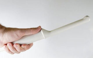
The examination is carried out using a special ultrasound sensor
When using the transvaginal method, a special attachment to the sensor is used. The device inserted into the vagina looks like a long rod. Diameter reaches 3 cm, length up to 12 cm.
To increase hygiene, a medical condom is used. The product is different from the usual one, which is used during sexual intercourse. There is no additional space for sperm.
A medical condom fits tightly around the sensor. The product is made from durable latex.
When to use
The method can be carried out as diagnostics and prevention. You should definitely resort to the method if there are appropriate indications. You should first consult with your doctor.
It is mandatory to undergo a vaginal ultrasound if there are pathological signs:
- the presence of spotting that is not menstruation;
- absence of pregnancy for 6 months, subject to regular unprotected sexual intercourse;
Ultrasound is performed for complaints of abdominal pain
- the presence of regular pain in the lower abdomen;
- the course of menstruation for more than 7 days;
- the duration of menstruation is less than three days;
- suspicion of an ectopic pregnancy.
Diagnosis is scheduled in the first trimester of the current pregnancy. In this case, the procedure is aimed at eliminating abnormalities in the development of the fetus and disturbances in the functioning of the female organs.
Vaginal examination is also mandatory as a preventive measure. In particular, doctors recommend resorting to the method annually upon reaching 35-40 years of age. Indications for transvaginal examination include:
- the need to study the length of the cervix;
- the need to study the shape of the ovaries;
- determination of the anatomical structure of the uterus;
- confirmation or refutation of likely present neoplasms of a malignant or benign nature;
One of the parameters that is checked by ultrasound is the shape of the ovaries.
- diseases of an inflammatory or infectious nature.
Diagnostics are also recommended as preparation before upcoming surgery.
What are the advantages and disadvantages
The need for the vaginal method is determined individually. The method has both advantages and disadvantages, which are presented in the table.
| Advantages | The main advantage is safety. This is due to the absence of ionizing radiation. Thanks to this, ultrasound of various organs is prescribed for both adults and children. There is absolutely no discomfort during the procedure. The diagnosis is not accompanied by pain. There is no need to take painkillers. The main thing is to set yourself up mentally. Thanks to the use of a medical condom, there is absolutely no risk of infection. In addition, the examination is considered quick and informative. You can combine the method with a biopsy. The diagnosis does not harm the pregnant woman or child. |
| Flaws | The main disadvantage is the impossibility of diagnosing virgins. In this case, the girl is recommended to use the transabdominal method. There is also a risk of obtaining inaccurate results if the girl has not previously followed the doctor’s recommendations. Ultrasound does not always help establish a final diagnosis. Sometimes other diagnostic methods are required. During pregnancy, the examination is informative only up to the 12th week. |
Only a doctor can say for sure whether an ultrasound examination is necessary for a particular patient.
Which authorities check
An ultrasound scan examines the pelvic organs. During the diagnosis, the doctor examines the condition and functionality of:
- ovaries;
- fallopian tubes;
The doctor also evaluates the condition of the bladder
- uterus;
- Bladder;
- rectum.
The doctor has the opportunity to confirm pregnancy in the early stages. The examination determines the size and location of the organs. The condition of the endometrium is assessed. Any violations may be a good reason to begin immediate treatment.
How to prepare properly
The accuracy of the results depends on proper preparation. Immediately before the examination, you should empty your bladder. For this purpose, visit the toilet. However, it should not be completely empty. That is why immediately after using the restroom you need to drink 500 ml of clean water without gas.
An important condition is the absence of gases in the intestines. For this purpose, you need to review your diet before 2 days before diagnosis.
Removed from the menu:
- fat;
- fried;
- pickled;
No need to eat fresh baked goods
- excessively salted or peppered;
- alcoholic drinks;
- confectionery;
- fresh baked goods;
- fatty dairy products.
Additionally, you may need to take medications that reduce gas formation. The best time for the procedure is 5-7 days of the menstrual cycle. It is at this time that it is most advantageous to assess the condition of the pelvic organs. If there is an urgent need, the study can be carried out at the time of menstruation.
It is not recommended to carry out cleansing enemas before diagnosis. Without preparation, the study can be carried out in the presence of dangerous pathological signs.
How is the procedure performed?
The patient should take a horizontal position. You need to lay your head towards the device. The doctor places a disposable condom on the device. Diagnosis takes place without any extraneous painful sensations.
From this video you can learn how a transvaginal ultrasound is performed:
The device is inserted into the vagina. If necessary, the doctor can press on the abdominal cavity. This allows for improved visualization. The indicators are displayed on the monitor.
The duration of the examination is 10-15 minutes. After completing the diagnosis, you need to take the conclusion to the attending physician.
Is the procedure dangerous?
Diagnostics is completely safe. In addition, as mentioned earlier, there are no unpleasant sensations during diagnosis. After the examination, there is no risk of any side effects.
It is thanks to complete safety that diagnostics can be prescribed even to minor patients. If necessary, the examination can be carried out repeatedly over a short period of time. The study is also recommended to monitor the effectiveness of the selected treatment. There are no complications after diagnosis. That is why the method is often recommended as a preventive measure.
In rare cases, after the study, a woman experiences abdominal discomfort. This symptom does not require the help of a doctor and disappears on its own.
What deviations are detected
Ultrasound is a universal procedure. Using a vaginal sensor you can detect:
- abnormalities in the structure of the uterus;
- fibroids;
Ultrasound helps in diagnosing ectopic pregnancy
- malignant processes;
- polycystic ovary syndrome;
- prolapse or bending of the uterus;
- endometrial hyperplasia;
- ectopic pregnancy;
- polypous formations.
If necessary, the woman is prescribed treatment or additional examination methods.
When is this method contraindicated?
Vaginal ultrasound has only relative contraindications. This method is not recommended:
- if the girl is not yet sexually active;
- during acute inflammatory processes in the body.
Transvaginal ultrasound is not performed if girls are not sexually active
There are no other contraindications. It is important that the girl prepares herself mentally for the upcoming examination and understands that the diagnosis is completely harmless.
Did the article help? Rate it
Source: https://mrt86.ru/prochee/uzi-transvaginalnoe-podgotovka-i-protsedura-obsledovaniya-vozmozhnye-rezultaty.html
What is transvaginal ultrasound and when is it used?
- It is superior in diagnostic reliability to any other type of ultrasound, since the sensor is located as close as possible to the organs being examined.
- The content of the article
Indications for transvaginal ultrasound
- Vaginal ultrasound has significantly expanded the diagnostic capabilities of gynecologists, obstetricians and urologists.
- It happens often indispensable for detecting diseases at their early stages, when changes in the organ are still minimal, and other diagnostics are uninformative.
- The study is indicated for the following symptoms:
- Discharge of blood in any quantity and any color outside of menstruation.
- Inability to become pregnant while having regular sexual activity for more than six months.
- Pain in the lower abdomen not associated with menstruation.
- Menstruation that lasts longer than 7 or less than 3 days.
- For early diagnosis of ectopic pregnancy (not informative before 3 weeks after missed menstruation).
- If pain or discomfort in the lower abdomen is felt during sexual intercourse.
- As an annual preventive examination.
Transvaginal ultrasound helps diagnose the following diseases and conditions
- ovarian cysts
- endometriosis
- pregnancy – both uterine and ectopic
- the presence of inflammatory fluid, pus or blood in the fallopian tubes (without differentiation of these fluids)
- uterine fibroids
- endometrial polyposis
- abnormal development of the internal genital organs
- hydatidiform mole - partial or complete
- malignant tumor of the uterus
- chorionepithelioma
- ovarian cyst rupture
- fluid in the lower abdomen and pelvis
- ovarian cancer.
A vaginal ultrasound will help assess the number and quality of maturing follicles. When a contrast agent is injected into the fallopian tubes, it helps to diagnose fallopian tube obstruction and its degree. This is an important test for your doctor who is helping you diagnose and treat infertility.
If the estimated gestational age is about 5 weeks or more, but there is a suspicion that the fetus is not developing, transvaginal ultrasound is the method of choice. It is in this case that the doctor will see on the monitor how the first contractions of the fetal heart occur (up to 5 obstetric weeks the procedure is not carried out for this purpose).
How to prepare for this examination
Vaginal ultrasound is performed only on women who have begun to be sexually active. The study is not carried out for virgins.
In their case, a transabdominal ultrasound will help to study the pelvic organs, that is, the same painless and safe procedure, but the sensor is moved along the abdominal wall.
If, due to severe obesity or flatulence, it is impossible to examine the necessary organs in a virgin, the doctor may recommend an examination through the rectum (transrectal ultrasound).
Conducting a transvaginal examination (tvUS) does not require preparation. The bladder, which needs to be filled before the abdominal examination, must be empty during this procedure. The doctor may even ask you to go to the toilet if you urinated more than an hour ago.
The only thing you need to prepare is to reduce the amount of gas in the intestines if you suffer from flatulence. To do this, an hour and a half before the procedure, you need to drink either 5 Espumisan tablets or a packet of Smecta (depending on tolerance).
Types of ultrasound diagnostics of the pelvic organs
Relationship between the time of the study and the menstrual cycle
It is very important to compare a vaginal ultrasound with the day of the menstrual cycle, because the results of the study depend on this. So, after ovulation, which usually occurs 12-14 days after the first day of the last menstruation, almost all genital organs undergo changes. This is necessary in order to be ready for conception and implantation of a fertilized egg.
A planned TVUS is usually performed at the beginning of the cycle, preferably after the end of menstruation on the very next day (5-7 days of the cycle), but it can also be done on days 8-12.
If endometriosis is suspected, it is recommended to undergo the procedure, on the contrary, in the second half of the cycle.
If you need to assess the dynamics of follicle maturation, it is carried out several times in dynamics (8-10, then 15-16, after 22-24 days of the cycle).
If spotting or bleeding not associated with menstruation occurs, it is carried out regardless of the day of the cycle - immediately after the occurrence of this symptom.
During pregnancy, transvaginal ultrasound can be performed only in the first trimester and according to indications. In the future, due to the risk of miscarriage, examination is carried out only through the abdominal wall.
How is the procedure performed?
Normally, the procedure is painless. A woman comes to the diagnostic room, where she will need to undress as if for a gynecological examination.
- The woman lies on the couch with her back or on the gynecological chair, spreads her legs apart and bends at the knees.
- A disposable condom is put on the transvaginal sensor (transducer), and the doctor applies a little special gel on top.
- It serves both as a lubricant and helps in conducting the ultrasonic wave.
- A transducer for vaginal examination looks like a rod with a diameter of about 3 cm, a length of about 12 cm. They often have a beveled handle, as well as a channel into which a biopsy needle can fit.
- The sensor is not inserted into the vagina to a very great depth; it does not cause pain. During the procedure, the sonologist can move the transducer up, down, and sideways. If this makes a woman uncomfortable, she should report it immediately.
- The abdominal sensor, unlike the vaginal one, has a wide emitting surface of a curved shape. In order to conduct a gynecological examination with this type of sensor, you need to fill your bladder.
How to decipher research data
Transvaginal ultrasound allows us to evaluate the following parameters in more detail than abdominal ultrasound:
- body and cervical dimensions
- location of the uterus
- uterine structure
- size and location of the ovaries
- ovarian structure
- places where the fallopian tubes exit (the tubes are completely visible only when they are filled with liquid, which is a contrast for ultrasound)
- follicle: size, number of mature and emerged
- free fluid in the abdominal cavity.
Normal echo picture of the pelvic organs
Uterus
- The position is anteflexio, that is, the organ should be inclined anteriorly. Retroflexio is not a pathology, but a developmental option. This is bad for pregnancy. It can cause not only infertility, but also the occurrence of ectopic implantation of the fertilized egg.
Also, due to this arrangement, a woman may suffer from chronic constipation.
- Contours of the uterus. Should be designated as “smooth” and “clear”. Their unevenness means inflammation of the uterine muscle or surrounding tissues. Blurredness indicates a benign or malignant tumor.
- Dimensions.
Length – 70 mm, width – about 60 mm, diameter – 40-42 mm. If these indicators are less, it is called an “infantile uterus”; if more, it is pregnancy, fibroids, or a malignant neoplasm.
- Echogenicity of the walls. It should be uniform.
If this is not the case, it may be a sign of a tumor.
- Endometrial thickness. This is what changes depending on the phase of the cycle:
-3-4 days of the cycle – thickness is minimal
-5-7 days – 3-6 mm
-11-14 days – 8-15 mm
-15-19 days – 10-16 mm
-before menstruation – 10-20 mm depending on the day of the cycle.-if there is the term “decidualization of the endometrium”, this indicates a probable pregnancy.
- Cavity structure. Normally, it is homogeneous, its edges are smooth and clear. Polyps, myomatous nodes, and cancerous tumors are referred to as “hyperechoic formations.” Heterogeneity of the structure or blurred contours is a sign of endometritis.
Cervix
- Dimensions: length – 3.5-4 cm, anterior-posterior size – 2.5-3 mm.
- The echostructure is homogeneous.
- Endocervix (cervical canal): 2-3 mm in diameter, filled with mucus of a homogeneous echostructure. If these sizes are larger or the structure is heterogeneous, this may indicate inflammation, cancer, and endometriosis.
Free liquid
A few milliliters of it in the space behind the uterus is normal only for a few days after ovulation (days 13-15 of the cycle). In other cases, the fluid is a sign of infection.
Ovaries
Transvaginal ultrasound is performed using this device
- Dimensions: 25x30x15 mm (width, length and thickness). The normal volume is 2-8 cm3. Their increase is a sign of polycystic lesions or inflammation.
- The contours are fuzzy, but lumpy.
- The echostructure is homogeneous, with small areas of fibrosis. If the foci of fibrosis are large, this indicates inflammation.
- There should be several follicles measuring 4-6 mm, one up to 20 mm in the middle of the cycle. If the size of this one (it is called dominant) follicle is more than 25 mm, they speak of a follicular cyst.
The fallopian tubes
They should be either not visible at all or barely noticeable. If they are visible without contrast, this is a sign of either inflammation or an ectopic pregnancy.
Echo signs of uterine pathology
- Myoma. The size of the body (less often the neck) is increased. The contours have been changed. There is a node in the muscle (myometrium), which is designated as the “hyperechoic zone”.
- Endometriosis: "bubbles" in the tubes, uterus or cervix.
- Polyps are designated as volumetric formations in the cavity, changes in the internal contours of the organ.
- Cancer can have a similar description to polyps, but usually there is also swelling. This diagnosis is confirmed only by the results of a biopsy (if possible).
- Endometritis: an increase in the size of the organ, as well as thickening and swelling of the endometrium.
- Tumor of the neck: increase in its size, deformation of the neck.
- Ovarian cyst: a fluid formation that has a diameter of more than 2.5 cm.
- Adnexitis (inflammation of the uterine appendages). In this case, the fallopian tubes are thickened, the ovaries are enlarged, their boundaries are blurred, and mobility is limited.
- Ovarian cancer: the organ itself is enlarged, and its contours are deformed.
The price of a transvaginal examination is about 1000-1500 rubles.
So, transvaginal ultrasound helps to make a timely diagnosis of inflammatory and oncological diseases of the female genital area. The examination also allows you to detect pregnancy in the early stages and establish its pathology. No preparation is required for the test; you will receive results immediately after the procedure.
Watch a video about the features of an ovarian cyst and its diagnosis using ultrasound.
ATTENTION! The information on the site is for reference or popular information only. Correct treatment and prescription of medications can only be carried out by a qualified specialist, taking into account the diagnosis and medical history.
Successful diagnosis and treatment, health and well-being! Your uzilab.ru.
02/27/2015 UziLab
Source: https://uzilab.ru/mochepolovoy-apparat/transvaginalnoe-uzi.html
Why is transvaginal ultrasound performed and what does it help identify?
Transvaginal ultrasound is a more reliable diagnostic method compared to others. This is explained by the close location of the device to the female genital organs. This medical examination is based on the delivery of ultrasound waves through a special sensor that is placed in the vagina.
This ultrasound examination is performed vaginally and is an instrumental method with which the doctor can make an accurate diagnosis and subsequently prescribe effective treatment for the disease.
In modern gynecology, this diagnostic method is called differently:
- intravaginal examination;
- intravaginal examination;
- vaginal examination of female reproductive organs.
The general name is transvaginal ultrasound.
Often, when performing ultrasound examinations, doctors use a special technology for visualizing blood flow - color Doppler mapping. Using color circulation, you can evaluate the resistance of blood vessels, their diameter and patency, as well as other pathological processes.
Intravaginal ultrasound examination of the genitals can be used as a separate diagnostic method or in combination with a transabdominal examination (through the walls of the abdominal cavity) or palpation.
Indications for use
The doctor may refer the patient to undergo a transvaginal ultrasound in the following cases:
- for pain in the lower abdomen;
- inflammatory diseases of the female genital organs;
- pathologies of the urinary system;
- implementation of artificial insemination;
- using hormonal contraception, installing an IUD or vaginal ring;
- irregular menstruation;
- suspicion of infertility;
- pain during sexual intercourse or immediately after it.
Intravaginal examination is also used to determine pregnancy. This method is more informative than a standard transabdominal examination and allows you to see a clearer picture of the uterus.
Advantages of the method
The intravaginal diagnostic method has its positive aspects:
- availability;
- harmlessness and safety of ultrasound for the patient;
- does not require extensive preparation;
- high level of image detail;
- carried out without filling the bladder;
- can be used for obese women (when other examination methods cannot be implemented).
In addition, thanks to a vaginal ultrasound, the doctor will be able to examine in detail the cervix (which is difficult to visualize during an abdominal examination).
Preparing for a transvaginal ultrasound
To carry out this type of procedure, no special preparation is required.
The patient is recommended to take the following actions:
- take a hygienic shower before the examination;
- take with you a disposable diaper, dry wipes and a special condom for intravaginal ultrasound (sold in every pharmacy);
- an hour before the procedure, you need to empty your bladder;
- It is not advisable to have sexual intercourse the day before the test.
Women who are concerned about increased gas formation should prepare in advance. It is better to do an ultrasound after taking medications, they will help reduce flatulence (Espumizan, Smecta).
On what day of the cycle is it done?
The most appropriate time for examination is the first days of the cycle after the end of menstruation. It is recommended to do an examination on days 5–8 of the menstrual cycle. The reliability of the sensor readings will depend on when the patient last ovulated. As a rule, it occurs 12–14 days after the critical days; subsequently, the condition of the reproductive organs changes noticeably.
If uterine endometriosis (proliferation of endometrial tissue) is suspected, the procedure is postponed to the second part of the cycle.
Transvaginal ultrasound is not recommended during menstruation. However, certain pathologies of the pelvic organs (polyps, fibroids, small cysts) can be seen only in the first days of menstruation.
How is the procedure done?
Before the examination, the woman must remove all clothing below the waist. In order for an ultrasound of the pelvic organs to bring less discomfort, you need to take care of your clothing (come to the examination in a skirt or dress).
The procedure is carried out as follows:
- The woman lies on the couch lying on her back. In this case, you need to bend your legs and spread them slightly at the knees.
- Next, the doctor puts a condom on the sensor and lubricates it with a special gel. It will help eliminate air between the internal organs and the device, and also promote better penetration.
- The reproductive organs are displayed on the screen, and the doctor reads out the data.
A nurse takes part in the implementation of the procedure; she records the results of the study in the protocol.
In the absence of sudden movements, a gynecological ultrasound is not painful and is not accompanied by other unpleasant sensations.
Photo gallery
Introduction of the ultrasonic sensor device (photo) Direct procedure
What does a vaginal ultrasound show?
An intravaginal examination may reveal the following conditions and diseases:
- malignant and benign tumors of the reproductive organs;
- uterine fibroids (a neoplasm in the muscle layer of the female genital organ);
- fluid in the pelvic area;
- endometriosis;
- complete or partial hydatidiform mole;
- blood, purulent formations in the uterine tubes;
- inflammatory processes.
Decoding the results
This procedure is carried out by a specialist who, upon completion of the manipulation, will provide the patient with the results and conduct a consultation to compare them with normal values.
Table of norms for ultrasound of internal organs
| Parameter | Normal echo pattern | Symptom of pathology |
| Ovarian size |
Volume: up to 4 cubic meters see Smooth and clear contours |
Increased size of the ovaries, as well as their uneven contours, may indicate polycystic disease, congenital malformation, or oncological processes. |
| Position of the uterus and its size |
|
|
| Surface of the cervix | Clear contours and smooth surface | Blurred contours indicate congenital anomalies or inflammation. |
| The fallopian tubes | Not rendered | Thickening of the walls, an increase in the diameter of the fallopian tubes, and fluid accumulation are observed during ectopic pregnancy or inflammation. |
| Free liquid | Present in small quantities behind the uterus during ovulation | A large volume of fluid is a sign of an inflammatory process. |
Ultrasound of the uterus and appendages
This study is a scan of:
- vaginal cavities;
- uterus;
- ovaries;
- fallopian tubes.
This diagnostic method allows you to identify the following pathologies:
- adnexitis (inflammation of the appendages and uterus);
- fibroids;
- leiomyoma (benign tumors);
- neoplasms on the cervix;
- ovarian cancer.
Ultrasound of the bladder
This study is used as an alternative to palpation or catheterization (insertion of a special medical instrument through the urinary system, which is carried out for various purposes).
Indications for ultrasound examination of the bladder may be as follows:
- frequent urination or retention;
- blood in the urine or detection of red blood cells;
- cystitis;
- various organ injuries.
Ultrasound of the pelvic organs
It is a comprehensive examination, during which the doctor, at his discretion, determines the need to examine a particular organ. Intravaginal ultrasound of the anatomical space can be performed with Doppler ultrasound. This method allows you to identify vascular (pelvic) disorders and show images of other pathological processes in more detail.
Transvaginal ultrasound during pregnancy
This examination method is widely used in obstetrics and allows you to display an image of the uterus and fetus on a screen monitor and take a picture. Transvaginal ultrasound examination reflects the developmental characteristics of the child. As a rule, the doctor prescribes a scanning procedure for the pregnant woman in the first trimester.
General indications for prescribing an intravaginal ultrasound in the early stages of pregnancy are the following:
- the need to establish the fact of fertilization;
- control over fetal development;
- study of the periuterine space;
- identifying possible threats to pregnancy.
Transvaginal or transabdominal?
Your attending physician will help you decide on the examination method. Thanks to its unique technology, intravaginal diagnostics are significantly superior to examinations carried out through the walls of the abdominal cavity. In addition, the specialist may refer the patient for a vaginal ultrasound if she has postoperative adhesions on her abdomen.
How often can I do it?
For preventive purposes, you should prepare for a vaginal ultrasound at least once a year.
As for the pregnancy period, this method of ultrasound examination can be carried out until the twelfth week. The doctor may prescribe a repeat procedure to determine the woman’s health or to exclude signs of a frozen fetus. Transvaginal examination is completely safe for the mother and child.
Contraindications and complications
Doctors should not be allowed to perform intravaginal ultrasound on girls who are not sexually active. In this case, it is recommended to conduct an examination of the pelvic organs in only two ways: transabdominal or transrectal (through the rectum). In addition, this type of examination is prohibited during the second or third trimester of pregnancy.
The implementation of the procedure is not accompanied by any complications. In case of discharge of various types, it is recommended to visit a specialist.
Post-procedure care
Many patients rush to douche after an intravaginal ultrasound. However, no additional hygiene procedures should be carried out without a doctor’s prescription.
How much does the procedure cost?
| Region | Price | Firm |
| Moscow | from 1300 rub. | "Clinic No. 1" |
| Chelyabinsk | from 700 rub. | "Diagno'Z" |
| Krasnodar | from 800 rub. | City Clinic |
| Saint Petersburg | from 900 rub. | SM-Clinic |
| Ufa | from 500 rub. | "Aimed" |
Video
Source: https://hromosoma.com/uzy/transvaginalnoe-uzi-30189/
Transvaginal ultrasound
Indications for transvaginal ultrasound
Indications for this examination method are suspicion of diseases of the pelvic organs, emergency conditions (for example, ectopic pregnancy), monitoring of treatment. Transvaginal ultrasound is performed for the following conditions:
- Diagnosis of pregnancy in the early stages;
- Pain in the lower abdomen;
- Diagnosis of the causes of infertility and monitoring of the follicular apparatus of the ovaries;
- Menstrual irregularities (delayed menstruation, bleeding in the middle of the cycle), pathological discharge from the genital tract;
- Detection of inflammatory gynecological diseases;
- Diagnosis of pelvic tumors, including uterine fibroids, endometriosis, ovarian cysts, etc.;
- Taking hormonal medications, having intrauterine contraceptives (IUDs) to monitor the condition of the endometrium and prevent complications;
- Early pregnancy, when traditional transabdominal access (through the abdominal wall) is of little information;
- Support of the IVF (in vitro fertilization) procedure;
- Determining the causes of urological diseases, urinary disorders, urinary incontinence and pathology of the urethra (urethra).
Transvaginal ultrasound of the pelvic organs is an ideal option for obese women, since conventional examination through the abdominal wall is not very informative.
Contraindications
There are no absolute contraindications to transvaginal ultrasound. In virgins, examination through the rectum (transrectal) is possible. Transvaginal ultrasound during pregnancy is justified only in the early stages (up to 11-12 weeks).
Preparing for a transvaginal ultrasound
For transvaginal ultrasound of the uterus and appendages, special preparation (filling the bladder) is not required. When visiting the ultrasound room, you will need a towel or diaper to lie on during the examination.
A prerequisite for transvaginal pelvic ultrasound is the absence of gas in the intestines.
To do this, 2-3 days before the study, you need to limit foods that cause increased gas formation (vegetables, fruits, bread, dairy products, confectionery), and it is also recommended to take certain medications that reduce gas formation in the intestines - enzistal, activated carbon. It is not recommended to perform cleansing enemas before the study. Transvaginal ultrasound of the uterus and appendages does not have to be performed on an empty stomach.
In emergency cases, transvaginal ultrasound can be performed without preparation, but its information content may be reduced.
Ultrasound examination of the gynecological organs is recommended to be carried out in the first half of the menstrual cycle (usually on days 5-7), since in the second half the uterine endometrium is in the secretory phase, which can lead to incorrect interpretation of the results. However, for endometriosis, it is recommended to perform a transvaginal ultrasound of the uterus in the second phase of the cycle. To assess folliculogenesis (formation and development of ovarian follicles), the study must be carried out on days 5, 9, 11-14 and 15 of the menstrual cycle.
Methodology
The patient lies down on the couch with her head towards the ultrasound machine. The doctor places a condom on the vaginal sensor, lubricates it with gel and inserts it into the vagina.
The examination is absolutely painless, with the exception of acute conditions during inflammatory processes. During the examination, the doctor may press on the abdominal area to better locate the organs.
The time for a transvaginal ultrasound of the pelvic organs is usually 15-20 minutes.
Complications of transvaginal ultrasound
When the transvaginal ultrasound procedure is performed correctly, there are no complications.
This examination is recommended to be performed even in healthy women for preventive purposes; before the age of 40, it should be done at least once every 2 years, and after 40 years, annually.
Found an error in the text? Select it and press Ctrl + Enter.
Source: https://www.neboleem.net/transvaginalnoe-uzi.php
Ultrasound of the pelvis with an internal sensor: vaginal and transvaginal - Medsi
Vaginal (transvaginal) ultrasound examination of the pelvis is carried out by inserting a special device equipped with a sensor into the vagina.
The device is a rod with a handle, which is made of plastic, about 10-12 centimeters long and up to three centimeters in diameter. A special groove may be built into it to insert a needle to take biopsy material.
The examination allows you to determine the presence of pathologies, neoplasms or diseases in the following female genital organs:
- Uterus
- Fallopian tubes
- Ovaries
- Cervix
It is considered the most effective for studying these parts of the reproductive system, as it allows one to identify various health problems of the patient in the early stages. A pelvic ultrasound with a sensor can show the presence of abnormalities even at a time when other studies do not show any problem areas.
How is the procedure done?
The study is organized as follows:
- The patient should remove clothing from the lower part of the body (from the waist down)
- She sits on a special couch in the same way as during a regular gynecological examination.
- The doctor prepares the sensor: puts an individual condom on it, lubricates it with a special gel for the procedure
- The physician then inserts the device shallowly into the patient’s vagina.
- To get a complete picture of the state of the organs, he can move the sensor from side to side
- All data is recorded and processed by a doctor
The gel is necessary to facilitate the penetration of the sensor (and thereby reduce the likelihood of negative sensations) and enhance the ultrasonic effect by increasing conductivity.
This type of examination lasts no more than 10 minutes. It is painless and gives the most complete picture even in cases where an abdominal ultrasound shows nothing or cannot be performed.
When is a pelvic ultrasound with a sensor necessary?
There are symptoms in which the doctor must refer the patient for a transvaginal examination:
- Pain in the lower abdomen (not related to the menstrual cycle)
- Suspicion of the presence of neoplasms
- Periods of menstrual bleeding that are too short, too long or absent
- Impossibility of pregnancy
- Bloody discharge that is not menstruation
- Presence of fallopian tube obstruction
- Nausea, vomiting and weakness with bloody discharge from the vagina
Doctors recommend using this type of examination for preventive purposes, since not every ailment may have symptoms at an early stage, just as pregnancy in the first trimester may not manifest itself with classic symptoms (nausea, etc.).
In this case, vaginal ultrasound is used for:
- Diagnosis of infertility
- The need to determine the presence of changes in the size of the ovaries and uterus
- Diagnosis of pregnancy
- Pregnancy monitoring (first trimester only)
- General monitoring of the condition of the uterus, fallopian tubes and ovaries
A pelvic ultrasound can be performed simultaneously with two sensors. In this case, an abdominal ultrasound examination is performed first, and then a transvaginal one. The use of two types of analysis at once is necessary to identify disorders in the high-lying pelvic organs.
What does a vaginal ultrasound show?
This examination allows you to evaluate the following parameters of the reproductive system organs:
- Dimensions of the uterus. In normal condition, it should be about seven centimeters in length, six in width and 4.2 in diameter. If it is significantly less or more, then this indicates the presence of pathology
- Echogenicity. The structure of the organs must be homogeneous, uniform, have clearly defined, clearly visible edges
- General picture of internal organs. The uterus should be slightly tilted forward. And the fallopian tubes may be slightly visible, but should not be clearly visible without the use of a contrast agent
Diagnosed diseases
Transvaginal ultrasound allows you to identify a number of diseases and problems in the reproductive system at an early stage. It allows you to detect:
- Fluid and pus in the uterus and fallopian tubes. The cause of their appearance may be infections, viruses, mechanical damage
- Endomentriosis is an excessive proliferation of cells in the inner layer of uterine tissue into other layers and organs. It can occur due to inflammatory processes, damage (surgery, abortion), the appearance of neoplasms, disruption of the endocrine system, too frequent use of certain medications and substances
- Myoma is a benign neoplasm in the tissues of the uterus or its cervix. May occur due to chronic diseases, frequent abortions, hormonal imbalances, constant stress, pathologies, excess weight, or with hereditary predisposition
- Cysts and polycystic ovaries are tumors filled with fluid. Occurs with endocrine disorders, chronic diseases of the genitourinary system
- Various polyps on the walls of the uterus are benign formations in the endometrium of the organ. They can reach several centimeters in diameter. Their appearance may be associated with polycystic disease, chronic diseases, mastopathy, fibroma
- Inflammation and enlargement of organs can occur due to both infection and injury.
- Hydatidiform mole - appears instead of a full-fledged embryo during the process of conception, filled with fluid. Occurs due to duplication of male chromosomes with the loss of female chromosomes, sometimes due to fertilization of an egg that does not contain a nucleus. This disease is rare
- Fetal development disorders during pregnancy
- Defects and pathologies in the development of the fallopian tubes: obstruction, spiral-shaped or too long tubes, blind passages, duplication of organs
- Ectopic pregnancy occurs when an egg implants outside the uterine tissue after fertilization. Occurs due to blockage of the fallopian tubes, congenital anomalies in them, as well as after an inflammatory process, abortion
- Cancer is a malignant tumor in various organs:
- Uterus
- Ovarian
- Cervix
- Chorionepithelioma is a malignant neoplasm that arises during or after pregnancy from chorion cells (the membrane of the embryo attached to the wall of the uterus)
Stages of preparation for the study
To conduct a pelvic ultrasound with a sensor, no special preparation is required, but there are several mandatory requirements:
- Unlike an abdominal examination, with a transvaginal analysis the patient should not drink liquid one to two hours before the examination.
- If she emptied her bladder more than an hour before the test, she needs to do it once immediately before the procedure
- With increased flatulence, the patient needs a drug that will help normalize the processes of gas formation in the gastrointestinal tract. She can consult with her doctor about the choice of medication
Doctors also recommend using such a study on certain days of the cycle, depending on which organ needs to be diagnosed and for what purpose:
- In the case of a preventive examination, it should be done in the first days after the end of menstruation
- If there is a suspicion of an increase in the endometrial layer in the uterus, then in the second half of the cycle
- When it is necessary to monitor the development of a disease or the progress of treatment, the study can be carried out several times in one cycle, at different stages.
- An ultrasound is performed urgently if there is bleeding that is not menstrual, regardless of the day of the cycle
- It is important to remember to maintain personal hygiene before the examination, use wet and other wipes.
- If you plan to conduct a pelvic ultrasound with two sensors, then you should pay attention to preparing for the abdominal examination.
- This includes:
- Follow a diet for at least three days before the test to reduce the likelihood of symptoms of flatulence and bloating
- The last meal should be completed by six o'clock in the evening on the eve of the test.
- It is recommended to do an enema after eating
- If there is still a risk of flatulence, you need to use special medications that reduce gas formation
- An hour before the test, drink at least 400 ml of water
The diet involves excluding a number of foods from the diet:
- Sweets
- Flour (bread, cookies, etc.)
- Legumes
- Cabbage
- Milk and dairy products
- Vegetables and fruits that have not undergone heat treatment
- Coffee and strong tea
- Carbonated drinks
- Fast food
- Fatty foods (meat, fish, oils)
You can eat porridge cooked in water, lean boiled beef, poultry and fish, and hard cheeses. It is recommended to drink lightly brewed, slightly sweetened tea.
It must be remembered that since you need to drink liquid before the abdominal examination, you must empty your bladder before the transvaginal analysis.
Contraindications
Vaginal ultrasound examination has a small number of contraindications:
- It is never performed if the patient is a virgin, so as not to violate the integrity of the hymen. In this case, such a patient can undergo a transrectal examination, in which a sensor is inserted into the rectum
- The study is prohibited from being carried out in the second or third trimester of pregnancy, because it may provoke premature contractions or uterine contractions before the expected due date.
- This test is not used if the patient is allergic to latex
- If the patient has epilepsy, since the examination requires that she lie still
Source: https://medsi.ru/articles/uzi-malogo-taza-vnutrennim-datchikom-vaginalnoe-i-transvaginalnoe/