In order for the examination to be effective and accurate, proper preparation for an ultrasound of the liver and gallbladder is needed. If the patient ignores the doctor’s recommendations, the data obtained during the diagnostic process will be distorted. Since the ultrasound protocol serves as the basis for prescribing further treatment, an incorrect diagnosis aggravates the problem.
Examination of the abdominal cavity using ultrasound is safe, because there are no age restrictions, the method is also used in pediatrics. There are some prohibitions for ultrasound of the liver and gall bladder during late pregnancy; this is not due to danger, but to the impossibility of obtaining reliable results.
Standards for ultrasound
When performing an ultrasound, the doctor examines the structure and size of organs and assesses their condition. Ultrasound of the gallbladder determines its mobility, wall thickness, duct lumen and contractile function. This technique allows you to refute or confirm the presence of stones, polyps and tumors in the gallbladder.
During an ultrasound of the liver, both lobes are examined, the emphasis is on monitoring the condition of the veins, showing the condition of the bile ducts.
Liver norms on ultrasound:
- contours are clear and even;
- the structure of the liver is homogeneous;
- width – from 23 to 27 cm;
- length – from 14 to 20 cm;
- diameter within 22.5 cm;
- left lobe – from 8 to 10 cm;
- right lobe – 10 – 12.5 cm;
- diameter of the hepatic duct – 5 mm;
- The width of the lumen of the liver vein is 1.5 cm.
The standard sizes for ultrasound scanning of the gallbladder in adults are as follows:
- gallbladder length – 10 cm;
- width – 50 mm;
- transverse size – up to 35 mm;
- gallbladder wall thickness – 4 mm;
- duct diameter – up to 8 mm;
- the diameter of the lobar ducts is 3 mm.
The given sizes of the liver and gall bladder are optimal for adults. Deviation from the described norms may indicate the development of liver pathologies.
What diseases can be detected
Deviations from the norms identified during liver ultrasound may indicate various diseases. With cirrhosis and hepatitis, the size of the affected liver increases, and echo signals weaken or increase.
The appearance of abnormalities also indicates tumor damage to the liver vessels and the development of atherosclerosis, the presence of primary tumors and their foci from other organs.
The method shows cystic neoplasms, abscess, fatty hepatosis and other existing changes.
With cholelithiasis (GSD), light spots appear in the cavity, confirming the presence of stones. However, small stones are not detected during gallbladder diagnostics.
Enlarged ducts indicate a similar process. If there are polyps in the gallbladder, round formations appear on its walls. If they are more than 2 cm in diameter, the organ is deformed and a tumor process is possible.
During ultrasound, congenital anomalies of the structure of the abdominal organs can be detected.
Indications for testing
The list of indications for ultrasound of the liver and gallbladder can be presented as follows:
- pain in the right hypochondrium;
- yellowness of the skin and sclera of the eyes;
- suspicion of the presence of tumor processes in the liver and other organs of the peritoneum;
- chronic pancreatic diseases;
- constant bitter taste in the mouth;
- long-term use of certain medications;
- alcohol abuse;
- abnormalities in blood tests when considering AST and ALT samples;
- abdominal organ injuries;
- chronic lesions of the gallbladder;
- history of urolithiasis.
In case of the listed pathologies, the liver should be examined regularly. You should not give up comprehensive control; it is better to immediately examine all the organs of the abdominal space.
There is no need to wait until the last minute; liver diseases tend to progress quickly, so it is better to undergo an ultrasound once. Simultaneously with an ultrasound of the liver, the pancreas should be examined; no additional preparation is necessary.
How does the procedure work?
For ultrasound of the liver and gallbladder, equipment is used, the frequency of which is set to 2.5–3.5 MHz. Due to these characteristics, it is possible to consider areas within the range of 1–3 mm. The depth reached by the waves is 24 cm.
The procedure itself is very quick, taking 15–20 minutes. The patient lies on the couch, lies on his back, and exposes his stomach. The doctor applies gel to the skin and begins the examination. First of all, the area of the right hypochondrium is examined. After a thorough study, other areas are studied, the method shows changes.
Unpleasant sensations during the diagnosis of the liver, pancreas and gallbladder are excluded; the patient can feel light pressure from the sensor.
The method is safe and harmless, but it is better if the patient with acute syndrome is accompanied by a friend or relative. When signing up for an ultrasound, it is better to choose comprehensive monitoring of the abdominal organs, then the pancreas will be examined along with other tissues.
How to prepare properly
In order for the information obtained during an ultrasound to be accurate, you need to properly prepare for the scan. Within 3 days, it is important to start following a special diet and exclude fatty foods, carbonated drinks and alcohol from your diet. Coffee is banned.
In some cases, it is recommended to take enzyme preparations and sorbents that help restore digestion and cleanse the liver. Your doctor will tell you how to properly prepare for an ultrasound examination of the liver, pancreas and gallbladder. Recommendations may differ, but you definitely need to prepare.
Diet
On the appointed day before the ultrasound of the abdominal organs, eating breakfast is prohibited. A light snack is allowed 6 hours before the diagnosis. Preparation for ultrasound examination of the liver, gallbladder and pancreas requires compliance with the following rules and regulations.
They begin to prepare 3 days in advance; before monitoring, the following are removed from the menu:
- bread and other baked goods, cookies and sweets;
- milk and all dairy products;
- vegetables that increase increased gas formation: cabbage, radishes, turnips and radishes;
- whole grain cereals and pasta;
- fatty hard and curd cheese;
- mushrooms and legumes;
- fruit and vegetable juices;
- Carbonated drinks and mineral water are prohibited.
The listed products can increase the release of gases, which complicates the course of ultrasound. The last meal, taken 6 hours before, should be light; overeating is unacceptable. Before examining the liver, pancreas and gall bladder, you can eat oatmeal in water without butter, add a hard-boiled egg.
Taking medications
In order for an ultrasound of the abdominal organs to be successful, it is necessary to prevent bloating and increased flatulence. In case of excessive predisposition to the appearance of these symptoms, even diet does not help. It is better to use medications before diagnosis that will help:
- White carbon, regular activated carbon or Enterosgel. The purpose of these drugs is to cleanse the entire gastrointestinal tract.
- To prevent the formation of gas in the abdomen, use the drugs Espumisan, Simethicone or Bobotik.
- You can minimize gas formation by drinking mint, chamomile tea or a fennel drink.
- Festal, Creon or Mezim helps normalize intestinal motility. You can take other enzyme preparations.
- There is no need to use choleretic drugs. Under their influence, ultrasound will not show the condition of the biliary tract.
You should discuss the possibility of using drugs from this list with your doctor. If the pancreas is additionally examined, you need to be more careful with taking medications. Many drugs interfere with its functioning.
Other recommendations
During the period of preparation for ultrasound examination of the intestine, the following rules must be observed, especially regarding nutrition:
- You must not smoke or chew gum within 2 hours. It's better to increase this time.
- People suffering from type 1 diabetes are usually allowed to eat a small piece of bread the day before and wash it down with a cup of fresh tea.
- Patients taking any medications on an ongoing basis should notify their doctor about this. You can drink them if they are vital. It is necessary to provide him with information before receiving a referral for an ultrasound of the organs.
- You should not exercise before examining the liver, gallbladder and pancreas.
- Overweight patients are prescribed an enema in the morning and evening. The amount of water infused or a special drug is prescribed by the doctor.
The recommendations listed are relative.
Do I need an enema?
An enema should be given if a person is prone to constipation and cannot go to the toilet on his own. For this purpose, you can use drugs like Fortrans and Microlax. If the ultrasound is scheduled for the morning, the procedure should be performed in the evening.
If the patient has emptied on his own, the contents of the intestines cannot be removed using a cleansing enema. The procedure is done carefully. You can use a classic Esmarch mug filled with warm boiled water.
What to take with you
When making an appointment for an ultrasound of the liver and gallbladder, listen carefully to the specialist, he will tell you what you need to bring with you. If an ultrasound is performed in a government institution, it is important not to forget and prepare:
- patient's medical record;
- own insurance policy;
- passport confirming identity (adults).
A large towel will come in handy to place on the couch. You will also need shoe covers or slippers. If an ultrasound is performed to determine liver function, the study is first performed on an empty stomach, and then under load. The doctor asks the patient to consume any foods and repeat monitoring. For a snack, you can take a few boiled eggs and sour cream with you.
Why do you need to follow the training rules?
Doctors recommend that their patients follow the rules for preparing for an ultrasound. If the rules are ignored, the intestines may become overfilled with gases. Because of this, diagnostic results will be inaccurate. The patient may be given an incorrect diagnosis, and the doctor will not be able to fully analyze the functioning of the internal organs and identify any pathologies.
Failure to comply with general recommendations may lead to incorrect determination of the treatment regimen. In this case, the effectiveness of the treatment will be lost.
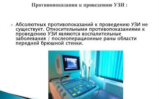
Contraindications for diagnosis
This technique has no strict contraindications. Ultrasound diagnostics is used for patients of any age category. Concomitant diseases do not limit this possibility.
The only temporary limitation is an acute purulent lesion of the skin at the diagnostic site or a burn. In this case, applying the gel can cause harm to the body, and due to the pressure of the sensor, the infection can spread.
Ultrasound of children under 3 years of age and pregnant women is performed with caution.
Ultrasound techniques for examining organs are popular among doctors due to their accuracy and safety for the patient. This is confirmed by articles and expert comments.
Examination of the abdominal organs: liver, gall bladder, pancreas, stomach, spleen, intestines, takes no more than 30 minutes.
Based on the examination protocol, a preliminary diagnosis can be made.
Source: https://puzyr.info/kak-provoditsya-uzi-pecheni-i-zhelchnogo-puzyrya-nyuansy-podgotovki/
What does an ultrasound of the liver show and how to prepare for it?
One of the most accessible, painless, and reliable instrumental methods of medical diagnosis is liver ultrasound. This study is prescribed for the purpose of visual inspection in real time of the condition of the organ, the features of its structure and operation.
Ultrasound is an ultrasound examination, the principle of which is to record a high-frequency wave reflected from the tissues of internal organs. Waves do not affect the human body.
The strength of wave reflection varies depending on the structure and density of the tissues of the internal organs. When computer processing of the reflected signal, a flat image is displayed on the monitor.
Studying the resulting picture allows us to draw conclusions about whether the condition of the liver corresponds to the normal variant.
Structure and function of the organ
The liver is the largest unpaired organ in humans. In a newborn child, this gland occupies 12 parts of the total volume of the abdominal cavity. In an adult man, the normal weight of the organ is up to 1800 g, in women (depending on the constitution) the norm is from 1300 g.
The liver is located in the right hypochondrium, has the shape of a conventional right-angled triangle, the acute angle is located in the upper part and directed towards the left side. The length along the upper line is about 18 cm, the width is about 13 cm. In the middle part of the organ from below there is a gallbladder and ducts.
The liver is composed of two unequal lobes, the ratio of which can be defined as 1:6. The larger lobe is located on the right, the smaller one is the sharp vertex of the conditional triangle. The parts of the organ are separated by a ligament. During an ultrasound examination, each lobe individually and the organ as a whole are measured and described.
The liver is a vital organ; its damage or developing pathology is a direct threat to human life.
Liver functions:
- Metabolism. All blood that “works” in the digestive organs passes through the liver. All nutrients are absorbed and processed in this organ. The organ supplies the bile necessary for digestion to the intestines. Processes and sends nutrients and medicines to body tissues.
- Depot of energy and vitamins. Here is a “mobile warehouse” of energy in the form of glycogen. A supply of vitamins A, D, B12, as well as cobalt, iron and copper is “stored”.
- Detoxification. The liver captures and removes from the body the bulk of toxic metabolic products and harmful substances that come from outside with food, medications, and harmful influences.
- Hematopoiesis and blood depot. The liver is the organ that produces blood in the fetus. When a person is born, this liver function stops working. However, with significant blood loss, the liver is able to “throw” a significant volume of blood into the bloodstream due to the narrowing of its blood vessels.
Indications for diagnosis
Ultrasound examination of the liver is prescribed if the following conditions and diseases are suspected:
- The patient exhibits “liver” symptoms: yellow sclera and skin, weakness, nausea, complete lack of appetite.
- The patient complains of abdominal pain in the area of the gland; palpation reveals an increase in size.
- History of chronic hepatitis.
- Diseases of the gallbladder and pancreas.
- Brown urine against a background of discolored feces.
- Abdominal injuries.
- Dynamic monitoring of the condition of the gland during chemotherapy and long-term drug therapy.
- For women – when prescribing oral contraceptives.
Ultrasound of the liver is performed:
- to clarify the diagnosis of previously obtained studies and suspected pathology in this organ;
- during a planned medical examination;
- for urgent indications.
Contraindications
This study has no medical contraindications. A conditional and temporary contraindication may be insufficient preparation for the diagnostic procedure. Without preliminary preparation of the body, the study may be uninformative.
The tissues of a healthy liver are dense, homogeneous, dark, red-brown in color. The main parameters that are determined by ultrasound of the liver:
- Contours and structure of fabrics. These indicators change during inflammatory processes, oncological pathologies, fatty degeneration or when affected by helminths. Normally, the liver is smooth (without bulges or depressions), the edges are sharp (with pathological changes - rounded), the left angle is 45 degrees, the lower right is 75. The structure of a healthy gland is homogeneous (homogeneous).
- Size. Normally, the organ is completely covered by the ribs of the right side. If the gland protrudes, this indicates its pathological enlargement.
- Grain. The liver is an organ whose tissues have a fine-grained structure. An increase in “grains” is a pathology.
- Color. A healthy liver has the color of fresh dried blood. With hepatitis, tissue color changes to gray.
- Echogenicity (the ability of tissues to absorb or reflect waves) is an indicator that indicates the presence of water in the organ. The greater the degree of absorption of high-frequency waves, the greater the “wateriness.”
Possible pathologies
When issuing a referral for an ultrasound scan to a patient, in some cases (for example, bad tests), a preliminary diagnosis is made, which should be confirmed or refuted.
Diffuse changes in the liver parenchyma
Often, when receiving ultrasound results, the patient sees “diffuse changes in the liver parenchyma.” This entry is not a diagnosis of the disease. It indicates the heterogeneity of organ tissues, which “give” different reflections of waves:
- A decrease in echogenicity indicates “sparse” tissue. Water retention may be associated with drug intoxication and acute hepatitis.
- Diseases of increased echogenicity: alcoholic (fatty) degeneration, oncopathology (cirrhosis), amyloidosis, chronic hepatitis.
Focal pathologies
Focal pathologies are clearly visible on the monitor screen when examining the liver. These include:
- Congenital cysts. A round formation that produces a dark, clearly defined spot on the screen.
- Echinococcal cysts. Dark, heterogeneous spots with gaps and darker inclusions. These inclusions confirm the parasitic nature of the neoplasm.
- Traumatic cysts. In the first days they are visible as a round formation with a characteristic dense rim. Further, when the blood clot dissolves, they turn into areas of reduced echogenicity.
- Polycystic disease is a serious congenital pathology that is combined with polycystic kidney disease; echogenicity is increased.
Tumors
Neoplasms on the liver give a variety of “pictures” on the monitor screen. It can be:
- Angiomas, lipomas - the sizes and contours are different, the echogenicity is increased, the tissue structure is compacted.
- Hepatoblastoma - shows clear contours, increased echogenicity.
- Metastases, malignant formations - due to increased blood flow, the formations are hypoechoic, the contours are unclear, the shape and size are different. The tumor grows into the vessels against the background of enlarged lymph nodes.
- Benign formations “give” a change in the shape of the organ, a change (curvature and compression) of large vessels and bile ducts, without growing into them.
Preparing for the study
Preparation for an ultrasound of the abdominal organs is aimed at ridding the patient of the formation of gases in the intestines, “calming” the stomach and making it as easy as possible. It is correct to start preparing a week before the test.
Necessary:
- Adjust your daily diet so that your diet excludes foods that cause increased gas formation. These are: cabbage, legumes, yeast bread, carbonated drinks, any raw vegetables.
- Eliminate foods that cause the liver to work hard. These are: fatty dairy and meat products; fried, smoked, chocolate; strong tea, coffee.
- You can drink no more than 1.5–2 liters of water per day.
You should eat in small (up to 400 ml total volume) portions, no earlier than every 3 hours. Low-fat foods, steamed, baked, stewed, boiled, are allowed. The diet consists of soups, cereals, low-fat fish, chicken, turkey, eggs in limited quantities (1 per day).
On the evening before the day of the liver ultrasound, dinner should be finished 3-4 hours before bedtime. It is forbidden to eat or drink in the morning - the study is carried out on an empty stomach. The minimum period of abstinence from food, drink and cigarettes is 8 hours.
Newborns should skip one feeding before the procedure. The fasting period should be at least 3 hours. Children aged 3 years are not fed for 4 hours before the test and are not allowed to drink for an hour. For older children, the fasting period is extended to 6–8 hours.
Medication preparation
Drug preparation for liver ultrasound is carried out as necessary. It consists of preventive or therapeutic use of drugs that improve the digestion process, prevent fermentation processes from developing, or reduce the amount of gases in the intestines.
If the patient has digestive problems, such as bloating, decreased intestinal motility, poor digestion of food, then the following medications and procedures are indicated:
- Polysorb, activated or white carbon, Enterosgel for bloating.
- Glycerin suppositories or cleansing enema for constipation.
- Mezim, Pancreatin, Festal - for poor digestion.
It is forbidden to take antispasmodics. Other necessary medications only in consultation with your doctor.
During the procedure, the patient lies on his back, with his knees slightly bent. The abdomen in the liver area is lubricated with a special gel, the purpose of which is to reduce the air gap between the sensor and the patient’s skin. The doctor moves the sensor over the patient’s abdomen and looks at the monitor screen. The procedure lasts 15–30 minutes. The results are issued in a few minutes, upon registration.
Data decryption
The average size of the liver in an adult is normal:
Whole organ:
- length from 13 to 18 cm;
- height 18.5–22.5 cm;
- thickness (posterior-anterior direction) 9–12 cm;
- vertical oblique cut up to 15 cm.
Right lobe:
- length 11–15 cm;
- thickness anterior-posterior direction 11–13 cm;
- oblique vertical cut up to 15 cm.
Left (small) lobe:
- height up to 10 cm;
- thickness up to 6 cm.
Normal liver size in children depends on age, constitution and individual development parameters. An ultrasound of the child’s liver can be performed from the moment of his birth.
Averages:
| Age | newborn | 1 year | 3 years | 5 years | 12 years |
| Right lobe (in cm) | 5,8 | 6,0 | 7,0 | 8,0 | 9,0 |
| Left lobe (in cm) | 3,7 | 4,0 | 4,4 | 4,8 | up to 5.5 |
| Vena cava (in mm) | ____ | 3,0–5,7 | 3,5–6,9 | 4,3–7,6 | 5,3–10,0 |
An examination of the gland will be incomplete without assessing its blood vessels. The organ has a complex, double blood circulation. The vena cava (incoming or portal) diameter is normally up to 15 mm, indicated as an echo-negative (not reflecting signal) band formation. The ducts of the portal vein should be located along the periphery of the organ. The diameter of the hepatic artery is no more than 6 mm.
The diameter of the common bile duct (common bile duct) is normally ½ the diameter of the vena cava. In adults, this figure normally ranges between 6–7 mm.
During pregnancy, indicators deviate from the generally accepted ones, which is the norm for a woman in her position. As a rule, an ultrasound scan of pregnant women is performed by a special doctor who specializes in these patients and knows their characteristics.
Deviations from the norm in liver size may be associated with the physiological characteristics of a person. Thus, a large physique leads to larger organ parameters.
Source: https://kiwka.ru/pechen/uzi-6.html
What does an ultrasound of the liver show: preparation for the procedure, interpretation, normal sizes, price
Ultrasound examination of the liver allows you to determine the characteristics of the organ. It is carried out in the presence of symptoms of certain diseases, as well as for preventive purposes for people at risk.
Ultrasound is a type of diagnostic procedure when, through the interaction of a device, it becomes possible to study the structure of the liver. The method is reliable and allows you to determine individual indicators and parameters.
Anatomy
In an adult, the weight of the organ ranges from 1300-1800 g. In newborns, it occupies almost half of the abdominal cavity.
The organ is covered on all sides by peritoneum. The exception is the gate and the back of the surface. The parenchyma is covered with a fibrous membrane.
The liver is located on the right under the diaphragm, has a triangular shape, it consists of soft pinkish-brown tissue. Average dimensions are approximately 18 cm in length and 13 cm in width. The organ consists of two lobes, which are separated from each other by a curved ligament. The right lobe is 6 times larger than the left.
The body is responsible for:
- Digestion. Plays an important role in the production of bile.
- Metabolism. All blood passes through the hepatic portal vein. It is responsible for the absorption of carbohydrates, lipids, proteins and their transformation into biologically useful materials.
- Detoxification. Hepatocytes control the quality of blood and remove toxic compounds from it.
Indications
Most often, the procedure is performed on older people, since serious pathologies of the organ may appear over the years. Indications for the procedure are:
- Yellowness of the skin, whites of the eyes and mucous membranes.
- Coloration of urine in a bright color with simultaneous discoloration of feces.
- Long-term treatment using serious medications.
- Radiation effects on the body and chemotherapy.
- Abdominal pain on the right side.
- Feeling of heaviness after eating.
- Severe abdominal injuries.
A procedure is also prescribed to clarify the presence and size of a focus of inflammation in the liver, which was discovered after other research methods. Sometimes there is a suspicion of an abscess in the organs.
Ultrasound allows you to determine the diameter and size of the organ, identify the presence of deviations from the norm and analyze the structure of the tissue.
Contraindications
The procedure is painless and does not imply an aggressive effect on the body through the introduction of special drugs and liquids. The examination is carried out on an outpatient basis.
There are no absolute contraindications to it, but the study is postponed if the patient was taking medications that can change the clinical picture. Infectious skin diseases may appear in the liver projection area. Then, to prevent the spread of the disease, the date of the ultrasound is postponed.
Contraindications include the patient's consumption of alcoholic beverages and food, which cause flatulence. In this case, the possibility of making an erroneous diagnosis increases.
Preparing the patient for the procedure
It is needed to obtain more accurate information about human health. At the time of the examination, there should be no gas in the intestines, so examination on an empty stomach or with a change in diet is recommended.
A few days before the ultrasound, fiber, cabbage, whole milk, legumes, fruits and bread are excluded.
Sometimes the doctor prescribes a sorbent and an enema. Drugs such as Smecta, Activated Charcoal, and Espumezan will help reduce the amount of gas in the intestines. It is possible to take enzyme preparations, for example, Pancreatin and Creon.
Video on how to prepare for a liver ultrasound:
Methodology
The duration of the procedure is from 15 to 30 minutes. The person is placed on a couch in a supine position. Sometimes the doctor may ask you to change your position.
After the procedure, you can immediately return to your daily activities and get behind the wheel of your car. In emergency situations, liver ultrasound is performed without preparation.
The specialist asks to expose the abdominal area. A conductive gel is applied to the liver projection area. The examination using an ultrasound probe begins from the right hypochondrium. If necessary, the entire peritoneum can be examined at once. The data is displayed on the screen of the device, and the doctor uses it to make his conclusion.
The dimensions of a healthy liver in adults are as follows:
- The thickness of the right lobe is 110-130 cm, length – 110-150 mm.
- The maximum vertical oblique size is up to 150 mm.
- The thickness of the left lobe is 50-70 mm, height – up to 100 mm.
When studying the dimensions, the uniformity, clarity of the contour, and the condition of the veins are immediately taken into account. The liver in men and women has the same parameters. In this case, the maximum size of the hepatic artery reaches a maximum of 13 mm, the portal vein - 11-18 mm, and the hepatic veins up to 10 mm.
In children, the examination takes into account age:
AgeRight lobeLeft lobe| 1-2 years | 60 | 33 |
| 5-6 years | 84 | 41 |
| 9-10 years | 100 | 47 |
| 13-18 years old | 100 | 50 |
But these data are for informational purposes only, since only a hepatologist or a general practitioner can judge deviations and features.
What does an ultrasound of the liver show: explanation
The structure must be assessed. The edge of the liver should be smooth. After this, the sizes of the shares are examined. After this, the parenchyma of the gland is analyzed for the presence of nodes, compactions and calcifications that form changes in the signal received by the device.
Based on the strengthening or weakening of ultrasonic waves, a functional diagnostics doctor recognizes the presence of liquid and solid formations.
Developmental anomalies
The hardware diagnostic method allows you to determine:
- Agenesis of the right lobe of the liver and the left. The latter is more common. When the disease occurs, one lobe or part of it is missing. Additionally, other diagnostic methods are used to make a diagnosis.
- Riedel's share. It is characterized by a change in the shape of the organ. The doctor may detect the formation of a tongue shape.
- Additional shares. They are located above the diaphragm or in the hernial sac. They are connected to the main organ by a fibrous cord.
- Cystic and polycystic diseases. the latter appear on the walls of organs during intrauterine development. Diseases may not manifest themselves for many years.
Diffuse parenchymal changes
With advanced processes, they indicate the presence of a serious pathological process. Anomalies and changes in liver tissue can occur in the event of disorders and severe damage to the organ.
Usually, with diffuse changes in the parenchyma, deformation or thinning of the walls of the parenchyma and surrounding tissues is formed. This leads to disruption of the integrity and normal functioning of the liver.
Any type of hepatitis, cirrhosis, an increase in the fat layer in the tissues, a sharp increase or decrease in body weight, and a long course of antibiotics can lead to such changes. Signs are aching pain on the right side of the abdomen, the appearance of a yellow color on the sclera, and a coating on the tongue.
This is a focal cavitary change in the liver, manifested by pain, asymmetry of the abdomen, and nausea. Using ultrasound, you can find such benign formations in various segments, lobes and ligaments of the liver. The diameter usually ranges from a few millimeters to 25 cm.
Cysts in the liver are found in 0.8% of the population. They occur more often in women than in men. This disease is often combined with cholelithiasis, liver cirrhosis, and polycystic ovary syndrome.
Congenital
This type is formed as a result of a violation of the development of the ducts. It turns out to be blocked, so bile cannot come out of it. Gradually, a cavity forms. The pressure increases so much that further liver flow becomes impossible.
Congenital cysts have their own capsule. This is how they differ from other types. Ultrasound can detect both single and multiple forms.
Fluid formations are usually anechogenic. If the fluid is heterogeneous, then the echogenicity may vary.
Echinococcal
There are two forms of the disease:
- The hydative form has the form of cysts.
- Alveolar – tumor-like formations.
Sometimes both types are combined. On an ultrasound, the doctor will see rounded limited areas of altered liver tissue that contain fluid. Additionally, to clarify the diagnosis, an immunological test is prescribed.
Traumatic
They have a spherical or oval shape and are free from echoes. Traumatic ones develop after a central or subcapsular rupture of the liver; they can appear after treatment of a liver abscess.
Such a benign formation appears with strong blows, falls, or rib fractures.
Traumatic cysts are differentiated from hematomas. The latter do not have a clear shape or roundness. Their structure is not uniform. With progression, an ultrasound examination reveals the structure of the formation, resembling a tumor.
Liver ultrasound can detect both benign and malignant tumors.
The first type is adenoma. It looks on the device as a simple formation with smooth contours.
Hemangiomas are formed from vascular tissue. On ultrasound, the picture is represented by a formation with uneven contours and a heterogeneous structure.
Possible detection of liver lipoma. This is a fatty tumor that is similar to hemanigoma and metastases. A rare formation is biliary cystadenoma. Ultrasound shows that the walls of the cyst have a rich blood supply and multiple papillary foci.
For malignant tumors, ultrasound confirms the presence of dense formations. The technique makes it possible to determine the presence of such dangerous diseases as carcinoma, angiosarcoma, and hepatoblastoma. With a primary lesion, the ultrasound picture is varied.
Suspicion of the presence of a tumor may be caused by:
- seals in the area of the portal vein branches,
- changes in the vascular pattern,
- increase in organ size,
- rounding the bottom edge
- effect of weak ultrasound conduction.
Due to the attenuation of ultrasonic waves, the image of the diaphragm becomes blurred.
Why is the organ enlarged?
The liver is said to be enlarged when its dimensions at the intersection of the organ with the right midclavicular line start from 12 cm, and the left lobe is located in the epigastric region. Such formations can be provoked by both formations and:
- infectious liver lesions,
- alcohol damage to organ cells,
- hepatitis,
- cirrhosis,
- lipid metabolism disorder,
- heart failure,
- parasites,
- cholelithiasis.
An increase can be assumed by the appearance of heaviness in the right side, emotional instability, changes in color and stool. Dangerous consequences include oncological processes, cirrhosis and the development of liver failure.
Liver enlargement is not a disease. This is a symptom of the disease, indicating that the organ ceases to perform its functions.
Normally, the internal structure of the liver is fine-grained and soft.
Pathological processes develop gradually.
Medium or coarse grain appears first. The latter indicates the appearance of hepatitis, severe obesity or the presence of diabetes mellitus.
Ultrasound can show a significant increase in segments of the liver structure and heterogeneity of the lymph nodes.
Elastography study
This method is used to assess the severity of fibrosis. With a regular ultrasound, the first stages of fibrosis, cirrhosis and hepatitis look the same. Previously, liver biopsy was used to make an accurate diagnosis. This procedure is expensive and has many side effects.
The elastographic technique makes it possible to make an adequate diagnosis. Transient ultrasound elastometry is performed through the intercostal spaces.
A special device has an ultrasound sensor with a source of low-frequency vibrations. They reach the desired tissues and are then transformed into electromagnetic waves. This method determines the speed of wave distribution, which depends on the elastic component.
With the method, two modes are performed simultaneously, thanks to which an ultrasound picture of the liver and color mapping that evaluates tissue density are visible. This research method gives a complete picture of the pathological process developing in the liver.
Where can I get tested?
A routine liver ultrasound can be performed both in a clinic and during treatment in a hospital. Typically, you should sign up for the procedure in advance at the reception desk or through the website of the selected institution. Diagnostics are also carried out in various medical centers, which are located in all major cities.
Price
The most expensive research method is ultrasound with elastography. In Moscow and St. Petersburg, 2-3 years ago it could be completed for 7 thousand rubles. Today prices are more affordable, so on average the procedure will cost 4 thousand.
Prices for classic ultrasound examinations start from 700 rubles.
Thus, liver ultrasound can be performed in almost any clinic. This is a harmless procedure that allows you to identify the presence of pathology in the organ. As the disease develops, it becomes sensitive and vulnerable to any influence. Liver ultrasound is performed for children, pregnant women and those who are contraindicated for MRI with contrast.
Source: http://gidmed.com/gastroenterologiya/diagnostika-gastro/uzi-pecheni.html
Ultrasound of the liver
Ultrasound is a classic diagnostic method that is used in medicine along with modern methods of detecting diseases: computed tomography and magnetic resonance imaging. In the abdominal examination program, liver ultrasound is performed most often.
Note: the liver is responsible for neutralizing (detoxifying) toxic substances, is a blood depot, synthesizes proteins and some carbohydrates, regulates the amount of lipids in the blood plasma, destroys hormones that have become unnecessary for the body, and performs many other functions. In addition, the liver is the only organ in the human body capable of self-healing.
General description of the ultrasound procedure
The method of ultrasound diagnostics of the liver is based on the property of ultrasonic waves to be absorbed by body tissues and partially reflected from dense organs.
The liver is a parenchymal organ with a dense structure, so ultrasound, reflected from its boundaries, is converted by the sensor into electrical impulses, which are processed by a special program and displayed on a computer monitor in the form of an image. The quality of the image depends on the angle of inclination between the ultrasound wave and the border of the organ.
To obtain the most informative picture, the doctor may ask the patient lying on his back to turn on his side, stand straight or on all fours, sit or bend over.
To improve the reception and transmission of ultrasonic waves, a water-based gel is used, which is applied to the skin of the patient's abdomen. In the absence of a gel, an examination is impossible - ultrasound is reflected from the surface of the skin, like from a mirror, without penetrating to the internal organs.
Indications for liver ultrasound
Who prescribes liver tests and why?
A liver ultrasound may be prescribed by a general practitioner, gastroenterologist, hepatologist, or oncologist in the following cases:
- the presence of patient complaints, clinical signs or laboratory test data that indicate the possibility of liver damage:
- yellowness of the skin and mucous membranes, whites of the eyes;
- staining urine bright yellow;
- increased blood bilirubin;
- dull pain, heaviness, discomfort in the right hypochondrium;
- nausea, vomiting, etc.;
- clarification of the results of other diagnostic methods;
- suspicion of a neoplasm;
- identification of liver metastases and determination of their location and quantity;
- long-term use of medications or alcohol abuse;
- diagnosed acute and chronic diseases of the liver and gall bladder;
- abdominal injuries and assessment of their severity;
- Ultrasound control during surgical interventions;
- medical examination;
- monitoring the effectiveness of surgical or conservative treatment.
Contraindications
Liver ultrasound can be performed at any age and with any concomitant diseases.
The only contraindication for a routine examination is purulent lesions of the skin of the abdomen. However, in emergency situations (with acute pain in the right hypochondrium), this condition will not be considered a contraindication.
Preparation for Ultrasound of the liver
The presence of gases in the intestines can affect the information content of the examination, as well as cause an incorrect diagnosis. Therefore, before ultrasound of the liver, it is necessary to properly prepare the intestines.
- 3-5 days before the procedure, you need to exclude from your daily diet foods that cause increased gas formation (cabbage, sweet fruits, dairy products, brown bread, bakery yeast products, carbonated drinks).
- It is necessary to change the food intake: meals should be fractional (4-5 times a day) and in small portions.
- The amount of liquid you drink should not exceed 1.5 liters per day.
For chronic indigestion and flatulence, it is recommended to take enzyme preparations (Festal, Penzital, Pancreatin, Mezim-Forte, Panzinorm, etc.).
To reduce gas formation, you should use activated carbon, Smecta, Espumisan, and chamomile infusion.
In case of persistent constipation and bloating, the day before and immediately before the liver ultrasound (1 hour before), it is necessary to give a cleansing enema. For healthy people, compliance with this condition is not necessary.
The ultrasound examination is performed on an empty stomach, so the last meal should be no earlier than 8 hours before the procedure.
If a liver ultrasound is performed for emergency indications, no preparation is required.
Important! Liver ultrasound is not performed within 2 days after fluoroscopy of the stomach with contrast and esophagogastroduodenoscopy. After laparoscopy (minimally invasive surgical intervention on the abdominal cavity), the examination is performed no earlier than 3-5 days later.
Methodology
The liver examination is performed on an outpatient basis. The duration of the procedure is from 15 to 30 minutes.
The patient is positioned on the diagnostic couch in a supine position. If necessary, you may need to change your body position.
Ultrasound diagnostics of the liver does not cause any discomfort to the patient and is completely painless. After the examination, you can immediately begin your usual activities, return to work, or get behind the wheel of a car.
Ultrasound of the liver does not cause side effects or complications.
Decoding the results
The doctor gives the conclusion to the patient immediately after the examination. This is a form with notes and a paper tape with a recorded image of the area under study.
- What does ultrasound of the liver show?
- Liver cirrhosis - in the initial stages, diffuse changes in the liver and an increase in its volume appear, and as the disease progresses, the liver shrinks due to large-scale sclerosis of hepatocytes.
- Acute and chronic hepatitis - an increase in the size of the liver according to ultrasound, as well as the heterogeneity of its structure indicate the development of an infectious process in the organ.
- Metastases in oncological pathologies are detected 20 times more often than primary malignant neoplasms, and in 90% of cases these are multiple foci.
Liver hemangioma is diagnosed by ultrasound in 14% of the population, and in women 4 times more often than in men. The formation is a network of many capillary vessels and can reach large sizes, occupying one of the lobes of the organ almost entirely.
Hepatoma (hepatocellular carcinoma) is the most common liver cancer, which is characterized by rapid growth and a particularly aggressive course.
Calcifications - deposits of calcium salts - are dense, single or multiple formations that form as a result of a previous bacterial infection (malaria, tuberculosis, amebiasis, etc.) or helminthic infestation (giardiasis).
Liver cysts are more often single, from a few millimeters to 10 cm, have a round shape, a dense capsule containing fluid inside.
Fatty degeneration - according to ultrasound, liver enlargement, areas of compaction, blurred contours, increased density may be signs of fatty degeneration of liver tissue.
Normal liver ultrasound parameters
During the diagnosis, the doctor pays special attention to the location, structure and homogeneity of the liver, the condition of the gallbladder and bile ducts, small and large blood vessels in the liver, records the size of the organ and its lobes, identifies pathological foci and/or foreign inclusions, and assesses the degree of their severity and prevalence, etc.
Liver sizes according to ultrasound in adults:
- anterior-posterior size of the right lobe – up to 12.5 cm;
- anterior-posterior size of the left lobe – up to 7 cm;
- the transverse size of the organ is from 20 to 22.5 cm.
- The lower corner of the liver should have a pointed shape.
- The contours are smooth and clear along the entire perimeter, the structure is homogeneous.
- All large blood vessels (portal, inferior vena cava, portal vein) should be clearly visible.
- Any deviation from these indicators serves as a reason for carrying out a differential (comparative) diagnosis of the detected changes using modern research methods or monitoring the dynamics of the patient’s condition within the time frame established by the attending physician.
Where is liver ultrasound performed?
You can undergo an ultrasound examination of the liver at any medical institution, either at your own request or for medical reasons.
However, in any case, the patient must have an official referral for a diagnostic procedure, which can only be written by a doctor.
Source: https://www.diagnos.ru/procedures/manipulation/uzi_pecheni