Diabetes mellitus is one of the most common diseases, affecting more than 5% of the world's population.
With diabetes, the patient's blood sugar level increases, which affects the condition of all blood vessels in the body, as well as the vessels of the retina .
Retinal damage caused by diabetes is called diabetic retinopathy, which is the main cause of blindness and disability.
The age of the patient plays an important role in the development of the disease. If diabetes mellitus was diagnosed before age 30, the incidence of retinopathy increases: after 10 years - by 50%, after 20 years - by 75%.
If diabetes began after 30 years of age, then retinopathy develops faster and can appear after 5-7 years in 80% of patients.
The disease affects patients with both insulin-dependent and non-insulin-dependent types of diabetes .
Stages of diabetic retinopathy
Diabetic retinopathy consists of several stages.
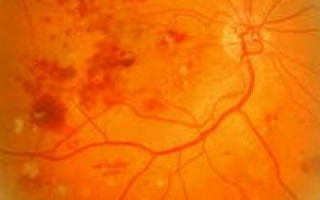
The initial stage of retinopathy is called nonproliferative , and is characterized by the appearance of microaneurysms that dilate the arteries, pinpoint hemorrhages in the eye in the form of dark round spots or streak-like stripes, the appearance of ischemic zones of the retina, retinal edema in the macular area, as well as increased permeability and fragility of the vascular walls. In this case, the liquid part of the blood enters the retina through thinned vessels, leading to the formation of edema. And if the central part of the retina is also involved in this process, then a decrease in vision .
It should be noted that this form of diabetes can occur at any stage of the disease, and represents the initial stage of retinopathy. If it is not treated, then the transition to the second stage of the disease occurs.
The second stage of retinopathy is proliferative , which is accompanied by impaired blood circulation in the retina, which leads to oxygen deficiency in the retina ( oxygen starvation , ischemia ).
To restore oxygen levels, the body creates new blood vessels (this process is called neovascularization ). The newly formed vessels are damaged and begin to bleed, causing blood to enter the vitreous body , the layers of the retina.
As a result, floating clouds appear in the eyes against the background of decreased vision.
In the later stages of retinopathy, as new blood vessels and scar tissue continue to grow, it can lead to retinal detachment and the development of glaucoma .
The main reason for the development of diabetic retinopathy is an insufficient amount of insulin , which leads to the accumulation of fructose and sorbitol , which contribute to increased pressure, thickening of capillary walls and narrowing of their lumens.
Symptoms of diabetic retinopathy
The main symptoms of retinopathy depend on the stage of the disease. Typically, patients complain of blurred vision, the appearance of dark floating clouds in the eye (gnats), and sudden loss of vision.
It is important to note that the sharpness of vision depends on blood sugar levels.
However, in the initial stages of retinopathy, visual disturbances are practically not observed, so patients with diabetes should undergo regular ophthalmological examinations to identify the first signs of the disease.
Diagnosis of diabetic retinopathy
The diagnosis of diabetic retinopathy is made based on the patient's complaints of decreased vision and examination of the fundus using an ophthalmoscope . Ophthalmoscopy allows you to identify pathological changes in the fundus. Ophthalmological examinations include determination of the level of intraocular pressure, biomicroscopy of the anterior part of the eye.
In addition, photographing of the fundus is carried out using a fundus camera , which allows you to document changes in the retina, as well as fluorescein angiography to determine the location of the vessels from which fluid is released and causes macular edema . Biomicroscopy of the lens is performed using a slit lamp.
Treatment of diabetic retinopathy
Treatment of retinopathy depends on the severity of the disease and consists of a number of treatment procedures.
At the initial stages of the disease, therapeutic treatment is recommended.
In this case, long-term use of drugs that reduce capillary fragility - angioprotectors ( dicinone , parmidine , predian , doxium ) is prescribed, as well as monitoring the maintenance of blood sugar levels. Sulodexide is also prescribed for the prevention and treatment of vascular complications in retinopathy . In addition, vitamin P , E , ascorbic acid , and antioxidants are used, for example, Strix , which contains blueberry extract and beta-carotene. This drug strengthens the vascular network, protects them from free radicals, and improves vision.
If the diagnosis of diabetic retinopathy shows serious changes, such as the formation of new vessels, swelling of the central zone of the retina, hemorrhages in the retina, then it is necessary to promptly begin laser treatment, and in advanced cases, abdominal surgery .
In case of swelling of the central zone of the retina ( macula laser coagulation of the retina is required . During this procedure, laser energy is delivered directly to the damaged areas of the retina through the cornea, anterior chamber aqueous humor, vitreous body and lens without incisions.
The laser can also be used to cauterize areas of the retina outside of central vision that are starved of oxygen. In this case, the laser destroys the ischemic process in the retina, as a result of which new vessels do not form. Also, the use of laser removes already formed pathological vessels, which leads to a decrease in swelling.
Thus, the main task of laser coagulation of the retina is to prevent the progression of the disease, and to achieve this, several (on average 3-4) coagulation , which are performed at intervals of several days and last 30-40 minutes. During a laser coagulation session, painful sensations may occur, during which local anesthesia may be used in the tissues surrounding the eye.
A few months after the end of treatment, fluorescein angiography is prescribed to determine the condition of the retina.
Cryocoagulation of the retina is performed if the patient has severe changes in the fundus of the eye, a lot of fresh hemorrhages, newly formed vessels, and if laser coagulation or vitrectomy is impossible.
If a patient with nonproliferative diabetic retinopathy develops a vitreous hemorrhage that does not resolve ( hemophthalmos ), then vitrectomy . It is advisable to perform this operation in the early stages, which significantly reduces the risk of complications from diabetic retinopathy.
During a vitrectomy, the doctor removes the vitreous and the blood that has accumulated there, and replaces it with a saline solution (or silicone oil). At the same time, the scars that cause ruptures and detachment of the retina are dissected and the bleeding vessels are cauterized with a laser ( diathermocoagulator ).
In the treatment of such a disease as diabetic retinopathy, a special place is occupied by the normalization of carbohydrate metabolism , because hyperglycemia contributes to disease progression. This happens by prescribing glucose-lowering drugs . Normalization of the patient’s diet also plays an important role.
Treatment of diabetic retinopathy should be carried out jointly by an ophthalmologist and an endocrinologist. With timely diagnosis and comprehensive treatment, there is every chance of maintaining vision and a full social and personal life.
The doctors
Medicines
Dicynone Doxium Sulodexide Ascorbic Acid Strix Trental
Prevention of diabetic retinopathy
Proper nutrition and regular exercise have a positive effect on the general condition of patients with diabetes. It is also important to have regular checkups with your eye doctor. Timely prevention of diabetic retinopathy and eye damage in diabetes mellitus is very important. Since in the later stages of the disease treatment is not effective. However, due to the fact that visual disturbances are not observed in the initial stages of retinopathy, patients seek help when extensive hemorrhages and changes in the central zone of the retina already occur.
Complications of diabetic retinopathy
The main complications of diabetic eye damage are tractional retinal detachment , resulting in hemophalmos , as well as secondary neovascular glaucoma , the treatment of which requires surgery.
Diet, nutrition for diabetic retinopathy
Diet for diabetes
- Efficacy: therapeutic effect after 14 days
- Timing: constantly
- Cost of products: 1300-1400 rubles per week
List of sources
- Vorobyova I.V., Merkushenkova D.A., Estrin L.G., Kalinina N.I., Ivanova D.P. Molecular mechanisms of the pathogenesis of diabetic retinopathy and maculopathy. // XI All-Russian School of Ophthalmology: collection of scientific papers. - M., 2012;
- Balashevich L.I. Ocular manifestations of diabetes / L.I. Balashevich, V.V. Brzhesky, A.S. Izmailov and others; Ed. L.I. Balashevich. - St. Petersburg: Publishing House St. Petersburg MAPO, 2004;
- Astakhov Yu.S., Shadrichev F.E., Lisochkina A.B. Diabetic retinopathy (patient management tactics) // Clinical ophthalmology. - M., 2004.
Source: https://medside.ru/diabeticheskaya-retinopatiya
How to identify and treat diabetic eye retinopathy - a dangerous complication of diabetes
19.07.2018
Diabetic (metabolic) retinopathy is a complication of diabetes mellitus. The disease is complex, severe, and dangerous due to its asymptomatic course. High levels of glucose in the blood and metabolic disorders lead to irreversible changes in the retina, and subsequently to the sudden onset of blindness.
It is very important to understand the causes, development, and symptoms of diabetic retinopathy, to diagnose the pathology in time, and to take immediate measures to treat this complication. The affected fundus of the eye at the stage of already obvious visual impairment cannot be restored.
What is diabetic retinopathy
Diabetes mellitus has an adverse effect on all human organs and systems, but the retina of the eye is most susceptible to pathological effects. This is due to the peculiarities of its structure and physiology. Diabetic retinopathy of the eye develops in the retina of both eyes, but with varying degrees of damage and severity of the process.
The retina is the main structure of the eye that allows us to see. All metabolic processes in the retina occur continuously through the microvascular network of the eye. It is this choroid that is affected by diabetes. Microvessels are located in the fundus of the eye, through which the retina is nourished, supplied with oxygen, and waste products are removed.
In diabetes, blood vessels become denser, thicken, lose elasticity, their permeability is impaired, and exchange through the walls deteriorates. This leads to poor microcirculation of the retina, which impedes its performance, contributes to a decline in visual functions, and dystrophic changes in the optic nerve. This is how diabetic retinopathy appears.
New vessels begin to grow (to compensate for the old ones), but they are very brittle and fragile, which leads to aneurysms, hemorrhages, and edema.
Often new vessels cover the vitreous body, which normally should be homogeneous and transparent. And if a new vessel bursts inside the vitreous body, hemorrhage occurs, which is called hemophthalmos. In this case, the blood screens, preventing the passage of light rays to the retina.
Also, new vessels, due to their thinness and single-layer walls, have greater permeability, which leads to blood plasma leaking into the outer or neighboring tissues. This causes swelling of the vessels themselves and the tissues they supply.
Complications of the disease
Complications of retinopathy in diabetes:
- Increased intraocular pressure, acute attack of glaucoma.
- Retinal edema, macular edema - the appearance of fog before the eyes, blurred images.
- Swelling, retinal detachment.
- Hemorrhage in the retina or other structures of the eyeball.
- Hemorrhages into the vitreous body - disrupts its transparency, accompanied by a veil before the eyes.
- Cataract.
- Partial or complete loss of vision.
- Diabetes mellitus affects all blood vessels of the body, so diabetic angioretinopathy is accompanied by general angiopathy (damage to all blood vessels), as well as an increased risk of strokes, heart attacks and thrombosis.
We invite you to watch a video that details the possible consequences and complications of the disease:
Classification of the disease
Diabetic retinopathy is classified according to stages of development. Depending on the severity or stage of retinal vascular damage, three main phases of the disease are distinguished.
Phases of retinopathy in diabetes mellitus:
- Non-proliferative diabetic retinopathy stage 1 - characterized by damage to retinal microvessels, aneurysms, pinpoint hemorrhages, and small exudative foci. There are no symptoms of diabetic retinopathy; the process can only be diagnosed by examining the fundus of the eye.
- Preproliferative diabetic retinopathy stage 2 – the number of damaged vessels increases, as well as the overall severity of the process. The vascular network becomes more tortuous, with the presence of blockages, loops, double visions or anomalies, and the volume of hemorrhage and edema increases. The clinical picture at this stage may be completely absent or may appear periodically, paroxysmally, simultaneously with surges in blood sugar levels.
- Proliferative (irreversible) diabetic retinopathy stage 3 – complete damage to the retinal vessels. Due to their inability to ensure normal metabolism, intensive germination (proliferation) of new vessels occurs in those structures of the eye where normally there should not be vessels. A pronounced clinical picture, intense, progressive vision loss.
Diabetic angioretinopathy only begins to manifest itself clinically at the 3rd stage. Unfortunately, at this stage nothing can be cured. The process can only be stopped or slowed down, while maintaining the level of vision at which the problem was diagnosed.
For a clearer picture of the disease and its forms, watch the video:
Reasons for the development of pathology
The main and only reason for the development of diabetic retinopathy is a chronic increase in blood sugar levels. This condition occurs in diabetes mellitus, when the body does not produce enough insulin.
All vessels are affected (angiopathy), and against this background local retinopathy (damage to the vessels of the retina) develops in diabetes mellitus. Besides diabetes, there are other risk factors that predispose you to developing diabetic retinopathy.
Aggravating reasons for the development of diabetic retinopathy:
- Metabolic disorders, obesity.
- Kidney disease.
- Hypertonic disease.
- Hormonal imbalances, changes during pregnancy, puberty or endocrine diseases.
- Genetic predisposition or presence of the disease in close relatives.
- Bad habits.
- The risk of the disease increases with the patient's age.
Symptoms
The main insidiousness of diabetic retinopathy is associated with the absence of symptoms until the stage of irreversible changes. Therefore, all diabetics should be examined by an ophthalmologist at least two or three times a year. A routine fundus examination will allow the doctor to assess the quality and health of the fundus vessels.
Diabetic retinopathy – the main symptoms and signs in diabetics:
- Floaters, dots, stars before the eyes, decreased clarity and visual acuity.
- Periodic sharp blurring of vision, floating clouds appear. Subsequently, patients associate this symptom with the moment of increased sugar levels.
- Blurring of the spot, disruption of the transparency of some areas of the field of view.
- The appearance of fixed black spots in the field of view.
- Hemophthalmos, a rupture of a vessel with hemorrhage into the vitreous body, is manifested by a sharp decrease in vision, as well as a red color of the white part of the eye.
Diagnostics
A very important stage in the treatment, prevention of complications and favorable resolution of diabetic retinopathy. Since it is impossible to independently identify pathology in the early stages, a diagnostic examination will help confirm or refute the presence of retinopathy in diabetes, as well as make an accurate diagnosis.
Methods for diagnosing diabetic retinopathy:
- Questioning the patient, collecting anamnesis of life and illness, identifying the main complaints and concomitant pathologies.
- Visual inspection.
- Determination of visual acuity.
- Ophthalmoscopy – examination of the fundus of the eye.
- Biomicroscopy of the eyes.
- Perimetry.
- Ultrasound and CT of the eyeballs.
- Measuring intraocular pressure.
- Additional laboratory examinations, consultation of related specialists, if necessary.
It is best if the diagnosis and treatment of diabetic retinopathy is carried out jointly by at least two specialists: an ophthalmologist and an endocrinologist.
Watch the diagnostic video:
Treatment
It is impossible to completely cure retinopathy in diabetes; the diagnosis is lifelong, since retinopathy is a complication of diabetes. Therefore, timely prevention and diagnosis of both diabetes and incipient vision problems is important.
The diagnosis of “diabetes mellitus” and “diabetic retinopathy” is not a death sentence. By following all the rules of nutrition, medication, and leading a healthy lifestyle, you can preserve your vision and avoid surgery and blindness. But this requires a lot of desire, as well as self-discipline, so everything is in your hands.
Diabetic retinopathy has four areas of treatment: reducing sugar levels, normalizing blood pressure, restoring metabolism, fighting and preventing complications. To achieve these goals, a conservative approach is used - diet, drug treatment, folk remedies, and surgical methods.
Diet therapy
Diet and proper nutrition are half the success of treating diabetes mellitus or its complications. The main goal of therapeutic nutrition is to ensure stability, uniformity in the supply of carbohydrates and their compliance with the physical activity performed. For diabetic retinopathy, nutrition should be completely balanced.
We do not recommend using other people’s diets or inventing anything yourself. Your doctor should prescribe proper nutrition based on your age, gender, weight, type of physical activity and type of diabetes.
Products that are strictly contraindicated:
- fast, easily digestible carbohydrates (sugar, confectionery, honey, fruits, juices);
- alcohol;
- foods high in fat (mayonnaise, butter, lard, cream);
- smoked;
- roast;
- salty;
- spicy.
Drug therapy
In the treatment of diabetic retinopathy, due to the deep anatomical location of the retina, eye drops are practically ineffective. More often, tablet forms of drugs are used, injections that are administered near the eye or into the eyeball itself, intramuscular, intravenous injections or droppers.
The main drugs used in the treatment of diabetic retinopathy:
- Angioprotectors are drugs that improve the condition of retinal vessels (Pentoxifylline, Doxium, Anginin, Parmidine).
- Anticoagulants - reduce the formation of blood clots (Etamzilat, Dicinon, Fraxiparin, Heparin, Flexal).
- Nootropic drugs to improve the condition of nerve cells (Cerebrolysin, Piracetam, Trental).
- Anti-inflammatory drugs (Ibuprofen, Dexamethasone, Prednisolone).
- A VEGF factor blocker is one of the main drugs for advanced diabetic retinopathy, with proliferation of defective vessels. This drug helps eliminate the formation of new vessels and the disappearance of already formed ones. The only drawback is its high cost. Not all patients have the opportunity to purchase it, and it must be administered periodically, but constantly. "Pegaptanib" or "Makugen" (cost 50,000 rubles), "Ranibizumab" or "Lucentis" (cost 47,000 rubles).
- Vitamins of group B, C, E, R.
- Drugs that improve metabolism in the retina: “Phosphaden”, “Emoxipin”, “Taufon”.
- If necessary, antibacterial therapy.
Watch a video about treating diabetic retinopathy without surgery:
Folk, home methods
We warn you that treatment of diabetic retinopathy with folk remedies should be agreed with your ophthalmologist and not at the expense of basic drug therapy. Self-medication can only aggravate or complicate the disease.
Common nettle is popular in folk medicine. It is eaten raw, with salads, juice or decoctions are made from it, and it is infused with collections. In second place is linden tea, which is very effective in lowering blood sugar levels.
At the pharmacy you can purchase vascular or diabetic preparations, tinctures based on herbs, without the addition of synthetic agents. Calendula tincture, blueberries, lingonberry juice, aloe leaves, persimmon, cranberries are useful.
Surgery
Surgical methods include laser coagulation of the retina. The type of operation depends on the area of the retina that is subjected to coagulation and the type of operation performed, namely:
- focal;
- panretinal;
- by lattice type.
Which type of coagulation and in which area to perform it is decided by the retinal surgeon who will perform the operation.
The essence of the operation is the local impact of the laser on certain areas of the retina in order to form a post-coagulation scar and stop bleeding and reduce swelling. Laser coagulation is also used in the prevention of retinal detachment.
The operation is used at the last stage of the disease; very rarely it is the only one, since the number of damaged vessels is constantly growing.
A complication of this operation is the negative effect in the form of destruction of visual cells at the sites of laser exposure; they are simply burned out, forming blind spots on the retina. So surgery is not a panacea, and it is wiser not to bring the situation to surgical intervention.
Watch a video about treating the disease with laser:
Disease prevention
For healthy people, prevention should begin with periodic scheduled blood sugar testing. If the level does not exceed the norm of 3.3–5.5 mmol/l, then everything is in order. When your fasting sugar level is higher than normal, you should consult an endocrinologist; these may be the first signs of diabetes.
The earlier retinopathy in diabetes is detected, the easier it is to fight it. If you have diabetes, you should not neglect preventive examinations with an ophthalmologist. You must clearly understand that the problem will catch up with you sooner or later, and timely detection and treatment can save your vision.
Save the article to bookmarks and share it on social networks. Write your treatment methods in x and be healthy.
How to identify and treat diabetic eye retinopathy - a dangerous complication of diabetes Link to main publication
Source: https://ozrenieglaz.ru/bolezni/drugie/diabeticheskaya-retinopatiya
Diabetic retinopathy
Diabetic retinopathy is a specific angiopathy that affects the vessels of the retina and develops against the background of long-term diabetes mellitus. Diabetic retinopathy has a progressive course: in the initial stages there is blurred vision, veils and floating spots before the eyes; in later cases there is a sharp decrease or loss of vision. Diagnostics includes consultations with an ophthalmologist and diabetologist, ophthalmoscopy, biomicroscopy, visometry and perimetry, angiography of retinal vessels, and biochemical blood tests. Treatment of diabetic retinopathy requires systemic management of diabetes and correction of metabolic disorders; in case of complications - intravitreal administration of drugs, laser photocoagulation of the retina or vitrectomy.
Diabetic retinopathy is a highly specific late complication of diabetes mellitus, both insulin-dependent and non-insulin-dependent types.
In ophthalmology, diabetic retinopathy causes visual impairment in patients with diabetes in 80-90% of cases. People with diabetes are 25 times more likely to develop blindness than other members of the general population.
Along with diabetic retinopathy, people with diabetes have an increased risk of coronary artery disease, diabetic nephropathy and polyneuropathy, cataracts, glaucoma, occlusion of the central and central veins, diabetic foot and gangrene of the extremities.
Therefore, the treatment of diabetes mellitus requires a multidisciplinary approach, including the participation of endocrinologists (diabetologists), ophthalmologists, cardiologists, and podologists.
Diabetic retinopathy
The mechanism of development of diabetic retinopathy is associated with damage to the retinal vessels (blood vessels of the retina): their increased permeability, occlusion of capillaries, the appearance of newly formed vessels and the development of proliferative (scar) tissue.
Most patients with long-term diabetes mellitus have some signs of fundus damage. When diabetes lasts up to 2 years, diabetic retinopathy to varying degrees is detected in 15% of patients; up to 5 years – in 28% of patients; up to 10-15 years – 44-50%; about 20-30 years – in 90-100%.
Risk factors
The main risk factors influencing the incidence and rate of progression of diabetic retinopathy include:
The development and progression of retinopathy can be promoted by puberty, pregnancy, hereditary predisposition, and smoking.
Taking into account the changes developing in the fundus, non-proliferative, pre-proliferative and proliferative diabetic retinopathy are distinguished.
Elevated, poorly controlled blood sugar levels lead to damage to blood vessels in various organs, including the retina.
In the non-proliferative stage of diabetic retinopathy, the walls of the retinal vessels become permeable and fragile, which leads to pinpoint hemorrhages and the formation of microaneurysms - local saccular dilatation of the arteries.
Through the semi-permeable walls of the vessels, a liquid fraction of blood leaks into the retina, leading to retinal edema. If the central zone of the retina is involved in the process, macular edema develops, which can lead to decreased vision.
In the preproliferative stage, progressive retinal ischemia develops due to occlusion of arterioles, hemorrhagic infarctions, and venous disorders.
Preproliferative diabetic retinopathy precedes the next, proliferative stage, which is diagnosed in 5-10% of patients with diabetes.
Contributing factors for the development of proliferative diabetic retinopathy include high myopia, carotid artery occlusion, posterior vitreous detachment, and optic nerve atrophy.
At this stage, due to oxygen deficiency experienced by the retina, new vessels begin to form in it to maintain adequate oxygen levels. The process of retinal neovascularization leads to repeated preretinal and retrovitreal hemorrhages.
In most cases, minor hemorrhages in the layers of the retina and vitreous resolve on their own.
However, with massive hemorrhages into the eye cavity (hemophthalmos), irreversible fibrous proliferation occurs in the vitreous body, characterized by fibrovascular adhesions and scarring, which ultimately leads to tractional retinal detachment. When the outflow pathways of the intraocular fluid are blocked, secondary neovascular glaucoma develops.
The disease develops and progresses painlessly and with few symptoms - this is its main insidiousness. In the non-proliferative stage, vision loss is not subjectively felt. Macular edema can cause a feeling of blurred vision and difficulty reading or doing close-up work.
In the proliferative stage of diabetic retinopathy, when intraocular hemorrhages occur, floating dark spots and a veil appear before the eyes, which disappear on their own after some time. With massive hemorrhages into the vitreous body, a sharp decrease or complete loss of vision occurs.
Patients with diabetes require regular examination by an ophthalmologist to identify initial changes in the retina and prevent proliferating diabetic retinopathy.
In order to screen for diabetic retinopathy, patients undergo visometry, perimetry, biomicroscopy of the anterior segment of the eye, biomicroscopy of the eye with a Goldmann lens, diaphanoscopy of eye structures, Maklakov tonometry, ophthalmoscopy under mydriasis.
The ophthalmoscopic picture is of greatest importance for determining the stage of diabetic retinopathy. In the non-proliferative stage, microaneurysms, “soft” and “hard” exudates, and hemorrhages are detected ophthalmoscopically.
In the proliferative stage, the fundus picture is characterized by intraretinal microvascular abnormalities (arterial shunts, dilation and tortuosity of veins), preretinal and endoviteral hemorrhages, neovascularization of the retina and optic disc, fibrous proliferation.
To document changes in the retina, a series of fundus photographs are taken using a fundus camera.
In case of opacities of the lens and vitreous body, instead of ophthalmoscopy, an ultrasound of the eye is used. In order to assess the safety or dysfunction of the retina and optic nerve, electrophysiological studies are carried out (electroretinography, determination of CFSM, electrooculography, etc.). Gonioscopy is performed to detect neovascular glaucoma.
The most important method for visualizing retinal vessels is fluorescein angiography, which allows recording blood flow in the choreoretinal vessels. An alternative to angiography is optical coherence and laser scanning tomography of the retina.
To determine the risk factors for the progression of diabetic retinopathy, a study of blood and urine glucose levels, insulin, glycosylated hemoglobin, lipid profile and other indicators is carried out; Ultrasound of renal vessels, EchoCG, ECG, 24-hour blood pressure monitoring.
In the process of screening and diagnosis, it is necessary to earlier identify changes indicating the progression of retinopathy and the need for treatment to prevent decrease or loss of vision.
Along with the general principles of treatment of retinopathy, therapy includes the correction of metabolic disorders, optimization of control over glycemic levels, blood pressure, and lipid metabolism. Therefore, at this stage, the main therapy is prescribed by an endocrinologist-diabetologist and a cardiologist.
The level of glycemia and glucosuria is carefully monitored, and adequate insulin therapy for diabetes mellitus is selected; Angioprotectors, antihypertensives, antiplatelet agents, etc. are prescribed. Intravitreal steroid injections are performed to treat macular edema.
Patients with progressive diabetic retinopathy are advised to undergo laser coagulation of the retina. Laser photocoagulation makes it possible to suppress the process of neovascularization, achieve obliteration of vessels with increased fragility and permeability, and prevent the risk of retinal detachment.
There are several main techniques used in retinal laser surgery for diabetic retinopathy. Barrier laser coagulation of the retina involves the application of paramacular coagulates in a “lattice” pattern, in several rows, and is indicated for non-proliferative retinopathy with macular edema.
Focal laser coagulation is used to cauterize microaneurysms, exudates, and small hemorrhages identified during angiography.
In the process of panretinal laser coagulation, coagulates are applied over the entire retinal area, with the exception of the macular area; this method is mainly used at the preproliferative stage to prevent its further progression.
When the optical media of the eye are clouded, an alternative to laser coagulation is transscleral cryoretinopexy, based on cold destruction of pathological areas of the retina.
In the case of severe proliferative diabetic retinopathy, complicated by hemophthalmos, traction of the macula or retinal detachment, vitrectomy is performed, during which blood and the vitreous itself are removed, connective tissue cords are dissected, and bleeding vessels are cauterized.
Severe complications of diabetic retinopathy can include secondary glaucoma, cataracts, retinal detachment, hemophthalmos, significant vision loss, and complete blindness. All this requires constant monitoring of patients with diabetes by an endocrinologist and ophthalmologist.
A major role in preventing the progression of diabetic retinopathy is played by properly organized control of blood sugar and blood pressure levels, and timely intake of hypoglycemic and antihypertensive drugs. Timely implementation of preventive laser coagulation of the retina helps to stop and regress changes in the fundus.
Source: https://www.KrasotaiMedicina.ru/diseases/ophthalmology/diabetic-retinopathy
Diabetic retinopathy: symptoms, stages, treatment
Diabetic retinopathy is a disease accompanied by damage to the blood vessels of the eyeball. Develops with prolonged diabetes mellitus. This severe complication causes blindness in people aged 20 to 74 years. What examination and treatment is required for this disease?
Pathogenesis and causes
The pathogenesis of diabetic retinopathy is quite complex. Among the main reasons are damage to the blood vessels of the retina: their excessive permeability, blockage of capillaries, the appearance of proliferative (scar) tissue and newly formed vessels. Such changes are caused by genetic features of the structure of the retina.
Metabolic changes that occur with elevated blood glucose levels play an important role in the development of the disease. In the presence of diabetes for up to 2 years, diabetic retinopathy is detected in 15% of patients; up to 5 years – 28%; up to 10–15 years – in 44–50%; from 20 to 30 years – in 90–100%.
Risk factors that influence the rate and frequency of disease progression include:
- level of hyperglycemia;
- duration of diabetes mellitus;
- chronic renal failure;
- arterial hypertension;
- having excess weight (obesity);
- metabolic syndrome;
- dyslipidemia.
Pregnancy, puberty, and bad habits also contribute to the development and progression of diabetic retinopathy.
Classification
There are several stages of the disease:
- initial;
- preproliferative;
- proliferative;
- terminal, or stage of final changes in the retina.
The initial stage of diabetic retinopathy is called non-proliferative. Occurs at any stage of the disease. It occurs in three phases.
- The first is vascular. It is characterized by phlebopathy and expansion of the avascular zone, the appearance of microaneurysms in areas of local capillary blockage.
- The second phase is exudative. It is distinguished by the occurrence of soft and dense exudates, muffs, edema in the macula (central zone of the retina), small single retinal hemorrhages.
- The third is hemorrhagic. It is accompanied by multiple hemorrhages, constriction of veins (they look like sausages), subretinal hemorrhages, the appearance of ischemic zones of the retina, and impaired permeability of the vascular wall.
If the non-proliferative form is not treated, it becomes pre-proliferative . At this stage, there are more changes in the retina.
During the examination, the ophthalmologist detects traces of multiple hemorrhages, ischemic areas (areas in which blood circulation is impaired) and fluid accumulations.
The pathological process affects the macula area. The patient complains of decreased visual acuity.
The proliferative stage of diabetic retinopathy is diagnosed in 5–10% of patients with diabetes.
Provoking factors for its development include posterior vitreous detachment, high myopia, optic nerve atrophy and blockage of the carotid arteries. The retina suffers from oxygen starvation.
Therefore, to maintain the required level of oxygen, new vessels are formed in it. This process leads to repeated retrovitreal and preretinal hemorrhages.
At the last (terminal) stage of the disease, massive hemorrhages occur in the vitreous body (hemophthalmos). Gradually more blood clots form. The retina stretches until it peels off. When the lens stops focusing light on the macula, complete loss of vision occurs.
Clinical picture
Diabetic retinopathy develops and progresses without characteristic symptoms. Decreased vision in the nonproliferative stage is not subjectively noticeable. A feeling of blurriness of visible objects can cause macular edema. There are also difficulties when reading at close range. Moreover, the sharpness of vision depends on the concentration of glucose in the blood.
In the proliferative stage of the disease, a veil and floaters appear before the eyes (the result of intraocular hemorrhages). After some time they disappear on their own. With massive bruising into the vitreous body, a sharp deterioration or complete loss of vision occurs.
Diagnostics
To screen for diabetic retinopathy, the patient is prescribed ophthalmoscopy under mydriasis, visometry, biomicroscopy of the anterior segment of the eye, perimetry, biomicroscopy of the eye with a Goldmann lens, Maklakov tonometry, diaphanoscopy of eye structures.
The ophthalmoscopic picture is of greatest importance for establishing the stage of the disease. In the non-proliferative phase, microaneurysms, hemorrhages, hard and soft exudates are detected.
In the proliferative phase, the fundus picture is distinguished by intraretinal microvascular abnormalities (tortuosity and dilatation of veins, arterial shunts), endoviteral and preretinal hemorrhages, fibrous proliferation, neovascularization of the retina and optic disc.
To document changes in the retina, a series of fundus photographs are taken using a fundus camera.
For opacities of the vitreous body and lens, an ultrasound of the eye is prescribed instead of ophthalmoscopy. To assess the impairment or preservation of the functions of the optic nerve and retina, electrophysiological studies are carried out: electrooculography, determination of CFSM, electroretinography. To detect neovascular glaucoma, gonioscopy is performed.
The most important method for examining retinal vessels is fluorescein angiography. It records blood flow in the choreoretinal vessels. If necessary, angiography is replaced by laser and optical coherence scanning tomography of the retina.
To identify risk factors for the progression of diabetic retinopathy, the levels of sugar in urine and blood, glycosylated hemoglobin, insulin, lipid profile and other indicators are examined. No less informative diagnostic methods include ultrasound of the renal vessels, 24-hour blood pressure monitoring, ECG and echocardiography.
Conservative therapy
In the initial stages of the disease, the main method of treatment is conservative. The patient is advised to take long-term medications that reduce capillary fragility - angioprotectors (Doxium, Parmidin, Dicynon, Predian). It is also necessary to maintain adequate blood glucose levels.
Sulodexide, ascorbic acid, vitamin P and E are prescribed for the treatment and prevention of vascular complications. Antioxidants (for example, Strix) provide a good effect. These preparations contain beta-carotene and blueberry extract. These beneficial substances improve vision, strengthen the vascular network, protecting against the effects of free radicals.
A special place in the treatment of diabetic retinopathy is occupied by the normalization of carbohydrate metabolism. This occurs by taking glucose-lowering medications. Conservative therapy also involves normalizing the patient’s diet.
People suffering from this disease are subject to medical examination. Based on the severity of diabetes, the period of incapacity for work is determined. The patient is contraindicated from work involving heavy visual load, vibration, tilting of the head and body, or lifting heavy objects. It is strictly forbidden to work in transport and in hot shops.
Surgery
If the diagnosis of diabetic retinopathy reveals serious disorders: hemorrhages in the retina, swelling of its central zone, the formation of new vessels, then laser therapy is indicated for the patient. In particularly difficult cases - abdominal surgery.
When new bleeding vessels and macular edema appear, laser coagulation of the retina is required. During this procedure, laser energy is delivered to damaged areas of the retina. It penetrates the cornea, vitreous body, anterior chamber humor and lens without incisions.
The laser is also used outside of central vision to cauterize areas of the retina that are starved of oxygen. With its help, the ischemic process in the retina is destroyed. As a result, new vessels stop appearing. This method also eliminates already formed pathological neoplasms. This leads to a reduction in swelling.
The main method of surgical treatment of diabetic retinopathy is laser therapy.
The main goal of laser coagulation is to prevent the progression of the disease. To achieve it, an average of 3-4 sessions are required. They last 30–40 minutes each and are held at intervals of several days.
Pain may occur during the procedure. Therefore, local anesthesia is performed in the tissues surrounding the eye. A few months after completion of therapy, a specialist evaluates the condition of the retina.
For this purpose, fluorescein angiography is prescribed.
If vitreous hemorrhage occurs with nonproliferative diabetic retinopathy, the patient requires vitrectomy.
During the procedure, the doctor removes the accumulated blood and replaces the vitreous with silicone oil (or saline solution). At the same time, scars that cause retinal separation and ruptures are cut with a laser, and bleeding vessels are cauterized.
This operation is recommended to be performed in the early stages of the disease. This significantly reduces the risk of complications.
If the patient has severe changes in the fundus of the eye, many newly formed vessels and fresh hemorrhages, cryocoagulation of the retina is performed. It is also necessary if vitrectomy or laser coagulation is impossible.
Prevention
The main way to prevent diabetic retinopathy is to maintain normal blood sugar levels. Optimal correction of lipid metabolism, compensation of carbohydrate metabolism, and blood pressure control are also important. Taking antihypertensive and hypoglycemic drugs will help with this.
Regular physical activity has a positive effect on the overall well-being of diabetics. Another way to a fulfilling life is proper nutrition. Limit the amount of carbohydrates in your diet. Focus on foods rich in natural fats and proteins.
For timely diagnosis, regularly consult an ophthalmologist. Do this when relevant complaints appear and at least once a year. Young people with diabetes are recommended to undergo examination at least once every 6 months.
Possible complications
Dangerous consequences of diabetic retinopathy:
- cataract;
- secondary neovascular glaucoma;
- significant decrease in vision;
- hemophthalmos;
- traction retinal detachment;
- complete blindness.
These conditions require constant monitoring by a therapist, neurologist, ophthalmologist and endocrinologist. Some complications can be eliminated with surgery.
The most effective treatment for diabetic retinopathy is to lower blood glucose levels and maintain normal levels. Eat right and visit your ophthalmologist regularly. Once a week, in the evenings, measure intraocular pressure. With timely diagnosis and comprehensive therapy, there is every chance to preserve vision.
Source: https://bezdiabeta.net/oslozhneniya/diabeticheskaja-retinopatija.html
Diagnosis and treatment of diabetic retinopathy - recommendations for patients to prevent complications
Diabetes mellitus is dangerous not only in itself, but also in the many negative consequences that it can lead to. One of its most common complications is diabetic retinopathy. This pathology is associated with the destruction of retinal vessels, which, in the absence of adequate and timely treatment, can lead to absolute blindness.
Previously, retinopathy was often diagnosed in patients who had crossed the 50-year mark. Nowadays there is a tendency towards its “rejuvenation”. Experts are not surprised by the presence of this pathology in patients with diabetes, whose age varies between 25-28 years.
The disease in question develops against the background of a violation of the structure of the blood vessels of the fiber, which is accompanied by:
The main reason for the development of this pathology is the long course of diabetes mellitus.
In general, if a patient lives with diabetes for about 25 years, there is an almost 100% guarantee that the disease will appear.
As the amount of sugar in the blood vessels supplying the organ of vision increases, their gradual degeneration occurs.
The main symptom of the initial stage of diabetic retinopathy is retinal swelling. This condition can lead to decreased visual function.
But often the errors are so insignificant that the patient does not pay attention to it. This is the main insidiousness of this disease: at the stage when it can be completely cured while maintaining vision, it practically does not manifest itself at all.
Degenerative phenomena extend beyond the retina and affect the veins: their diameter increases. Frequent hemorrhages are normal.
In addition, a characteristic feature is the appearance of cotton wool-like zones, which, in essence, are retinal infarcts.
At this stage, therapeutic measures can help stop the destructive process, but it will not be possible to restore the damaged areas. This means that lost vision cannot be restored.
As a result of the accumulation of blood clots in the vitreous body, the patient complains of the formation of the so-called. black spots before his eyes, causing him to rub his eyelids or blink frequently. These spots can be seen more clearly by looking at the light source.
In such a condition, you should immediately seek help from a medical facility. Lack of timely and adequate treatment at this stage can cause absolute blindness!
Source: https://www.operabelno.ru/diagnostika-i-lechenie-diabeticheskoj-retinopatii-rekomendacii-pacientam-po-profilaktike-oslozhnenij/