Greetings to everyone who is reading an article on the topic of MRI of the hip joints. Here you can find information about how the procedure itself goes, how to prepare for it, and whether it is better to choose computed tomography or magnetic resonance imaging.
Principle of MRI scanning
Sometimes, to identify pathological processes occurring in the hip joint, it is not enough to conduct only ultrasound and radiography.
In this case, an MRI procedure is prescribed, which makes it possible to identify the smallest damage to the joint, determine the condition of the tendon-ligamentous and cartilaginous apparatus, and muscle structures.
Using MRI, high-quality, three-dimensional images are obtained, on the basis of which a correct diagnosis can be made in 95% of cases.
- The principle of MRI scanning is based on nuclear magnetic resonance, which produces three-dimensional images of the scanned area.
- This diagnostic method is the most informative and promising. The advantages of MRI include:
- no radiation exposure to the body;
- possibility of repeated carrying out;
- identification of the smallest disorders in the joint that are not diagnosed by other means;
- refusal of invasive painful research methods;
- creating clear and three-dimensional images with the ability to zoom in on the area of interest;
- examination of all components of the joint in any plane;
- if necessary, a detailed examination of the area of the joint can be carried out;
- detection of tumor metastases in bones at early stages.
MRI examination of the hip joint allows you to obtain a layer-by-layer image of all its structures, including bones, tendons, joint ligaments, soft tissues, and blood vessels.
This diagnostic method is perfect for identifying various types of injuries and foci of pathologies. The procedure is absolutely painless, no recovery is required after it, and MRI can be performed repeatedly.
A magnetic tomograph creates a three-dimensional image of all joint structures in any plane and produces a three-dimensional image with some magnification.
This diagnostic method makes it possible to distinguish the most minor pathological processes occurring in tissues that are not capable of diagnosing alternative methods of examining the joint. Usually, when examining the hip joint, an x-ray is prescribed, which is an excellent way to see bone deformation and bone growths (osteophytes).
But, unfortunately, X-ray examination does not reveal changes in cartilage and synovial fluid.
And MRI allows you to abandon painful methods of examination, when synovial fluid is taken for analysis using a needle. Images obtained using MRI are always clear, which simplifies diagnosis. During the scan, the doctor has the opportunity to examine in detail those areas of the joint in which pathology is suspected.
It is this diagnosis that allows you to draw up a clear surgical plan. In this case, a scan is performed before the actual surgical intervention. The procedure does not have a negative effect on the human body, so it can be prescribed to children pregnant in the second half of pregnancy. The main condition is to remain still during the procedure.
MRI is one of the most informative ways to detect metastases in bone structures at the earliest stages, when X-ray examination does not show anything.
This is the main advantage of MRI over other diagnostic procedures, such as CT, tomography, X-ray, ultrasound.
The price of a hip MRI varies depending on the clinic. So in Moscow clinics the procedure can be performed from 3170 rubles. The question of how much a magnetic resonance scanning procedure costs depends on the distance of the clinic from the city center, its equipment, and the professionalism of the doctors.
First, you should study the addresses of all clinics where MRI of the hip joint is performed, read reviews and choose the most suitable one in terms of price and quality.
When choosing a clinic, you should know that in Moscow the price for a study using a high-field and low-field tomograph differs by 10-20%. The procedure in St. Petersburg costs approximately the same, so if you only need to identify an injury or mechanical, gross damage, then you should not spend money on a high-field tomograph.
If there are suspicions of vascular changes or systemic disorders, then you need to use a high-field tomograph, the power of which is more than one and a half Tesla.
The need for MRI and contraindications
The magnetic resonance imaging procedure is performed quite often.
If we talk specifically about the hip joint, then such a diagnostic procedure is prescribed in cases where there is:
- osteoarthritis of the hip joint;
- rheumatic joint lesions;
- joint dysplasia;
- systemic lupus erythematosus;
- spondylitis;
- Bekhterev's disease;
- arthritis;
- rupture of the joint capsule;
- fracture or crack of a bone;
- sprained muscles and ligaments;
- dislocation of the hip joint.
The procedure should definitely be done in case of aseptic necrosis of the femoral head. MRI is used to identify metastatic formations and their spread in the joint in cancer.
Scanning is carried out in case of pinched tendons, joint nerves, septic arthritis, osteomyelitis .
When not to do an MRI
Despite numerous indications for the procedure, MRI has contraindications. It cannot be performed on people who have metal devices installed in their bodies.
These include pacemakers, cerebral vascular clips, prostheses, neurostimulators, various implants (electronic, iron magnetic, metal). Research is not carried out if a person does not have the ability to control his body.
These could be various diseases of the nervous system, early childhood or old age, mental illness, the first half of pregnancy, heart failure, fear of closed spaces.
All contraindications should be clarified with the doctor who prescribes MRI diagnostics.
Hip MRI procedure
Magnetic resonance scanning shows the condition of all components of the joint. These are ligaments, tendons, synovial fluid, cartilage, muscles, articular heads of the femurs.
Sections are made in oblique and transverse planes. If the need arises, the doctor combines all the images into a three-dimensional volumetric image.
This comprehensive approach makes it possible to assess and characterize all the structures of the joint, to identify foci of injury and inflammation in it. During the scan, the diagnostician sees all the bones of the hip joint and adjacent soft tissues.
Tomography also shows how blood vessels function.
Preparation
Preparation for the procedure directly depends on the patient’s health condition and the use of contrast. It is necessary to make sure that there are no allergic reactions to the injected contrast agent. A study is carried out in advance to exclude renal failure and pregnancy at the very beginning. These two indications do not allow the procedure to be performed with contrast.
A few hours before the scan you should stop eating.
The doctor should be warned about medications taken, allergic reactions to certain medications, and existing chronic diseases.
Before the procedure, remove all jewelry, glasses, and watches. If you are afraid of closed spaces or you are too nervous, you should ask your doctor for a sedative.
Progress of the procedure
How is an MRI done? It's quite simple. The patient changes into disposable medical clothing and lies down on the couch.
He is given a contrast agent if necessary. If this is not provided, then the couch is simply pushed into the tomograph. The tomograph chamber is closed, it is equipped with ventilation and lighting. The patient communicates with the doctor using a microphone.
During the procedure, the patient hears a slight crackling sound from the device; there should be no further discomfort.
The entire scanning procedure lasts from 15 minutes to half an hour, and if contrast is used, the duration increases to an hour.
The entire cycle with decoding of the results takes approximately two hours. After the scan, the person can immediately return to their normal lifestyle. The doctor deciphers what the scan shows and gives the patient the result. The exception is complex cases when the transcript results are given the next day.
With photographs and transcripts, you need to contact a highly specialized specialist, depending on the results obtained.
If there is arthritis and arthrosis, contact a rheumatologist; if the tendons are pinched, you need to visit a traumatologist; if there is a complex injury, you may need the help of a neurosurgeon. If malignant tumors are detected, you should visit an oncologist.
CT or MRI: which is better?
To choose the right treatment plan, you must first identify the cause of the disease as accurately as possible.
For this, MRI or CT is used. And here the question arises: what is better to choose? It is worth saying that these diagnostic methods differ in the principle of operation. Thus, with MRI, a strong magnet and exposure to an electromagnetic field are used, and with CT, X-rays are used.
When choosing a diagnostic technique, several parameters are taken into account.
- Efficiency for the patient. CT is not used often, MRI can be used as often as necessary.
- Speed: The CT scan is faster, which is important because you must remain still during both procedures. Its duration is only a few minutes per joint, the procedure does not require preparation, with its help even microscopic changes are detected, with high image clarity.
- Contraindications: MRI is not performed in the presence of endoprostheses, pacemakers, metal particles in implants, or implanted electrical devices. There are no such contraindications for CT scanning. CT scanning is recommended for metal implants or metal pins installed in the human body.
- Type of pathological process. A CT scan is usually sent for traumatic injuries, and an MRI is sent for suspected changes in soft tissues and nerves.
A CT scan of the hip joint is prescribed if it is necessary to determine the diameter of the femoral head and the distance between it and the acetabulum, to establish the exact localization of bones in complicated fractures, and to identify foreign bodies.
MRI can detect inflammation and ruptures of muscles, ligaments, and degenerative changes in bones.
Are MRIs done to determine abnormalities in the arteries and veins? Yes, but in this case, magnetic tomography without contrast is used. MRI with contrast is necessary for more informative results. It is prescribed to detect neoplasms and abscesses.
Contrast is administered intravenously. Its active ingredient is gadolinium (metal). It is practically safe for the human body.
The contrast is generally well tolerated by people and side effects are very rare.
Disadvantages of procedures
The disadvantages of CT include high cost, a large list of contraindications, exposure to X-ray radiation, and it is impossible to perform the procedure on people whose weight is more than 130 kg.
As a contrast, iodine-containing drugs are used, which can cause allergies. The contrast is excreted by the kidneys, creating additional stress on them.
It is not recommended to perform it more than once a month due to the small, but still radiation. The disadvantages of MRI include the duration of the procedure, the inability to carry out the procedure in the presence of implants introduced during endoprosthetics and tattoos made with paint based on metal components.
You need to remain motionless for a long time.
CT and MRI have their advantages and disadvantages, so a specialist must decide which is better in each specific case, depending on the purpose of the study, the patient’s health, and the symptoms manifested.
For the elderly and children, MRI is preferred as it is safer. Sometimes, if the answer does not satisfy the doctor, the patient may be sent after a CT scan to an MRI or vice versa.
The procedures are not carried out simultaneously, but one after the other is quite possible if this is necessary for diagnosis. To summarize, my dear readers, we can say that MRI in the presence of pathological processes in the hip joint and lumbar region is a very important diagnostic procedure, with the help of which it is possible to detect the most minor changes in such an important joint.
Timely MRI and CT will allow us to identify a disease such as coxarthrosis of the hip joint and begin treatment that will slow down the progression of the pathology.
All the best to you, take care of yourself!
Source: https://tazobedrennyjsustav.ru/lechenie/mrt-tazobedrennyh-sustavov.html
Indications and contraindications for MRI of the hip joint
Content:
- Benefits of MRI of the hip joint
- Indications
- Contraindications
- Diagnostic cost
- Addresses of clinics and centers where you can get an MRI of the hip joint
MRI is a highly accurate method for diagnosing the hip joint, which is quite popular due to its affordability and degree of information content.
Practice shows that a qualified doctor, analyzing an image obtained by X-ray diagnostics of the hip joint, is able to examine its structure in detail, identify existing pathologies, and assess the course of physiological processes.
Tomography of the hip joint takes several thin slices of a single three-dimensional image. The doctor carefully examines them, identifying, if necessary, even the vascular network and individual vessels, nerve trunks.
A procedure of this kind is considered indispensable when carrying out preparatory measures for an operation and in planning the stages of surgical intervention. Therefore, MRI of the hip joint is very often used in surgery and traumatology.
Advantages of the method
Tomography does not expose the body to radiation, so the procedure can be repeated many times. The device is capable of creating accurate images of structures with the required magnification and in different planes.
This area has certain structural features. Thus, the hip joint is the only one in the entire body whose structure contains a blood vessel.
It is surrounded by many tendons and ligaments, which are often damaged as a result of injury to this area.
MRI with high accuracy allows you to identify existing tissue disorders that are inaccessible to other diagnostic methods.
In addition, tomography allows you to abandon invasive diagnostic methods, for example, arthroscopy, when a thin endoscope is inserted into the joint cavity, thanks to which you can see its structure from the inside.
Indications
Currently, MRI of joints is the leader among technologies that allow diagnosing diseases in the pelvic area, relegating radiography and computed tomography to the background. Indications for MRI of the hip joint:
- rheumatic diseases of the hip joints: ankylosing spondylitis, rheumatoid arthritis, ankylosing spondylitis, systemic lupus erythematosus;
- coxarthrosis (i.e. deforming osteoarthritis of the joint);
- infectious arthritis (chlamydial arthritis, Reiter's disease, etc.);
- suspected accumulation of blood in the hip joint – hemarthrosis;
- suspicion of fractures/cracks of the pelvic and hip bones, rupture of the joint capsule, sprains/ruptures of ligaments, tendons and muscles, other industrial, sports and household injuries;
- dysplasia - abnormalities in the development of the joint;
- joint dislocation;
- swelling and stiffness of tissues in the joint area, lower limb;
- chronic pain;
- aseptic necrosis (death) of the femoral head;
- pinched nerves, tendons;
- metastases (foci of inflammation or tumor process) in the area of the joint itself;
- tenosynovitis and osteomyelitis;
- septic arthritis;
- various osteochondropathy (for example, Calve-Perthes disease).
Contraindications
It is not recommended to conduct an examination if there are metal elements in the body: a pacemaker, vascular clips, an insulin pump, etc., since the magnetic field that arises during the operation of the tomograph disrupts the functioning of these elements and causes ferromagnetic metals to heat up and move.
The presence of titanium prostheses, as well as other metal structures, modern dental implants, as well as braces (polymer, ceramic, plastic) are not a contraindication to MRI, since they are made from materials that are not attracted by a magnet.
- For MRI of the hip joints and pelvic bones, performed with a contrast agent, contraindications are lactation and pregnancy - the drug can adversely affect the fetus and penetrates into a woman’s milk.
- The procedure is not recommended for renal failure (in chronic form), since the removal of the substance from the body is impaired.
- Intolerance to the contrast agent itself, against which an allergic reaction may occur, is also a limitation to MRI.
Cost of the procedure
Regarding the monetary side of the issue, the price of an MRI of the hip joint procedure seems quite high for many. Thus, the cost of an MRI for examining 1 hip joint in Moscow clinics starts from 4 thousand rubles.
, for examination of 2 joints + use of contrast - up to 13 thousand rubles. Many consider the cost of the procedure to be a significant disadvantage of this method.
But experienced traumatologists and surgeons recommend MRI for diagnosing joints for its effectiveness and the lack of influence of X-ray radiation on the body.
Addresses of clinics and centers where MRI of the hip joint is performed
Addresses of clinics and centers in Moscow
Currently, there are 31 diagnostic centers in Moscow, in one of which anyone can undergo a tomography of the hip joint. Here are some of them:
Within half an hour after the study, experienced specialists from the European Diagnostic Center will interpret the images. Thus, within one day you will undergo the study and find out the exact diagnosis, which is very convenient. You don't have to wait, as all the research results will be sent to you by email for free.
You can make an appointment by calling 8 (499) 391 75 78 or online through the form on the website edc.ru (in this case, you will receive a 5% discount ), after which a representative of the center will contact you and clarify all the details - explain how to prepare to the procedure, agrees on the exact time of the study, etc.
Address: Moscow, st. Nagatinskaya 1 building 25, Nagatinskaya metro station (3 min walk)
- Multidisciplinary medical Center "Capital". m. Smolenskaya, m. Kropotkinskaya.
Address: Bolshoi Vlasyevsky Lane, 9
This center operates 24 hours a day. Located ten minutes walk from the metro. Children over twelve years of age and adults can undergo the necessary examination. Registration for MRI is carried out by phone: +7 (495) 255-12-74. Cost - 5500 rub.
- Honey. Center "PrimaMedica" metro Prospekt Vernadskogo, metro Novye Cheryomushki, metro Kaluzhskaya.
Address: st. Academician Chelomeya, 10B.
Multidisciplinary diagnostic center. Located ten minutes away by public road. transport from the metro. Examination of adults. Registration for MRI is carried out by phone: +7 (495) 966-38-13. Cost - 4600 rubles.
- Diagnostic department Med. center on Botkinsky. Begovaya metro station, Dynamo metro station.
2nd Botkinsky proezd, 8.
This center provides MRI examinations for adults. From Art. Begovaya metro station can be reached by bus No. 847 - stop. “2nd Botkinsky Proezd” or from the station. m. Dynamo - bus No. 12ts - stop. “Hospital named after. Botkin. Registration for MRI is carried out by phone: 8 (495) 785-61-44. Cost – 4200 rub.
- Diagnostic center "MRI-City". m. Proletarskaya, m. Paveletskaya.
st. Derbenevskaya, 1, building 5.
Children over 12 years of age and adults can undergo the necessary examination. Located ten minutes walk from the metro. Registration for MRI is carried out by phone: +7 (495) 236-70-53. Cost – 8000 rub.
Addresses of clinics and centers where MRI is performed in St. Petersburg
There are several centers in St. Petersburg where you can undergo MRI of the hip joint, including:
MRI Center of Professor V.A. Fokin
metro station Petrogradskaya (5 min. walk). Tel.: +7 (812)385-77-16. Cost – 5000 rub.
Located five minutes. walk from metro station "Gorkovskaya".
Tel.: (812) 385-51-45. Cost – 5000 rub.
- Center for MRI and CT diagnostics.
Maly Ave. VO (metro Vasileostrovskaya). Tel.: 8 (812) 385-77-56. Cost – 4200 rub.
Source: https://vashaspina.ru/pokazaniya-i-protivopokazaniya-k-mrt-tazobedrennogo-sustava/
MRI of the hip joint - indications and alternative methods of examination
Currently, the number of diseases of the musculoskeletal system has increased significantly.
The hip joint is the largest in the human body, and it bears the main load, as a result of which the likelihood of developing articular pathologies increases.
To diagnose pathological manifestations in the hip area, MRI of the hip joint is used - the most informative method that allows you to make the correct diagnosis in a short time.
MRI of the hip joints
Magnetic resonance imaging is very popular due to the high accuracy of the results - in a short time the doctor receives an accurate three-dimensional image of the desired object.
In addition, MRI of the hip joint is an absolutely harmless method of examination - it is based on the chemical effect of electromagnetic waves on human tissue.
The technique is widely used in traumatology and rheumatology.
Advantages of MRI over other examination methods
- The main advantage of MRI of the hip joint is the ability to diagnose various pathologies that are invisible by other examination methods, for example, with X-rays or CT.
- The study is non-invasive and does not cause pain to the person.
- High accuracy of results and minimal error percentage.
- Possibility of carrying out in childhood.
- Diagnosis of cancer in the early stages, when there are no clinical manifestations yet and the tumor is not visualized by other research methods.
All these points make the MRI procedure the most preferable for diagnosing pathologies of the hip joint.
In addition to the above, the method allows:
- Localize foci of inflammation.
- Visualize nerve trunks and blood vessels separately.
- Examine all structures of the hip joint.
Indications for MR imaging of the hip joint
MRI of the hip joint is indicated for any pain in the pelvic area of unknown origin. In addition to pain, there are certain already diagnosed diseases and conditions for which MRI is mandatory:
- Pain in the pelvic area of unknown origin.
- Cracks and fractures in the joint area that are not visible on x-rays.
- Stiffness and limited mobility in the joint.
- Arthritis of the hip joint associated with systemic lupus erythematosus and rheumatism.
- Traumatic injuries - rupture of ligaments and tendons, accumulation of blood and fluid in the joint cavity.
- Habitual dislocation.
- Bursitis, osteoporosis, arthrosis, myositis.
Osteoarthritis of the hip joint
- Tumors affecting soft and bone tissue in the joint area.
- Infectious diseases.
- Degenerative processes in the joint.
- Damage or compression of nerve trunks.
- Examination before surgery, as well as to monitor the effectiveness of treatment.
- Inability to perform CT and X-rays.
This is not a complete list of indications for MRI of the hip joint - the method is indispensable in diagnosing many articular pathologies. The main reason for prescribing MRI is the diagnosis of cancer, identifying the exact boundaries of the tumor, and the prevalence of metastases.
The patient does not require special preparation for magnetic resonance imaging of the joint. There is no need to follow a diet or take special medications.
Description of the study
Before an MRI session of the hip joint, the patient must prepare suitable clothing - it should be comfortable, not restrict movement and not contain metal jewelry. To obtain an accurate image, the patient must remain completely still, so it is better to worry about your own comfort.
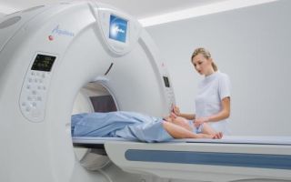
Girl undergoing MRI examination of hip joints
You should take with you documents related to the disease for which the examination is prescribed: a medical record, a referral for an MRI, the results of previous examinations.
Before scanning, be sure to remove all jewelry and take out keys, phones and bank cards from your pockets - they can be left in a special safe.
It is very important before going for a scan to make sure that there are no contraindications - the presence of metal objects in the patient’s body is an absolute contraindication to MRI.
Comparison of MRI, CT and radiography
Radiography is a research method that allows you to obtain x-ray images of structures on special paper or film.
X-ray is the most accessible method and is used for the primary diagnosis of diseases of the hip joint in cases where there are clear clinical symptoms. Like all examination methods, x-rays have positive and negative sides.
Unlike MRI, radiography and CT are accompanied by a certain dose of ionizing radiation, and the resulting images do not provide an idea of the functional state of the hip joint.
In addition, it is impossible to examine the desired areas layer by layer, and also to obtain information about the condition of soft tissues - they are not visualized by x-ray. X-ray is good for initial examination, and MRI is done to clarify the diagnosis in controversial cases.
A CT scanner looks very similar to an MRI machine.
Computed tomography (CT) is an examination method based on the effects of X-rays - a circular scan is carried out with the conversion of electrical signals into an image using a special computer program. CT shows bone tissue more clearly than MRI, but it cannot be performed frequently - the ionizing radiation that underlies CT and X-rays has a harmful effect on the human body.
MRI is more informative when examining cancer and determining the condition of soft tissues, and CT is more informative when diagnosing traumatic lesions.
Which method is better? The choice depends on the purpose of the examination and the indications - in some cases an X-ray will be sufficient, in others an MRI or CT scan will be required to clarify the diagnosis.
Source: https://diagnostinfo.ru/mrt/pozvonochnik-sustavy/mrt-tazobedrennogo-sustava.html
MRI of the hip joints: indications and interpretation of results
MRI of the hip joints is a modern radiation research method with enormous imaging capabilities.
Theoretically, magnetic resonance imaging can be used to diagnose deforming arthrosis, arthritis, hip dysplasia, ankylosing spondylitis, aseptic necrosis of the femoral head, etc.
In many clinics, doctors prescribe MRI to patients for diagnostic purposes.
However, conducting such research does not always make sense. Let's figure out why.
Magnetic resonance imaging is an accurate and highly informative, but expensive research method.
It allows you to obtain a three-dimensional layer-by-layer image of all structures of the hip joint, which is not always necessary.
In most cases, doctors only need to do x-rays and ultrasound to make a diagnosis. Such an examination will cost the patient less, which is extremely unprofitable for clinics and hospitals.
Advantages and disadvantages of the study
It is important to know! Doctors are shocked: “An effective and affordable remedy for joint pain exists...” Read more...
The advantages of MRI include the high visualization capabilities of the method and the absence of radiation exposure to the body. During the study, a magnetic field is used, which does not harm a healthy person in any way. Magnetic resonance imaging is allowed even for children and pregnant women.
The main disadvantage of the study is its high cost. In Moscow, prices for MRI of one hip joint range from 3500-6000 rubles. It is much more profitable for a person to have an ultrasound scan, which costs more than half the price (1800-2200 rubles).
Indications for MRI of the hip joint
In surgery and traumatology, MRI is used mainly in preparation for hip joint surgeries. Magnetic resonance imaging allows you to get an idea of the spatial arrangement of bones, ligaments, nerves and blood vessels. This helps doctors plan surgery correctly.
“Doctors are hiding the truth!”
Even “advanced” joint problems can be cured at home! Just remember to apply this once a day...
>
The study can be carried out when the diagnosis could not be made based on the results of radiography. However, in this case, arthritis, synovitis, osteoarthritis and many other diseases of the hip joint can be diagnosed using conventional ultrasound. Ultrasound examinations are much cheaper, which is a huge advantage over MRI.
If you are not planning to have surgery, do not rush to spend money on magnetic resonance imaging of the hip joint. Instead, consult another professional. Most likely, he will be able to make a diagnosis and prescribe treatment without conducting a study.
Who should not have an MRI?
Many people believe that MRI cannot be done in the presence of joint endoprostheses, hernia meshes, metal dental implants, braces, bone wires, rods and other fixation devices. Actually this is not true. Metal structures made from modern alloys interact very weakly with a magnetic field. Thanks to this, MRI is safe when available.
It is not recommended to examine the patient if there are several large prostheses. Magnetic resonance imaging is strictly prohibited for persons with installed electronic implants.
These include:
- stents and clips on vessels;
- pacemakers;
- artificial heart valves;
- insulin pumps;
- artificial lenses;
- cochlear implants.
Before the examination, do not forget to inform the doctor about the presence of any implants and severe chronic diseases. This will help the specialist understand whether you can be sent for an MRI.
What does an MRI of the hip joints show?
Magnetic resonance imaging is used to detect pathological changes in bones, articular cartilage, bursa, ligaments, nerves, vessels and periarticular tissues. If interpreted correctly, the study allows a diagnosis to be made without any additional diagnostic measures. Let's see what tomography of the hip joints shows in various diseases.
Patients who received a hip replacement with a diameter of more than 35 mm with a metal-to-metal friction pair require careful annual monitoring for the first 5 years. MRI allows us to identify pathological changes in periprosthetic soft tissues and bones. They indicate a high risk of osteolysis, endoprosthesis instability and infectious complications.
Table 1. MRI signs of common hip diseases
| Disease | Its signs on tomograms |
| Aseptic necrosis of the femoral head | At the first stage of the disease, a local hypodense area with a possible border of moderate sclerosis is identified in the head of the femur. At the following stages, necrotic defects, areas of sequestration, cysts, etc. appear on MRI images. Aseptic necrosis of stages II-V can be diagnosed even using radiography |
| Deforming osteoarthritis | At stages I-II of the disease, articular cartilage is destroyed in humans. Pathological changes are detected using ultrasound or MRI. During studies, they are visualized as defects in cartilage tissue. Subsequently, bones become involved in the pathological process, and conventional radiography helps to diagnose the disease. |
| Ankylosing spondylitis | MRI can reveal pathological changes in the subchondral bone, characteristic of the initial stages of coxitis. They cannot be detected using ultrasound. Subsequently, the disease causes thickening of the hip joint capsule and accumulation of fluid in the synovial cavity. These changes can be detected using more accessible methods than this |
| Coxarthritis | It manifests itself as swelling and thickening of the joint capsule, inflammation of the hip joint, and accumulation of fluid in the synovial cavity. These changes can be detected using MRI or ultrasound. Indirect signs of arthritis can also be detected on radiographs |
| Hip dysplasia in children | MRI for dysplasia reveals inversion of the cartilaginous lip, hypertrophy of the transverse ligament, deformation of the joint capsule and its compression by the iliacus tendon. Magnetic resonance imaging allows you to obtain a spatial image of the structures of the hip joint, which is very important in planning the treatment of pathology |
| Femoral neck fractures, hip dislocations | They are diagnosed using conventional radiography, which reveals a violation of the integrity of the bones or their displacement. The study is used to assess the condition of the soft tissues, nerves and vessels of the hip joint. This allows us to understand how serious the patient’s condition is |
Source: https://sustavlive.ru/diagnostika/mrt-tazobedrennogo-sustava.html
MRI of the hip joint
Pathologies of the musculoskeletal system are difficult to diagnose due to their complex visualization. Based on patient complaints, the doctor will only make a preliminary diagnosis, which requires confirmation by hardware diagnostic methods. An MRI of the hip joint is just such a method that will help identify problems with the musculoskeletal system and soft tissues.
Features of the procedure
MRI of the hip joint is performed in cases where other diagnostic methods are not informative, or there is a need for a more detailed examination of the elements of the articulation. Using MRI, you can detect even the slightest structural damage that ultrasound and x-rays cannot show.
Using a magnetic resonance imaging scanner, it is possible to make a layer-by-layer image of the elements of the joint - to examine cartilage and bones, tendons and ligaments, soft tissues and blood vessels. MRI is safe, it is a non-invasive technique that gives informative results. In 95% of cases, MRI helps clarify the diagnosis.
The principle of the study is based on the capabilities of nuclear magnetic resonance, which allows you to obtain accurate, high-quality images of the hip area. These pictures are three-dimensional, so they more than replace x-ray examination in two projections - MRI allows you to see the joint in more detail.
The MRI of the hip joint shows all elements available for visualization. These are primarily bones and ligaments, soft tissues. Also in the MRI image you can see structural disorders of cartilage tissue and vascular pathologies.
REFERENCE. After an MRI, many doctors refuse arthroscopy as a diagnostic method, since the image shows the damage that caused the patient’s complaints, so there is no point in performing arthroscopy.
The magnetic resonance imaging procedure has many advantages for diagnosing pathologies of the hip joint over other research methods. It is thanks to them that MRI has become such an indispensable procedure. The advantages of the procedure are as follows:
Joint and bone doctor
- screening will show even the slightest abnormalities in the hip joint that CT or ultrasound will not see;
- By choosing magnetic resonance imaging, doctors can refuse a more harmful diagnostic method, such as x-rays, which negatively affects human health;
- the capabilities of MRI are comparable to those of arthroscopy, so in many cases doctors opt for a non-invasive research method;
- A huge advantage of MRI is the absence of radiation exposure, which allows tomography to be done several times in a short period of time. This is important if necessary to diagnose the patient’s condition before surgery, as well as take several photographs at different stages of recovery, for example, after endoprosthetics;
- MRI makes it possible to create a three-dimensional image, which, even at high magnification, retains a clear picture and the pathology of the joint can be examined;
- the image can visualize any structure - not only a bone element, but also blood vessels, nerve endings, and soft tissue areas;
- extensive imaging capabilities make it possible to see malignant tumors and metastases even at an early stage, when ultrasound and x-rays are unable to detect such pathological neoplasms;
- Surgeons actively use the results of magnetic resonance imaging before performing surgery on a joint. The clarity of the picture helps to draw up a plan for the operation, anticipate possible complications and be prepared for them;
- The test can be carried out even on children who are unable to move at the time of screening. Conventionally, MRI is performed on children from the age of seven, but if necessary, the procedure is also performed on younger patients;
- If necessary, MRI is also permitted for pregnant women from the second trimester.
Indications
Magnetic resonance imaging is recommended for patients who suffer from various diseases. Most often, MRI is performed for injuries and DOA of the hip joint.
Deforming osteoarthritis of the hip joint (DOA), or coxarthrosis, is a progressive pathology of the cartilage tissue of the hip joint, in most cases developing due to high load on the joint.
Leads to thinning of hyaline cartilage; as a result of pathological processes, bone elements take an incorrect position.
To reduce noise, patients wear special headphones
MRI is also prescribed for patients with the following diagnoses:
- Reiter's disease is an autoimmune pathology affecting the hip joint. Develops as a result of penetration of chlamydia into the genitourinary organs;
- articular dysplasia - the disease manifests itself in impaired development of the hip joint. It is usually determined from birth. According to the MRI results, the patient's femoral head is incorrectly oriented in relation to the acetabulum;
- Bechterew's disease - if the hip joint is affected, a diagnosis of rhizomyelic form of ankylosing spondylitis is made. At the initial stage of the disease, patients feel only morning stiffness in the muscles, and at a later stage, the image shows a narrowing of the joint space of the hip joint;
- rheumatoid arthritis - at an early stage, the synovial membrane is involved in the disease, hyaline cartilage suffers less, but as the disease progresses, secondary deforming arthrosis ultimately develops;
- systemic lupus erythematosus - the disease leads to the death of the femoral head;
- dislocation of the hip joint - can be congenital or acquired, which is rare. When a dislocation occurs, the head of the femur comes out of its anatomical position, and it moves to the side, the limb is shortened;
- a crack or fracture of a bone - usually accompanied by a dislocation. An MRI clearly visualizes the type of fracture - cut or depressed. In this case, you can see not only damage to the head of the bone, but also to the femoral neck, which happens with major damage - a car accident, a fall from a height, etc.;
- hemarthrosis - bleeding into the joint, which is clearly visible in the picture;
- damage to the integrity of the joint capsule - usually with severe damage, rupture of the joint capsule and outpouring of internal contents may occur;
- sprain of ligaments, thigh muscles - damage to the ligaments is clearly visible on MRI - it can be a rupture, tear or severe sprain. Similar processes are diagnosed in muscles;
- chronic pain of unknown origin - referred pain, neuralgia, usually not associated with damage to the hip joint itself. On an MRI, the picture will show normal;
- tenosynovitis - inflammation of the tendon and its synovial membrane surrounding the tendon;
- osteomyelitis is a purulent lesion of bone tissue that covers not only its surface, but also the bone marrow. To assess the extent of damage and diagnose the disease, it is better to do an MRI;
- necrosis of the femoral head - the last stage of osteomyelitis and other diseases leading to necrosis;
- pinching of nerve endings - possible with hemarthrosis, dislocation, damage to soft tissues;
- Calve-Perthes disease – tissue necrosis of the epiphysis of the femur caused by aseptic processes;
- neoplasms - benign tumors are formed as a result of the proliferation of cartilage tissue. Often, MRI diagnoses chondroblastoma, chondroma (can degenerate into malignant), osteochondroma;
- bone cancer - this can be Ewing's sarcoma (in adolescents) or chondrosarcoma. The tumor process affects various parts of the bone and leads to pathological cell proliferation. MRI visualizes what other elements of the hip joint are involved in the pathological process;
- metastases - the occurrence of daughter tumors that have penetrated through the blood or lymph into the hip joint and affect the bone or soft tissue.
IMPORTANT! Magnetic resonance imaging is used before planning surgery and to monitor treatment.
The procedure is often performed on patients with an endoprosthesis in order to monitor the survival of the implant and prevent its rejection in time. If it is necessary to take several images within a short time, discounts may be provided to certain categories of patients.
Contraindications
Magnetic resonance imaging of the hip joint can be done in almost all patients. But there are some caveats to this screening:
- MRI is not performed on patients who have had joint replacement made from special alloys;
- The procedure cannot be performed if the body contains structures made of ferromagnetic alloys, which can heat up and move under the influence of a magnetic field;
- they refuse to undergo a contrast MRI procedure if you are allergic to the injected contrast agent - gadolinium;
- Tomography should not be performed on patients with pacemakers, insulin pumps, or heart valve implants;
- tattoos on the body whose inks contain metals are contraindications;
- MRI is not performed in the first trimester of pregnancy.
Magnetic resonance imaging can be performed on patients whose endoprosthesis is made of titanium. This metal is not ferromagnetic, so the procedure will not have any side effects. The procedure can be performed on people with metal crowns on their teeth, veneers, dentures (screws, pins), and braces.
If it is necessary to introduce contrast, there are limitations. MRI with contrast is not performed on pregnant women in any trimester, women who are breastfeeding, or patients with chronic kidney disease, which may affect the excretion of gadolinium. In most cases, patients respond normally to contrast, but a local irritation reaction may occur at the site of contrast injection.
Preparation and execution
In order for the magnetic resonance imaging procedure to go smoothly, you need to properly prepare for it. This is not difficult to do, since MRI does not impose particularly complex requirements for the procedure and no lengthy preparation is required.
The MRI procedure can be performed even on children
Here are a few rules for those who are candidates for the procedure:
- Patients should be examined in comfortable clothing that does not restrict movement. Clothing should not have metal elements. The belt on trousers with a metal buckle must be removed before MRI;
- doctors will remind the patient that it is also necessary to remove watches, jewelry, hearing aids, metal hair clips, etc.;
- if possible, you should provide the doctor with the results of a previous study;
- if there is an allergic predisposition, this will also be reported before the procedure.
Patients who suffer from claustrophobia can find out the type of device in advance. They may ask to be screened in an open MRI scanner. Small children are first given sedation, and it is also given to mentally ill patients who cannot remain in one position for a long time.
The entire MRI procedure takes about half an hour, and an MRI with contrast takes a little longer. Before starting the procedure, the doctor places the patient on the table and explains the rules for conducting the study. Then the patient's body is placed in a magnetic field, and the procedure begins.
There is no unpleasant sensation during the procedure, but warmth is felt in the hip joint area. The only irritating factor is small noises surrounding the patient, but to eliminate them, the doctor may suggest headphones in advance.
IMPORTANT! If unpleasant sensations occur, the patient gives a special signal and the diagnosis is stopped.
After an MRI, the results are released the next day. They can be obtained in digital format on disk or flash memory, as well as on film. The MRI result is accompanied by a specialist interpretation. Based on the results of the MRI, the doctor will give a recommendation as to which specialist should be contacted for further treatment. This could be a surgeon, orthopedist, or oncologist.
The MRI procedure is the safest diagnostic procedure available today. It allows you to identify pathologies of the hip joint that require additional diagnostics. The unique technique is highly accurate, and the MRI results allow the patient to be given the correct treatment.
Source: https://elemte.ru/diagnostika/mrt-tazobedrennogo-sustava