Checking the mammary glands using ultrasound has become an indispensable procedure in the fight and early diagnosis of many diseases. The mammary glands are very susceptible to mastopathy - this is a disease that causes benign tumors to appear in the breast. In this article we will look at which day of the cycle to do an ultrasound of the mammary glands for mastopathy.
Let us note that ultrasound diagnostics is a modern, painless, harmless, non-invasive and highly informative procedure. For prevention it is indicated for absolutely everyone.
General information
Mastopathy is considered a dyshormonal disease, as a result of which pathological compactions of breast tissue begin to form.
Women aged 18 to 45 years are susceptible to mastopathy, with the peak of the disease occurring between 30 and 45 years.
Breast health is very dependent on the two main hormones in the female body, estrogen and progesterone, which the body produces to regulate the menstrual cycle.
With hormonal imbalances or pathological diseases that lead to a lack of progesterone and excessive production of estrogen, processes of proliferation of mammary tissue begin in the mammary gland.
The production of prolactin can also provoke mastopathy. This hormone is released in the body during pregnancy and lactation, but due to improper functioning of the secretory organs, it can be produced without pregnancy.
Although mastopathy is considered a benign formation, if left untreated or not detected in a timely manner, it can easily develop into malignant.
To prevent and timely detect tumors, mammologists recommend undergoing an ultrasound of the breast at least once a year. And the older a woman gets, the risk of mastopathy increases significantly.
After 50 years, it is necessary to undergo an ultrasound if nothing is bothering you, and if there are symptoms, then you need to see a doctor immediately. Tumors are not always benign. With age, the risk of getting cancer for women increases greatly, and if a tumor is detected in a timely manner, the recovery rate rises.
Symptoms
The disease is accompanied by classic symptoms:
- pain in the breast area;
- a sharp violation of the symmetry of the mammary glands (movement of the nipple or change in the size of one breast);
- the presence of hard areas on the chest;
- swelling of the axillary lymph nodes;
- increased body temperature and deterioration of the general condition of the body;
- discharge of purulent or bloody fluids from the gland.
The woman herself should regularly palpate her breasts. After each menstruation, you should carefully examine both breasts for pain or small hard lumps.
If these symptoms are present, women should immediately contact a mammologist, and only then decide on what day to go for an ultrasound.
Choosing the right day for an ultrasound
Ultrasound examination process
On what day of the menstrual cycle is it better to do a breast ultrasound?
The result of the study directly depends on when to do ultrasonography of the mammary glands. It is very important to choose the right day of the menstrual cycle when you can do an ultrasound.
Each woman has an individual cycle, so it is difficult to say exactly on what day to do an ultrasound of the mammary glands for mastopathy. The fact is that the mammary gland is very sensitive to hormonal changes.
During the period of egg maturation, the level of the female hormone estrogen rises in a woman’s body. Under the influence of estrogen, the breasts increase slightly in size, blood flow increases and the alveoli expand. This suggests that the mammary gland is preparing for a possible pregnancy.
But it is during this period that an ultrasound examination may give inaccurate results and may not reveal small cysts or lumps due to the expansion of the milk ducts and the gland itself.
Therefore, mammologists advise undergoing an ultrasound on the following days:
- for women who have a 28-day cycle, it is better to be examined 5-12 days after the start of menstruation;
- if the cycle exceeds 28 days, then the dates are slightly shifted from 7 to 14 days;
- If there are interruptions in menstruation for various reasons, you can use a test to check estrogen levels or donate blood for analysis. And visit the ultrasound room when its level is minimal in the woman’s body;
- If a woman is in menopause, then it makes no difference on what day to schedule an ultrasonography examination.
To accurately determine the day when ultrasound data will be as accurate as possible, it is better to agree with a mammologist.
In 5% of women, mastopathy is detected during pregnancy. In this case, the date of the ultrasound must be strictly agreed upon with the gynecologist and mammologist.
Stages of the procedure
The ultrasnography procedure is absolutely painless and is not contraindicated for pregnant and lactating women. Besides the fact that you need to correctly set the date for the inspection, no additional preparation is needed.
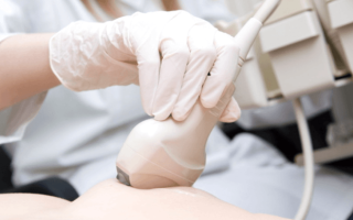
The examination itself is very simple and quick. The patient lies on her back, the doctor lubricates the mammary glands and chest with a special gel for greater ultrasound conductivity and moves the transducer over it.
There may be a slight vibration and cooling sensation from the gel, but there should be no squeezing, burning or pain during the procedure. All information is recorded on the screen. During the examination, sonographic and echographic results are generated.
If there is a suspicion of mastopathy, then first examine healthy areas of the breast so as not to mistake the norm for pathology or deviation.
- In addition to the glands themselves, the doctor examines 4 more areas: subclavian, supraclavicular, prothoracic and axillary.
- The doctor may ask you to lie on your side or put your hands behind your head - this is normal, because in order to describe the condition of the breast in detail, it is important to examine it from all sides.
- If malignant neoplasms are detected, the internal organs located near the chest must be examined: liver, lymph nodes, lungs, stomach, ovaries for the presence of metastases.
Ultrasound examination is suitable for examining breasts with implants. Ultrasound makes it possible to get to the breast tissue bypassing a foreign body.
An ultrasound of the breast can be done in all public clinics or private offices.
Interpretation of breast ultrasound
During the examination, the doctor pays attention to several factors:
- condition of the milk ducts;
- condition of glandular tissue;
- breast wall density;
- size of adipose tissue in the mammary gland.
For each of these indicators there is a normal level. For glandular tissue, the norm is considered to be no larger than 14 mm. Although with age this figure increases to the critical point of the norm of 20 mm, this happens after 40 years. The doctor pays special attention to the milk ducts; they should not be greatly expanded or deformed.
And echogenicity should be at a standard frequency. If the echogenicity is low, then there is a high probability of the presence of future or existing small foci of mastopathy.
Can it be cured?
- This unpleasant and rather dangerous disease can be cured in the early stages, so there is no need to ignore the ultrasound examination procedure, because only with its help can one clearly say about the presence of the disease, the size and shape of the seals.
- If nodules and cysts are detected early and their size does not exceed 0.5 cm, then they can be easily cured thanks to proper drug treatment.
- In the case of an advanced form of mastopathy or when large tumors form, they can only be removed through surgery.
Treatment of mastopathy is mainly aimed at balancing hormonal levels, because you need to fight not the formations, but the causes of their occurrence. Doctors often prescribe local hormonal drugs, sedatives and immunostimulants.
During pregnancy, the gynecologist, together with the mammologist, selects individual medications that will cure mastopathy and will not harm the fetus.
conclusions
To avoid such an unpleasant and dangerous disease as mastopathy, every woman is obliged to undergo an ultrasound examination of the mammary gland on time, and in no case ignore painful sensations in the breast area. The sooner you see a doctor with mastopathy, the faster, easier and without obvious consequences the treatment will be. Health must be protected and monitored carefully.
You can learn more about the rules of the procedure and the timing of its implementation from the video material.
It is important to know! In women who have not given birth under 25-30 years of age, fibrocystic disease (mastopathy) does not cause much concern, but closer to 30, especially during pregnancy and after childbirth, 80 percent of women develop a complication of mastopathy. Along with women who have not given birth, many mothers who devote almost all their time to their baby forget about their health or think that this problem is trivial and will go away on its own. Expectant mothers are in an even more difficult position - during pregnancy and breastfeeding, many pharmaceutical drugs are prohibited. Did you know that mastopathy, if not treated in time by preventing the disease, can cause breast cancer. Read about a completely natural remedy for mastopathy (fibrocystic disease), compatible with breastfeeding and pregnancy here...
Read more.. "
Source: https://ogrudy.ru/mastopatiya/na-kakoj-den-cikla-delat-uzi-30.html
On what day of the cycle should ultrasound of the mammary glands be done in women with mastopathy and what can be seen from echo signs
Ultrasound examination (ultrasound) is one of the most effective diagnostic techniques in the study of various organs, including the mammary glands.
It is the most gentle non-invasive type of examination, which does not cause patients any discomfort, does not expose them to dangerous doses of radiation and takes a short period of time - no more than half an hour.
A properly performed ultrasound can give the doctor a clear idea of the state of the structure of the glandular and fatty tissue of the breast. Therefore, this study is mandatory when a woman complains of pain or discomfort in the breast area.
Indications and contraindications for ultrasound
For pain not associated with pregnancy and lactation, it is recommended to undergo an ultrasound of the mammary glands
Experts recommend annual examinations for women after 30 years of age. After 40, ultrasounds should be done twice a year. This is urgently necessary for timely diagnosis for the following symptoms:
- Breast injuries.
- Changes in the shape of the mammary glands, a noticeable difference in their size or noticeable compactions (nodules, bumps) in their structure.
- Nipple discharge not associated with pregnancy or breastfeeding.
- Enlarged and inflamed lymph nodes in the armpits (lymphadenitis).
- Deformation or retraction (retraction) of the nipple.
- Changes in the color, structure, shape of the areola, the appearance of spots, pimples, and scales on it.
- Pain in the chest not caused by pregnancy or approaching menstruation.
The described symptoms may indicate a number of pathologies, including breast cancer. But the most common diagnosis revealed during examination is mastopathy.
According to medical statistics, this disease occurs in a third of the fair sex between the ages of 30 and 40, and between the ages of 40 and 60 this figure reaches 50 percent.
Nodular mastopathy of the mammary gland
Experts distinguish several types of benign pathology of the mammary gland, determined using ultrasound:
- fibrocystic mastopathy (FCM);
- focal mastopathy
- diffuse mastopathy;
- nodular mastopathy;
- mixed type, including a number of characteristics simultaneously.
The number of contraindications to the procedure is very limited, mainly due to individual reasons, for example, recent surgery. Temporary restrictions for ultrasound examination include early pregnancy.
There is no specific data indicating the danger of this procedure for the unborn child in the first trimester of pregnancy, but the woman’s body during this period requires a particularly careful attitude, so the study is done only if absolutely necessary.
It is not recommended to conduct an ultrasound examination of the structure of the mammary gland in the acute period of mastitis, during inflammation and suppuration of the glandular tissues.
In this condition, it is not possible to identify signs of mastopathy using ultrasound. The doctor recommends treatment and waiting until the inflammatory process subsides completely.
There are no permanent contraindications for breast ultrasound.
Preparation and carrying out the procedure
The ultrasound procedure of the mammary glands for mastopathy is recommended to be carried out immediately after the end of menstruation
Special preparation in the form of dietary restrictions, work and rest schedules is not required before the procedure, however, there are rules regarding the timing of its implementation. It is recommended to do an ultrasound of the mammary glands for mastopathy in women on specific days of the menstrual cycle, determined by the doctor, since the degree of information content of the results depends on this. If this rule is violated, the examination picture may turn out to be incomplete or insufficiently accurate. This is explained by the fact that this pathology is directly dependent on the level of production of the hormone estrogen, which reaches a maximum during menstruation, when the mammary glands swell. Against this background, large nodes become clearly visible. This condition lasts 5-7 days after the end of menstruation, gradually fading away, therefore, if mastopathy is suspected, it is best to perform an ultrasound of the mammary glands immediately after their end.
The timing of the ultrasound also depends on the course of the disease. Often, to exclude or confirm a malignant form of neoplasms in the mammary gland, examination must be done several times in different phases of the menstrual cycle.
For a patient, an ultrasound scan for suspected fibrocystic mastopathy does not present any difficulty.
- A woman needs to undress to the waist and stand straight, spreading her arms to the sides, or lie on the couch on her back and straighten up.
- The mammary glands are treated with a special gel that has the ability to conduct ultrasound. Regardless of which gland is suspected of having pathological changes, both breasts are treated and examined.
- The doctor moves a special device over the breast, which transmits an image of the structures of the breast tissue onto the monitor screen, allowing a diagnosis to be made.
At the end of the examination, the chest is wiped with a clean towel, after which the patient can get dressed.
Interpretation and description of the echographic result, performed by an ultrasound diagnostic specialist, usually takes no more than a week. The examination protocol is given to the woman along with an image and a conclusion from the attending physician made on the basis of ultrasound and other diagnostic procedures.
Character of the echogram
Nodular mastopathy of the mammary gland
The mammary gland is formed from three components: glandular tissue, which has a lobular structure, fibrous tissue, passing between the lobules and connecting them into a single whole, and fatty tissue, which performs a protective function. After 55 years, involution begins - the reverse development of the mammary glands, during which there is an increasing predominance of adipose tissue in the structure of this organ, which gradually crosses the boundaries of the norm and replaces the glandular tissue. Externally, the breast does not change - changes can only be detected using hardware diagnostics. Involution, recorded in elderly women during ultrasound, is not a disease, but a natural physiological process.
The doctor judges the condition of tissues by their echogenicity - their ability to reflect ultrasound:
- Most healthy breast tissue is characterized by isoechoicity, i.e. medium degree of reflection - they look like light gray spots;
- denser connecting septa are hyperechogenic, have good reflective properties and appear on the ultrasound monitor screen as white spots;
- hypoechoic adipose tissue, which has a loose structure, is characterized by a weak degree of reflection of ultrasonic waves, therefore it has a dark gray color on the screen;
Fibrofatty transformation - anechoicity indicates the presence of fluid in the tissues that completely absorbs ultrasound without reflecting it. Displayed in black on the screen. Normal anechoic structures include blood vessels and milk ducts during lactation.
Echosigns of fibrocystic mastopathy:
- Increased thickness and density of glandular tissue.
- Fibrosis (presence of connective tissue) in the milk ducts.
- Dilation of the milk ducts.
- The presence of cysts that have an anechoic liquid structure on ultrasound.
An echo sign of FCM is a discrepancy between the condition of the breast tissue and the patient’s age. If an ultrasound detects involutional changes in the breasts in a woman under forty, this may indicate that the patient has fibrocystic mastopathy.
When recording the results of mammography and ultrasound, the so-called BI-RADS scale is used, created to standardize descriptions. It has seven categories - from 0 to 6:
- BI-RADS 0 – it is impossible to clearly determine the conclusion, additional research is required;
- BI-RADS 1 – no pathological changes are observed;
- BI-RADS 2 – benign changes detected;
- BI-RADS 3 – suspected benign changes requiring dynamic monitoring;
- BI-RADS 4 – suspicious changes, biopsy required;
- BI-RADS 5 – definite malignant neoplasm;
- BI-RADS 6 – is set when assessing the results of chemotherapy and/or surgery.
A woman who has been diagnosed with echographic signs of FCM of the breast should be closely monitored by oncologists.
The decision on how to treat benign fibrocystic mastopathy is made by doctors depending on what changes were detected and at what stage of development. Single or a few small cysts can be removed using hormonal therapy.
At the same time, during the treatment process, the patient must periodically undergo an ultrasound examination so that the doctor has the opportunity to observe her condition over time.
If the size of the cysts can be reduced or stabilized, the treatment is considered successful. If conservative treatment does not bring positive results, and the disease progresses, the question arises about the need for surgery to remove the detected tumors.
Source: https://NogoStop.ru/grud/exograficheskie-priznaki-fibrozno-kistoznoj-mastopatii-molochnoj-zhelezy.html
Ultrasound of the mammary glands: how often can it be done, preparation, procedure and interpretation
Breasts emphasize the beauty and stateliness of a woman, but, unfortunately, they are often the cause of worry and anxiety due to possible diseases.
Indeed, diseases of the mammary glands are very common and can lead to extremely serious and dangerous consequences.
But timely diagnosis will help to “catch” the disease at a very early stage, which gives a chance for a complete recovery even if cancer is detected.
Ultrasound of the mammary glands: benefits
Features of the examination method
Many women are often frightened by the breast ultrasound procedure, how often it can be done, they do not know and are afraid that it is painful, time-consuming and dangerous to health. In fact, this is an absolutely painless and safe examination, which is carried out quite quickly using a special device and takes very little time.
The advantage of such a survey is its sufficient simplicity and accessibility, as well as high information content. An ultrasound machine can show even small tumors at various levels of the breast, even those that are located quite deep.
In addition, the surrounding tissues and lymph nodes are examined, so information about the condition of the organ is voluminous and comprehensive.
Based on the information received, the specialist can determine the presence of pathological changes in the gland, identify their nature (benign or malignant), measure the size and record the number and location of tumors.
No special preparation is required for the breast ultrasound procedure. However, it is preferable to contact a specialist on days 5–10 of the menstrual cycle. Such requirements are associated with hormonal changes in the structure of the gland tissue.
Examination procedure
Prescription and examination of the mammary glands
An ultrasound examination of the mammary glands shows the structure of the organ itself, the condition of the surrounding tissues, blood vessels and lymph nodes. A timely study makes it possible to identify the inflammatory process, the appearance of neoplasms or pathological processes at the earliest stages, when the diameter of the tumor, for example, is only 5 millimeters.
Ultrasound also allows you to differentiate the nature of the neoplasm and find out whether it is benign or malignant. In the case of breast cancer, detecting it in the early stages makes it possible to avoid the appearance of metastases and preserve the patient’s chance of survival. At the zero and first stages, in the case of a small cancerous tumor, it is even possible to perform an organ-preserving procedure.
When performing an ultrasound, the lymph nodes and tissues surrounding the mammary glands are also examined.
Such a detailed study makes it possible to exclude metastases and pathological processes, since enlarged lymph nodes symbolize the presence of an inflammatory process in the woman’s body.
The procedure itself is quite simple and absolutely painless for a woman:
- It is performed in a supine position with your arms raised above your head. In this position, the mammary glands occupy the most convenient position for performing the examination.
- To improve contact and sliding of the device sensor, a special colorless gel is used.
- The process itself proceeds sequentially, the breast is examined from top to bottom, without missing a single centimeter of the mammary gland.
When scheduling an ultrasound of the mammary glands, how often this procedure can be done is what worries women most often. The doctor will always answer one thing - the frequency of examination depends on the state of health. The slightest ailment requires an immediate visit to the doctor, since delay often affects health and can sometimes cost life.
This type of study is prescribed in the following cases:
- For preventive purposes - all women over 35 years of age, as well as in adolescence during the formation of the mammary glands, especially if various developmental abnormalities are noted. Such examination is also mandatory for women during menopause.
- When planning pregnancy to monitor the condition of the glands.
- If you have breast implants.
It is believed that the ultrasound procedure is harmless, but still, without vital reasons, it is better to do it no more than four times a year.
How often can you do it
A woman’s health largely depends on how often she visits specialized doctors. The most important are visits to the gynecologist and mammologist.
The frequency of breast ultrasound is once a year. If there is a suspicion of the presence of a disease, discomfort or various signs of neoplasms in the mammary gland, you should consult a doctor immediately.
The following symptoms may be the reason for your visit:
- Pain in the mammary gland.
- Discomfort.
- Sudden breast asymmetry or the appearance of neoplasms.
- Inverted skin and nipples.
- Changes in the color and texture of the skin on the gland.
- Rashes and spots.
- Enlarged lymph nodes.
If at least one of the listed symptoms appears, contacting a medical institution is mandatory, regardless of how long ago the ultrasound was performed.
Interpretation of ultrasound
Possible diseases of the mammary glands
- Having learned everything about ultrasound of the mammary glands, how often this procedure can be done and what its benefits are, a woman will not avoid visiting a doctor.
- An ultrasound examination is quite informative, and to obtain additional data, a specialist will prescribe additional examinations and tests.
- Ultrasound can reveal many different problems associated with the mammary gland, but most often it is necessary to diagnose the most common female problems - mastopathy, cysts and cancer:
- Mastopathy in its various forms is quite common, causing women a lot of suffering and discomfort. It can be found not only in different forms, but also in varying intensities. Sometimes a woman does not even feel pain, and the process is already in full swing. In other cases, mastopathy is accompanied by severe pain, fever, severe discomfort, swelling of the gland and other painful and extremely unpleasant manifestations. Mastopathy requires treatment and observation by a doctor, since this process can become complicated and develop, creating a danger for the patient. The selection of drugs is individual for each patient; it takes into account her age and state of health, the level of the process.
- A cyst is a benign neoplasm that consists of cavities filled with fluid. They are dangerous due to ruptures, degeneration, inflammation and suppuration. The cyst requires constant monitoring and treatment, and in some cases, surgery.
- The most dangerous disease that ultrasound can detect is breast cancer. Malignant neoplasms are one of the main causes of death among women. Early detection of the process avoids the appearance of metastases and increases the chances of survival. That is why all doctors insist on regular annual ultrasound examinations of the mammary glands, and, if necessary, more frequent visits to the doctor.
Source: https://DiagnozLab.com/uzi/breast/uzi-molochnyh-zhelez-kak-chasto-mozhno-delat.html
How often can you do an ultrasound of the mammary glands for mastopathy?
The question of what day of menstruation to do an ultrasound of the mammary glands for mastopathy is asked by many representatives of the fairer sex. This is the most popular, inexpensive and effective method for identifying diseases. But in order to get the most accurate result, you should follow some rules for conducting this study.
Features of the procedure and its positive aspects
With the help of such a study, you can obtain the result of the condition of the internal tissues of organs, as well as the quality of blood flow in them. For this purpose, sound waves are used, which do not cause any harm to the human body.
Ultrasound has the following advantages:
- excellent tolerability by all patients;
- the procedure is carried out quite quickly and painlessly;
- no additional medical procedures are used during the study;
- there are no contraindications to the procedure;
- can be done even during pregnancy.
An equally important advantage is that thanks to ultrasound it is possible to check the condition of both healthy and affected areas.
How often is such a procedure allowed?
In order to diagnose mastopathy, ultrasound is simply a necessary procedure. After identifying such a disease, the doctor prescribes treatment, which may differ depending on the complexity of the disease.
Therefore, in order to observe changes and adjust medication intake, repeated testing is mandatory.
How often you can do an ultrasound of the mammary glands for mastopathy is determined by the doctor, based on each specific case.
The number and frequency of ultrasound appointments is affected by:
- girl's age;
- the amount of estrogen and also progesterone;
- symptoms of the disease;
- the presence of concomitant illnesses.
Also, the number of procedures is affected by pregnancy, breastfeeding and the presence of surgery in the breast area.
On which day of the cycle is it better to do an ultrasound of the mammary glands when diagnosed with mastopathy?
Despite the safety, this procedure has its own characteristics. The accuracy of the data is affected by what day of the cycle the breast ultrasound was performed for mastopathy.
Before the procedure, you should remember that ultrasound is recommended to be done from the fourth to tenth day of the cycle. If the period is more than twenty-eight days, then the procedure can be carried out from 11 to 14 days.
It is at this time that the ultrasound will give the most accurate result. If the procedure is postponed to another day, then errors should be expected. This is due to the fact that the female body begins to prepare for ovulation.
The mammary glands swell and thus begin to transmit the signal from the device poorly.
Since the 1st day of menstruation coincides with the beginning of the cycle, ultrasound can also be performed during critical days.
To be completely confident in the accuracy of the data, it is better to do an ultrasound of the mammary glands for mastopathy after establishing the amount of estrogen and progesterone. Blood donation is carried out on the 3rd or 4th day of menstruation.
If you feel pain in the breasts or an attack that occurs during the treatment period, the doctor prescribes the procedure regardless of the day of the cycle. As for pregnant women and those who are breastfeeding, ultrasound is also done at any time of the month.
The procedure is not performed in cases where progressive changes are observed that do not allow distinguishing between mastitis, mastopathy and neoplasms. In this case, other types of tests are prescribed.
Knowing on which day of the cycle it is better to perform an ultrasound of the mammary glands for mastopathy, you can get the correct result. This will allow the doctor to prescribe the correct treatment, which will speed up recovery.
Source: https://uziman.ru/myagkie-tkani/kak-chasto-mozhno-delat-uzi-molochnyh-zhelez-pri-mastopatii
Ultrasound of the mammary glands
Ultrasound of the mammary glands is a reliable and painless way to diagnose the mammary gland, allowing the doctor to detect diseases accompanied by the appearance of lumps and other neoplasms in the tissue of the mammary glands. With its help, you can recognize the causes of deviations that have arisen, evaluate pathological changes, or simply make sure that everything is in order.
Who needs to undergo ultrasound of the mammary glands?
Ultrasound examination is done in two cases:
- In the first, pathology is detected - an examination is prescribed when the doctor suspects some kind of formation in the mammary glands, for example, a cyst. Based on the research results obtained, the ultrasound specialist writes a conclusion, which states whether there are pathologies in the breast or not. If a pathology is detected, the doctor prescribes appropriate treatment. In principle, a neoplasm can be detected in advance by palpation or mammography.
- In the second case, ultrasound is performed as a preventive measure - the examination is necessary in order not to miss the onset of the disease. During the research process, ultrasound doctors study the structure of the glands and try to identify their features. Such research is done for all teenagers (boys and girls), women in an interesting situation, and young mothers who are breastfeeding.
A breast ultrasound is mandatory if you have previously had surgery to enlarge the breast or replace a missing part of the mammary gland. Such a study helps to monitor not only the general condition of the mammary gland after surgery, but also how the implanted prosthesis/silicone implant is located.
If a woman is over 40, then she needs to undergo an ultrasound examination of the breast, since it is at this age that the risk of a malignant tumor appearing in the breast increases. In order not to miss the onset of symptoms of this terrible disease, you need to be examined at least once every 12 months.
How to prepare for research
No special preparation is needed for ultrasound examination of the mammary glands. Just before the procedure you need to carry out hygienic procedures to cleanse the skin and armpit area. There is no need to follow a diet. A woman should have a normal diet.
On what day of the cycle should I have an ultrasound?
Due to the fact that the female breast is a hormonally sensitive organ, before starting to study the mammary glands, it is necessary to take into account the phases of the menstrual cycle, because on different days of the cycle not very noticeable structural changes in the breast can be traced. It is recommended to undergo ultrasound diagnostics of the breast in the first phase of the menstrual cycle (days 4–10).
An ultrasound examination done in the second phase (15–20 days) may not give entirely accurate results.
This criterion does not apply to women undergoing menopause. But it is still advisable to agree with your doctor on the day when you can do an ultrasound.
How is the examination carried out?
A woman, having come for an ultrasound, enters the office and begins to undress to the waist. Before the procedure, she must remove all jewelry and lie on her back.
The doctor then applies a special gel to the breast to contact the sensor, after which he takes the transducer and moves it over the chest. The computer turns sound waves into images on a monitor. The study lasts no more than 20 minutes.
If during the examination some anomaly is determined, then the ultrasound will take much more time.
How are ultrasound results interpreted?
The basis for deciphering ultrasound indicators is sound waves that are reflected from organ tissue. If the breast tissue has a high density, then the term “hyperechoic structure” is used, and if it is low, then “hypoechoic structure”.
Standard indicators when deciphering the study:
- the echogenic structure of the skin surrounding the gland from the inside and outside is uniform;
- low-echoic structure of fat lobules, penetrated by echogenic lines and certain loci;
- elliptoid-shaped fat deposits;
- hyperechoic mammary zone, with small inclusions of adipose tissue with a hypoechoic structure.
In women, normal interpretation of data after an ultrasound examination of the breast looks like this:
up to 30 years - the mammary glands mainly contain glandular tissue, there is almost no connective tissue and there is no dilation of the ducts; from 40 to 50 years - the breast consists of glandular tissue, the connective tissue is moderately expressed, the ducts are not dilated, no focal pathology is observed;
after 50 years, glandular tissue is replaced by adipose tissue, connective tissue and skin become thinner. The breasts are dominated by adipose tissue.
What diseases can ultrasound of the mammary glands detect?
Diseases of the mammary glands that can be detected by ultrasound examination:
Mastitis, infiltrates and abscesses are inflammations of the breast of various origins and severity. They mainly occur in nursing mothers when pathogenic bacteria penetrate the breast tissue through cracks in the nipple or due to stagnation of milk. If no measures are taken at an early stage, they may become a reason for surgical intervention.
Mastopathy is a disease of the mammary gland, which is manifested by the appearance of single or multiple lumps in it. This disease can be caused by a hormonal imbalance in the body. If it is not detected in a timely manner and treatment is not started, then mastopathy may worsen over time, which means that surgery will be required.
Benign neoplasms (fibroadenoma or intraductal papilloma) depend on the hormonal background of the disease. A timely study allows such tumors to be cured.
Malignant tumors, including oncological ones - an ultrasound examination confirms/refutes the doctor’s suspicion of the presence of such a tumor, determines its size and location in the chest. After the examination, the doctor selects a treatment regimen.
How often should a breast ultrasound be done?
Doctors recommend the following frequency of examinations:
- up to 30 years - once every 24 months;
- from 30 to 45 - once every 12 months;
- after 50 - once every 6 months.
The frequency of the examination is explained by the fact that women at different ages have different hormonal status. The main thing is to “catch” possible changes in time while they are still successfully amenable to conservative treatment.
- Patients who have a history of cancer in their family should be attentive to their health, because it has long been known that the hereditary genetic factor has a great influence.
- To understand the question of whether mammography or ultrasound is better, you need to find out which method is more accurate and provides more information.
- So, let's figure out which method is the most informative.
At first glance, ultrasound examination has higher priorities. But there are cases when, in addition to ultrasound, it is advisable to perform a mammogram. Often, a mammologist prescribes ultrasound of the mammary glands for young girls, and mammography for women over 40. The fact is that photon flows are capable of ionizing matter, thereby activating the mutation process in cells in the female body.
If a representative of the fairer sex has a hereditary genetic predisposition to cancer, then mammography can push the tumor to progress. However, medical statistical studies have proven that only young patients are at risk.
After 40 years, women's breast tissue becomes denser. Therefore, after performing an ultrasound, the doctor will receive little information about the disease, because the tumor visually merges with the surrounding tissues, which makes it very difficult to recognize.
Women over 40 should have a breast x-ray every 12 months. Between mammograms, doctors recommend doing an intermediate ultrasound, which helps monitor any changes in the mammary gland.
After all, in 12 months the tumor can increase greatly, but this is not visible on x-rays.
As for the danger of radiation exposure during mammography, there is no need to worry, since a single diagnosis per year will not cause harm to health, just like fluorography.
Now let's figure out which method is more accurate.
If the doctor has prescribed a puncture/biopsy, then he will definitely also prescribe an ultrasound scan, because mammographic images most often cause errors (it determines the approximate location of the tumor).
Although, if you look at MRI from the other side, then thanks to it you can study the condition of the ducts much more deeply using a procedure such as ductography.
Having filled the ducts with a contrast agent, the specialist can examine the existing papillomas in the image.
Doctors use cystography with great success - they fill the cyst with air with a syringe, and take an x-ray, the image of which clearly shows not only the wall of the bladder, but also its entire contents.
This method makes it possible to identify modifications of the papilloma located inside the formation. That's why you shouldn't find out which is better: mammography or ultrasound. After all, each diagnostic method has its pros and cons.
Therefore, it is better to trust your doctor and follow all his recommendations.
Where to do an ultrasound of the mammary glands
Any examination is a set of measures that are focused on studying the patient’s condition, despite his social status and position in society.
If the patient has financial difficulties, then he has the right to have an ultrasound examination free of charge (twice within 12 months) if he has a medical insurance policy.
Most representatives of the fairer sex choose private centers to undergo any diagnostics, despite the fact that their services are not cheap.
Reasons why women prefer private clinics:
- Doctors have a convenient work schedule; appointments are available by appointment;
- individual approach to each patient;
- there is no queue;
- professional medical care;
- doctors' reputation;
- highly qualified doctors;
- friendly and helpful staff.
As for the last point, it plays an important role today.
When women need to undergo examination, they go to private clinics. And it doesn’t matter to them how much the procedures will cost, the main thing is the good-natured disposition of the medical staff. And ladies also choose private organizations because they provide examinations such as ultrasound screening, Doppler, mammological examinations, double ultrasound vibrations and much more.
In addition, in private clinics you can take all tests in the same room. The main thing is to have all the necessary documents with you: passport, insurance policy and resolution from previous research sites (if any).
State clinics are not distinguished by the integrity of their doctors and ethical standards. They have long queues, and there is always a “mixture” of population strata and social structures in the corridors. Although it is worth noting that public clinics do identical ultrasound examinations of the mammary glands as private ones, but for a much lower fee or for free.
Very important! The closer a private clinic is located to the center, the higher the cost of the services it provides. City clinics in this regard do not depend on their geographical location.
As for the work schedule (by appointment) at the clinic, it has a similar form of patient reception as for private ones. All public medical institutions require exactly the same package of documents as private ones. True, such medical institutions will not prepare you for research when private clinics prescribe a course of vitamins.
Source: https://www.mammologia.ru/diagnostika/uzi/
Breast ultrasound for mastopathy: when to do and how often to diagnose?
Since this problem occurs in many women in the age range of 30-45 years, doctors recommend periodic regular examinations.
One of these types of diagnostics is ultrasound of the mammary glands. When to do an ultrasound of the mammary glands for mastopathy, we will consider further.
The most important advantage when performing ultrasound in women suffering from mastopathy is the health safety of this examination method, as well as painlessness and a high degree of reliability of the information obtained.
- Also, quick results.
- An ultrasound can determine whether there are lumps, cysts or other formations in the glands.
- Does not require special training to carry out.
- Ultrasound can be performed on different days of the cycle.
- This type of examination does not always give an accurate result.
- Therefore, if any suspicions arise during examination for the presence of tumors, the doctor prescribes additional diagnostic measures to check the woman for the absence or presence of cancer.
- For this purpose, mammography, MRI, CT or biopsy may be prescribed.
Also, if you have mastopathy, you need to take tests. Which? Read more about this here.
On what day of the cycle should an ultrasound of the mammary glands be done for mastopathy? The day of the examination is individually selected for each woman in order to obtain the most accurate results during the procedure.
The female body can undergo various hormonal changes throughout the month.
At the beginning of the monthly cycle, a woman’s breasts are as relaxed as possible, and closer to the middle, hormonal changes occur in the body.
Active production of estrogen begins, which after a few hours can be replaced by progesterone. Due to this, changes occur in the mammary glands and diagnostics may show slightly different results.
Therefore, when performing ultrasound in women, it is important to take into account the menstrual cycle:
- If the cycle is short.For some women, the cycle length is 3 weeks, i.e. Menstruation begins after 21 days and lasts up to 7 days.
For such women, an ultrasound scan is recommended after the bleeding has completely stopped. And only on the 5th day after completion, you can conduct an examination of the mammary glands with an ultrasound of mastopathy.
- During a normal cycleIf the cycle is 28 days, then diagnostics can be carried out immediately after bleeding. In most cases this will be on the 7th day.
But if your periods were suddenly protracted and lasted for a longer period of time, doctors recommend an examination on the 10th day.
Otherwise, you may receive inaccurate information. Even if a woman suddenly had scanty periods and lasted only 3-4 days, an ultrasound should be performed only a week after the bleeding began to stop.
- With a long cycle.
If a woman's cycle is 35 days or more, doctors recommend being examined on the 10th day after the bleeding stops. It is during this period that you can obtain reliable information about the condition of the mammary glands.
- During menopause and menopause.Women during menopause may still experience bleeding.
But they can be intermittent, which does not indicate a full menstrual cycle. Therefore, doctors do not recommend starting from the end of bleeding, since they can occur spontaneously and not regularly. In this situation, an ultrasound is performed at any time as soon as the bleeding has stopped.
The same can be said about menopause.
If there is any suspicion of mastopathy and palpation is performed, the doctor may send the woman for examination.
An ultrasound is performed regardless of the cycle if there is a suspicion of a malignant tumor in the breast.
How often can you do an ultrasound of the mammary glands for mastopathy? Since ultrasound is a harmless procedure, it can be done as much as required for a given specific situation. It is recommended to start checking your mammary glands once a year at the age of 18.
- If a woman has reached 35 years of age, she also needs to undergo this type of diagnosis once a year..
- If the ultrasound reveals any pathologies, the doctor may refer you for additional examination - mammography.
- Women over 40 years of age should be examined at least 2 times a year.
- But in this case, it is still recommended to carry out mammography, since at this age adipose tissue in the glands begins to grow, and the information may not be entirely reliable.
At the age of 50, a woman undergoes hormonal changes , so it is recommended to carry out examinations at least 2 times a year, and preferably every 3 months. This is due to the fact that during this period the risk of mastopathy and tumors increases significantly.
Preparing for diagnosis
- No special preparation is required to carry out this type of diagnosis.
- After examination by a gynecologist or mammologist, if the presence of lumps, cysts or tumors is suspected, the patient is prescribed to undergo examinations to establish a more accurate diagnosis.
- No medications, food or liquid consumed that day will affect the test result.
- The woman should have directions in hand and take a sheet and wet wipes with her.
Before the procedure, the patient must undress to the waist. She lies down on her back on the couch.
The doctor applies a special gel to the woman’s chest to ensure good adhesion between the sensor and the skin.
The doctor uses a transducer (sensor) to move around the body, and an image is displayed on the screen that allows you to assess the condition of the mammary glands. Not only the breast is examined, but also the lymph nodes in the armpits, the area above the collarbone and the chest area.
The procedure takes about 15 minutes. The result of the examination can be seen immediately after the ultrasound.
Echo signs of mastopathy
- Ultrasound allows you to monitor the condition of a woman’s mammary glands, identify lumps and other formations, and determine whether there is any pathology.
- The normal state of the mammary glands may be different for each patient, since an important indicator is the structural features of each woman’s breasts, age, hormonal balance in the body, and tissue density.
- If the mastopathy is diffuse , the screen will display many small lumps that are distributed evenly over the entire surface of the breast.
If cystic mastopathy is suspected , the breast tissue will show many cavities that are filled with fluid.
In this case, after the examination, the woman undergoes a biopsy to accurately determine the contents of the capsule.
With a fibrous component, connective tissue compactions will be displayed. When examined, a fibroadenoma will appear as a compaction with limited contours.
If the examination shows a deformation of the breast, if the nipple is retracted, and there are seals in the tissues of the glands, there is a possibility of cancer. Cancer cells may begin to grow in the lymph nodes, which will also appear on the screen. In this case, after receiving ultrasound readings, the woman undergoes a puncture.
To prevent mastopathy from developing into cancer, and to avoid various complications, women and girls should regularly examine their mammary glands. This will allow timely, effective treatment to begin.
For more information on the benefits of breast ultrasound, watch the video below:
You can find more information on this topic in the Diagnosis and Symptoms section.
Source: https://nesekret.net/mastopatiya/diagnostika-i-simptomatika/uzi