A high degree of myopia is called complex or severe. The degree of impairment is measured in diopters on a minus scale. High myopia - deviation from -6 diopters and above. People with a high degree of myopia see well only those objects that are located directly near the face.
Mechanism of development of myopia
Usually the development of myopia is caused by the influence of external and internal factors, although in rare cases congenital high myopia occurs. The causes of this disorder are said to be a genetic predisposition, or premature or difficult birth. Congenital myopia can be detected even in infants, although most often its signs become noticeable in children under 6 years of age.
Risk factors:
- heredity;
- visual stress and strain;
- poor visual hygiene;
- congenital defects of the structure of the eyeball;
- poor nutrition;
- the influence of bad ecology.
Most often, myopia is acquired. High myopia is the result of ignoring symptoms, inaccurate diagnosis, lack of treatment or ineffectiveness of treatment. At the first symptoms of a disorder, you should immediately contact an ophthalmologist.
Symptoms of myopia
In a healthy eye, the image of visible objects is focused by the cornea and lens onto the retina, and information about it is transmitted to the brain through the fibers of the optic nerve. In myopia, when the shape of the eyeball changes from spherical to oval, the distance between the cornea and the retina increases, so the focus no longer falls on the desired point.
Since the brain does not receive enough information about the visible object, it cannot fully process it and provide normal vision. The world around us appears blurry. Symptoms of a high degree repeat the symptoms of mild myopia: headaches, rapid visual fatigue, eye strain.
Why is high myopia dangerous?
For most people, myopia tends to stabilize between the ages of 20 and 30. After this, vision can be improved with laser surgery or other surgery.
Progressive myopia increases the risk of developing dangerous complications, although a stable form of the disease can also have negative consequences. This is due to the fact that even in the absence of myopia progression, the patient’s eyeball remains elongated.
Complications of complex myopia:
- Retinal detachment is a consequence of elongation of the eye and thinning of the retina. Any stress can cause a tear or detachment when the retina separates from the choroid. Without immediate medical attention, permanent vision loss may occur.
- Glaucoma – increased intraocular pressure. This phenomenon can also cause blindness if not adequately treated.
- Retinal dystrophy is degenerative processes in its central part, which ensures the clarity of the visible image.
- Cataract is a clouding of the lens, which significantly reduces the quality of vision. Overmature cataracts can lead to blindness.
Often high myopia is combined with astigmatism. Since laser correction is contraindicated for refractive error of -15 diopters, a combined operation is performed to correct both pathologies. Myopia is corrected with intraocular lenses, and astigmatism is corrected with laser correction.
Complications of myopia are diagnosed in patients at any age. If you have a high degree of refractive error, you should regularly visit an ophthalmologist and monitor your eye condition in order to identify defects and retinal breaks in time.
Distortion of objects, flashes and darkening in the field of view are a reason to urgently consult a doctor and undergo an examination. It is noteworthy that degenerative processes can develop even after surgical treatment of myopia, so surgery does not eliminate the need to visit an ophthalmologist.
Conservative treatment of high myopia
Modern ophthalmology offers patients several ways to treat severe myopia, the main of which is still optical correction. Even when using contact lenses, glasses are needed to be worn in the evening and in the morning, when the eyes need a break from the lenses.
High myopia requires wearing glasses with very strong lenses that thicken from the center to the periphery. Lenses with high optical power are thick and heavy; not all frames fit them, but only wide ones, which are not convenient to use.
There are lenses made of a special highly refractive material, the refractive index of which is higher than that of plastic and glass. The higher the refractive index, the thinner the lens will be. The glasses needed by people with high myopia are very thin and have sufficient optical power to correct vision.
However, the weight of a mineral glass lens directly depends on the refractive index. Although such lenses are half as thin as regular ones, they can be the same in weight. Only lenses made of highly refractive polymers are light and thin at the same time.
Lenses made from highly refractive materials are more effective if they have an anti-reflective coating that lets in a lot of light and eliminates glare. This coating makes the lenses as transparent as possible.
High myopia can be corrected with contact lenses. Modern optical systems correct refractive errors down to -16 diopters. There are many different types and types of contact lenses, so you should trust your eye doctor with your choice.
Surgical treatment of high myopia
There are several ways to surgically improve vision for myopia. The choice depends on the state of the visual system, the degree of impairment and the presence of contraindications.
Any operation to correct myopia is performed only when vision is stable.
You need to understand that most operations to treat myopia help eliminate the symptoms of the disorder, while the elongated shape of the eyeball and fundus defects remain. Therefore, even after treatment you need to visit a doctor.
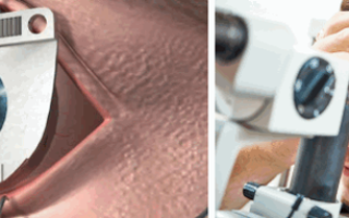
Laser correction of complex myopia
The most popular treatment method for moderate and high myopia is laser correction. This operation makes it possible to improve vision with myopia to -13 diopters. An ophthalmic surgeon uses a laser to reshape the cornea, vaporizing some of the tissue that distorts the focus of light on the retina.
At the moment, there are three main methods of laser vision correction: PRK, LASEK and LASIK. The choice of technique depends on the degree of myopia and the anatomy of the eye of a particular patient. The doctor determines the best method only based on the results of a comprehensive diagnosis.
Methods of laser correction of myopia:
- Photorefractive keratectomy (PRK) is a treatment method for low myopia based on laser changes in the curvature of the cornea.
- Laser subepithelial keratomileusis (LASEK) is the formation of a flap from the corneal epithelium, which, after laser correction, is fixed as a natural contact lens. After this treatment, the manifestations of corneal syndrome (lacrimation, photophobia, discomfort) decrease.
- Laser intrastromal keratomileusis (LASIK) is a combination of laser and surgical treatment. The operation is indicated for high myopia. First, the doctor cuts off the thinnest layer of the cornea, performs laser correction in the deeper layers and puts the flap back.
There is another technique - Super LASIK. This is an individual correction that takes into account all the features and parameters of the eyes, so it allows you to achieve maximum results. In other words, this is the development of a plan and analysis of all the details of the operation for a specific patient. After Super LASIK correction, it is possible to obtain consistently high vision.
Implantation of phakic lenses
Phakic intraocular lenses can be implanted into the anterior or posterior chamber of the eyeball. The operation is indicated if the natural lens has not lost its transparency and elasticity.
IOLs work similarly to contact lenses, only they are placed inside the eye to create a single optical system. There are anterior chamber and posterior chamber IOLs, as well as lenses that are fixed to the iris or pupil.
Posterior chamber lenses are usually used to correct myopia.
The advantage of phakic intraocular lenses is the ability to correct myopia down to -25 diopters. The operation is performed through a 1.6 mm incision. Since no stitches are required, the patient is sent home almost immediately after the procedure.
Refractive lens replacement
Lensectomy is performed for myopia up to -20 diopters, when there are additional symptoms of cataracts or there are contraindications to excimer laser correction. The eye's own lens is removed and an artificial one is installed in its place.
The operation is performed using the phacoemulsification technique, when the lens is destroyed and removed from the eye. This intervention requires drip anesthesia, which is well tolerated by most patients at any age. The surgeon performs all manipulations through a 1.6-1.8 mm incision, which eliminates the need to apply stitches and hospitalize the patient.
Intraocular lenses are selected individually, taking into account the degree of impairment, concomitant pathologies, the person’s age and type of activity. Multifocal IOLs will provide good vision at any distance, and aspherical IOLs will help you see clearly in the dark.
Keratoplasty for complex myopia
This operation is designed to restore the shape and function of the cornea by completely or partially replacing it. Keratoplasty can eliminate many congenital and acquired pathologies of the cornea. Donor or artificial material is used as a graft.
The graft is implanted into the thickness of the cornea, on the front layers or instead of a certain layer. According to this principle, keratoplasty is divided according to the size of the area being replaced (local, total, subtotal) and by layers (through, anterior and posterior layer-by-layer).
Keratoplasty is performed under local anesthesia. The ophthalmic surgeon removes a specific area of corneal tissue and applies a graft of the appropriate size. The new tissue is attached to the periphery of the cornea, and the doctor checks the uniformity using a keratoscope. For the operation, only the maximum smooth tissue of the correct shape is selected.
High myopia and childbirth
Women who have progressive refractive errors should regularly visit an ophthalmologist during pregnancy and after childbirth. The doctor may recommend eliminating myopia before delivery, since intraocular pressure increases during delivery, increasing the risk of retinal detachment and vascular rupture.
The state of a woman’s visual system largely determines the actions of obstetricians and gynecologists. Only with the permission of an ophthalmologist can such patients give birth on their own; otherwise, the period of pushing is limited.
However, this can negatively affect the condition of the mother and child, since doctors have to restrain the natural process and delay the birth.
Therefore, most often, women with progressive high myopia are recommended to have a cesarean section to avoid retinal detachment and eye bleeding.
https://www.youtube.com/watch?v=4WhiKQOURO8
After childbirth, you need to visit an ophthalmologist in the first days to check the retina and detect complications in time. Examination of the visual system does not harm the mother and does not affect lactation.
Limitations for complex myopia
High myopia often becomes a contraindication to military service and donation. A person may have difficulty obtaining a driver's license, lifeguard license, and similar statuses.
High myopia can cause blindness, which is the basis for receiving a disability group:
- first for partial or complete blindness;
- the second for poor vision, which makes it difficult to perform social and everyday functions;
- the third with a moderate decrease in visual acuity and the need for social protection of the person.
Complex myopia itself significantly increases the risk of retinal detachment, and alcohol abuse, visiting a bathhouse or sauna, attractions, surges in blood or intracranial pressure, and heavy physical and emotional stress can aggravate the situation. Therefore, people with a high degree of myopia need to exercise caution and avoid hazardous activities.
Having complex myopia, you need to maintain visual hygiene and constantly monitor the condition of the retina.
When myopia stabilizes, surgical treatment is recommended, since severe visual impairment reduces the quality of life and imposes many limitations.
High myopia is a constant risk, so it is always better to take care of your eye health.
Source: https://BeregiZrenie.ru/blizorukost-dalnozorkost/miopiya-vysokoj-stepeni/
Surgical treatment of myopia: types of surgery and possible complications
Myopia is a pathology that significantly reduces a person’s quality of life. People with myopia are forced to constantly use glasses or contact lenses. The problem can be solved once and for all by surgical treatment of myopia, in which the refraction of the eye is completely restored.
Indications for surgery
Surgical vision correction is a cosmetic operation, so the absolute indications include only the desire of the patient who wants to resort to a radical method of getting rid of myopia.
Surgery may be indicated in severe cases of myopia, when vision deteriorates by more than 1 diopter per year and there is a possibility of complete loss of vision. In such cases, experts offer surgical treatment (as the only way to avoid blindness if conservative treatment methods do not produce a positive result).
Contraindications
In some cases, myopia provokes the formation of pathology of the eyeball, in which surgery becomes impractical (due to high risks):
- deformation of the eyeball;
- ambioplapia;
- strabismus;
- retinal hemorrhage and retinal detachment;
- disturbance of blood supply;
- cataract.
In addition to specific contraindications, there are general contraindications:
- diseases of the endocrine system;
- autoimmune diseases;
- age under 18 years (the eyeball is not yet fully formed);
- pregnancy and lactation period.
Types of operations for myopia and their implementation
The choice of a specific method of surgical intervention depends on the wishes of the patient, the availability of indications and the condition of the patient’s optical system.
- Radical keratotomy of the anterior type (for pathological deviations in vision from 0.5 to 6 diopters).
- Myopic keratomyelosis (with pathological deviations in vision from 6 diopters and above).
- Extreme laser surgery (laser vision correction for pathological deviations of vision up to 6 diopters).
- Excision of the lens (the operation is technically complex and is used in cases where the patient has contraindications to other methods of correcting myopia).
Anterior radial keratomy
The patient is under local anesthesia. The working area is prepared with anesthetic and antiseptic drugs.
Then the specialist marks the locations of the planned incisions (the thickness should not exceed 90% of the total volume of the cornea). The surgeon then creates up to 12 incisions using a diamond knife.
Pressure inside the eyeball provokes swelling of the cornea in the area of dissection and its further thinning.
The duration of the operation is 20–30 minutes.
Contraindications for surgery:
- pregnancy;
- diabetes;
- progressive myopia;
- thinned cornea;
- mental disorders;
- inflammatory processes in the eye area.
Myopic keratomileusis
The patient is under local anesthesia. An anesthetic drug and 7% polyglucin, which has a blood replacement effect, are injected into the eyeball. The eyelids are fixed with a blepharostat to provide free access to the working area. When the pupil dilates, the specialist performs manipulations to measure intraocular pressure. Then marking lines are applied.
A flap is excised from the corneal tissue and folded back, after which another layer of the cornea is removed. Tissue dissection is performed with a laser or other device with a similar operating principle.
The formed flap is returned to the anatomically correct position (along the marking lines) and a continuous suture is applied. After this, a control measurement of intraocular pressure is carried out. An antibiotic is injected into the conjunctiva.
As a result, the cornea becomes flatter.
The duration of the operation is 10–20 minutes.
ATTENTION! The method has contraindications similar to those for anterior radial keratotomy.
Extreme laser correction
The operation follows the same scenario as for myopic keratomileusis. But the tissue to be removed is vaporized with a laser. The area of the removed cornea is calculated using computer programs.
The laser destroys bonds in tissues at the intermolecular level, without affecting the lens and other elements of the optical system. The accuracy of the laser is very high, thereby minimizing the risk of complications resulting from injury. The duration of the operation is 15–60 minutes.
Lens excision (removal of the clear lens)
The operation is associated with great risk and is prohibited if:
- the presence of inflammation in the eyeball;
- retinopathy and retinal detachment processes;
- insufficiently large size of the eyeball (or anterior chamber of the eye).
The operation consists of two stages: removing the patient's tissue and replacing it with a prosthesis. The intervention is performed under local anesthesia.
During the operation, the patient follows the surgeon’s instructions (focuses his vision and does not blink for a certain time). The doctor creates a tunnel incision on the membrane of the eye.
Through this access, an ultrasonic rod is inserted inside, destroying the lens. The breakdown product of the lens is sucked out.
A prosthesis (artificial lens) is inserted into the empty lens capsule. The lens body unfolds, after which the position of the prosthesis is adjusted by the surgeon. The puncture does not require stitches. The eye is treated with a medical solution and a bandage is applied.
Postoperative period
The patient may experience pain for two days after surgery. To reduce pain, the doctor prescribes painkillers. Temporary photophobia and uncontrollable tear production may occur. Vision may become blurred for a week, especially when looking at nearby objects.
Immediately after the operation, the doctor prescribes therapy (oral medications and eye drops). The patient will have to refrain from using gadgets and watching TV. It is recommended to wear sunglasses until your eyes are no longer sensitive to light.
If surgery for myopia was performed with lens replacement, the patient must strictly follow the doctor’s recommendations:
- do not tilt your head (and do not bend over in general) for several days after the procedure;
- sleep lying on your back or on the side opposite to the operated side;
- avoid thermal effects on the body (you cannot visit steam rooms);
- avoid any stress until the completion of the rehabilitation period.
All types of operations imply rapid restoration of vision (myopia disappears almost immediately after the intervention, when the optical system adapts to the changes).
Possible complications
After surgery, patients may encounter both temporary complications (pain, lacrimation) and serious pathological processes, the catalyst of which is surgery.
Complications of anterior radical keratotomy:
- inflammation and infection of the eyeball;
- adhesions at the border of the cornea and iris;
- suppuration in the vitreous body;
- high probability of corneal rupture due to injury;
- change in refraction to farsightedness (solved through additional interventions).
ATTENTION! Myopic keratomileusis has consequences similar to those of radical keratotomy.
Complications of extreme laser correction:
- erosive processes on the cornea;
- clouding of certain areas of the cornea;
- keratitis (inflammation of the cornea);
- relapse or myopization;
- impaired astigmatism;
- dryness of the mucous membrane of the eye.
Complications after lens removal:
- cataract formation;
- astigmatism;
- displacement of the prosthesis;
- swelling of the cornea and increased pressure inside the eye.
ATTENTION! Patients over 40 years of age are at risk.
Overcorrection of vision is a complication that often appears immediately after surgery and goes away on its own, without medical intervention. But in some cases, vision shifts toward farsightedness gradually and requires repeated corrective surgery or the use of glasses.
Source: https://MedOperacii.com/glaza/operaciya-pri-blizorukosti.html
Treatment of myopia
- With nearsightedness (myopia), a person sees objects closer to the eye better, while distant objects appear blurry and indistinct.
- Myopia should not be confused with farsightedness, in which, on the contrary, distant objects are clearly visible, but close objects are not.
- In a myopic eye, the relationship between the optical power of the cornea (the main structure of the eye that refracts light rays), the lens and the length of the eye is disrupted.
In myopia, light rays are collected in front of the retina, and not on it as in a healthy eye.
As a result, a blurry image is transmitted to the brain.
- In a normal eye, light is focused on the retina; the owner of such a healthy eye sees objects clearly at any distance
- Because of its elongated structure, a nearsighted eye focuses light in front of the retina, causing objects in the distance to appear blurry.
There are three degrees of myopia: weak, medium, high. The stronger the degree of myopia, the lower the distance visual acuity.
With myopia above 4 diopters, a person begins to see objects at close range poorly, and working on a computer and reading becomes difficult.
Our myopia specialists
- There are stationary and progressive myopia.
- In childhood and adolescence, myopia increases over time, which is associated with the growth of the body and the eyeball, in particular.
- This kind of myopia is called progressive.
- If the progression of myopia exceeds 1 diopter per year (that is, the eye grows significantly faster than normal), it is necessary to treat progressive myopia.
- The goal of this treatment is to stop the progression of myopia and prevent the occurrence of retinal tears, detachment, and other unwanted diseases.
Later in this section we will talk about stationary myopia.
You can find out more about the treatment of progressive myopia in the section of the site dedicated to it.
Laser vision correction using the Femto-LASIK method
- The leading method of treating myopia is laser vision correction using the Femto-LASIK (Laser-Assisted in Situ Keratomileusis) method.
- Femto-LASIK is recommended for patients of any degree of stationary myopia, in the absence of contraindications that are identified during the examination.
- The leadership of this method of vision correction is due to its high safety, efficiency and reliability, the absence of pain after surgery and the achievement of high visual acuity within the first day after correction.
- Detailed information about vision correction using the Femto-LASIK method is available in the “Vision Correction” section.
- In our center, operations using the Femto-LASIK method are performed on the Technolas Z100 Zyoptix excimer laser system using the Victus femtosecond laser produced by Technolas Perfect Vision (Germany).
- Most LASIK operations in the world are performed on the Technolas system; the Zyoptix Z100 is an advanced model of this laser.
- The operation lasts approximately 15 minutes:
- First, using the Victus femtosecond laser, a so-called flap is formed on the surface of the cornea.
- Next, using the excimer laser directly, a microscopic layer of tissue is evaporated, the volume of which is designed specifically to obtain the corneal curvature necessary for vision correction.
- That is, we can say that the surface of the eye turns into a lens, which creates the desired correction.
- A superficial flap on the cornea is formed in order to increase the safety of the operation, speed up the healing process, and improve the results obtained.
- LASIK allows you to see clearly almost immediately after the procedure, and unpleasant sensations (similar to those that occur when cleaning an onion) remain for only a few hours.
- The Technolas Z100 Zyoptix system for laser vision correction uses innovative dual “scanning/flying spot” technology, which allows the most accurate and, accordingly, the most safe and effective treatment of corneal tissue.
- A special tracking system constantly monitors and follows the slightest fluctuations of the eye, fully controlling each point of influence of the laser beam, with the help of which ideal precision and accuracy of corneal profile correction are achieved, which, accordingly, gives an excellent correction result.
- The Technolas Z100 Zyoptix excimer laser system also has special software that allows laser correction of farsightedness and presbyopia with a high degree of safety and efficiency, which is not available with a number of similar devices.
- The core of this program is the direction of laser exposure to the periphery of the cornea using the double “scanning/flying spot” method, which achieves an increase in the optical power of the cornea in its center.
- An advantage of the Technolas Z100 Zyoptix laser model is also the use of the Planoscan software package, which allows the laser to work in conjunction with a diagnostic station, including Orbscan and Zywave devices, with the help of which the doctor receives data on the smallest anatomical features of the patient’s cornea and transmits this data directly to the laser.
- Thus, the treatment takes into account the smallest details of the structure of each patient’s eye, thereby achieving the best treatment result.
- Laser vision correction using the LASIK method is carried out on an outpatient basis: the patient is under observation in the clinic for an hour, and then can safely go home.
- The very next day you can start work, watch TV, use a computer, drive a car and continue to lead a normal lifestyle.
- There are only a few exceptions:
- Do not rub your eyes, take a steam bath or swim in a pool for 2 weeks
- Do not drink alcohol for 1 week after surgery
You should visit your doctor for postoperative examinations once a week for the first month after surgery.
In order to protect yourself during the postoperative period, you must:
- To protect against infections, instill Tobrex antibiotics for 1 week.
- to improve healing and prevent unwanted dry eye from antibiotics, instill Oquis 0.3% for 1-2 months.
Implantation of an intraocular lens
- Sometimes (if the patient is about 50 years old, with high myopia of more than -12 diopters), it is advisable to use a different approach to vision correction - phacoemulsification of the lens with intraocular lens implantation (that is, replacing the native lens with an artificial one).
- New generation artificial lenses - Acrysof Restor from Alcon (USA) have several focuses, which allows the patient who has such a lens implanted to see near and far without glasses.
- The advantage of this method is that by the age of 50, most people’s own lens becomes cloudy - a cataract occurs, which still has to be removed using phacoemulsification and the lens also replaced with an intraocular lens.
- And at an earlier age, surgery is safer, since the transparent lens is easier to replace, and the risk of complications is lower, also due to the fact that a younger body is more resilient.
- More information about phacoemulsification can be found in the section on cataract treatment.
Methods of the past and others
PRK, LASEK, EPI-LASIK
- In the 80s, ophthalmologists Pollikaris (Greece) and Burrato (Italy) developed a method of laser vision correction, in which the radius of curvature of the cornea is changed using a laser, as a result of which the image moves to the retina.
- When performing an operation using this method, the laser beam acts on the surface layers of the cornea and “evaporates” the cornea until the required curvature is achieved.
- As a result of such exposure, the surface layer of the cornea, which performs important protective functions - Bowman's membrane, is inevitably damaged.
- Due to such damage, the recovery period is delayed, creating the potential for various complications to arise, and various side effects arise, for example, a halo around luminous objects, deterioration of twilight vision.
- The LASEK and EPI-LASIK methods can be called a variation of the PRK method, since their use also causes damage to Bowman's membrane and, in general, does not eliminate the disadvantages of the PRK method.
Radial keratotomy
Another way to change the radius of curvature of the cornea is surgically.
Operations of this type began in the 70s at the Eye Microsurgery MNTK under the leadership of S.N. Fedorov.
During this intervention, called “keratotomy” (from the Greek “keratos” - cornea, “tomia” - cut), non-penetrating radial incisions were made on the peripheral part of the cornea.
Keratotomy is an outdated method, much less effective and safe than LASIK.
"Super LASIK" and others
Among laser vision correction services, there are also other names, in particular, “Super LASIK”, “REIK”. These are the names that clinics use for their modifications of certain aspects of the LASIK method.
It should be noted that practically every surgeon modifies various aspects of the operation in his own way, trying to achieve the best result, but the leading core of laser vision correction today remains the LASIK method, and the greatest role in its use is played by the skill of the surgeon and the quality of the equipment.
Treatment of progressive myopia
Conservative treatment of progressive myopia
Conservative (non-surgical) treatment includes various types of visual simulators for the external and internal muscles of the eye to improve their blood circulation.
These procedures include electrical stimulation of the eye muscles, reflexology, which improve blood circulation in the eye and vision. Such procedures are comfortable and safe for the patient.
Surgical treatment of progressive myopia
- Sometimes conservative treatment is not enough, and myopia continues to increase by one diopter or more per year.
- In such cases it is necessary to carry out scleroplasty surgery (“scleroplasty”) to prevent the development of severe eye diseases associated with a high degree of myopia.
- The essence of sclera-strengthening surgery is the implantation of a special biomaterial on the back surface of the eye, as a result of which it becomes more difficult for the eye to grow in length, creating an obstacle to the progression of myopia.
Sclera-strengthening operations in many cases make it possible to achieve almost complete stabilization of myopia.
This procedure is tolerated very easily by patients, is painless and has the highest level of safety due to its non-penetrating nature.
First, anesthetic drops are instilled into the eye, then a small pocket is cut in the mucous membrane of the eye, through which an implant is placed behind the eyeball with a special tool.
With such interventions, it is important to use safe implants. We use the biomaterial "Xenoplast", which, today, is the safest scleroplastic implant, since it is completely sterile and biocompatible.
The patient can go home immediately after the operation; this procedure does not affect the general condition of the body.
But unfortunately, in a relatively small group of patients, myopia, even after scleroplasty, slowly but steadily grows, reaching more than 10 diopters.
This condition is called “myopic disease” . It is characterized by severe degenerative changes (thinning) in the inner membranes of the eye, accompanied by degeneration, hemorrhages, ruptures, and sometimes retinal detachment.
Patients suffering from myopic disease must undergo an ophthalmological examination annually (or even more often as recommended by a doctor) with a thorough examination of the fundus of the eye and strictly follow all the instructions of the ophthalmologist.
When thinning of the retina and breaks occur, it is necessary to carry out preventive laser strengthening of the retina to prevent retinal detachment.
Source: https://vostok-prozrenie.ru/treatment/myopia
Myopia treatment
Myopia is a refractive error that is accompanied by a decrease in the quality of “distant” vision. When diagnosed with myopia, or nearsightedness, a person sees objects that are close up well, but has difficulty viewing objects at a far distance.
In the absence of correct treatment and the cause of the disease persists, myopia progresses, and vision becomes steadily worse.
At the initial stages of the development of the disease, the decrease in visual acuity is compensated by accommodation, but the refractive system gradually loses its compensatory capabilities and complications of myopia develop.
Ultimately, advanced myopia can lead to total blindness.
Myopia can be a consequence of anatomical defects (irregular size, shape) of the eyeball. Also, myopia develops against the background of defects in the refraction of light rays and even due to insufficient eye hygiene. In some cases, myopia is caused by injuries, lesions and contusions of the eyeball.
Myopia is divided into 3 types:
- Axial or axial. It occurs due to excess length of the anteroposterior axis of the eyeball and is not accompanied by defects in the refraction of light rays.
- Lenticular. It develops against the background of an increase in the refractive power of light rays by the lens of the eye due to systemic diseases (for example, diabetes) or taking certain medications.
- With damage to the cornea. The cause of the disease is excessive curvature of the corneal surface, which is accompanied by an increase in the refractive power of this part of the optical system of the eye.
From the point of view of the mechanism of occurrence of the disease, true and false myopia are distinguished.
In the first case, there are pathological changes that are organic in nature and affect the eyeball, corneal surface or the lens itself.
True myopia can be congenital (hereditary) or acquired. In the absence of correct treatment, the disease progresses and is accompanied by serious complications.
False myopia is a type of myopia that occurs in children or young patients due to overstrain of the accommodating apparatus. When viewing nearby objects, the ciliary muscle contracts and causes an increase in the refractive power of the eye lens.
If the ciliary muscle remains in a state of contraction for a long time, metabolic processes and neuroregulation are disrupted in the optical system. When trying to view objects in the distance, in a state of spasm of the ciliary muscle, the refractive power of the lens does not change, and the image looks fuzzy and blurry.
This condition is called spasm of accommodation and false myopia.
With false myopia, there is no damage to the eyeball or optical system, and the condition is temporary and goes away on its own, after eliminating the causative factor. The list of reasons that can provoke false myopia includes stress on the eyes, non-compliance with sleep and nutrition, as well as physical fatigue.
In children, myopia can be congenital or physiological. Congenital occurs mainly in premature babies who were born after less than 37 weeks of development in the womb. The child is born with an elongated eyeball.
As a result, the rays are focused in front of the retina, and the newborn is diagnosed with myopia.
Congenital myopia most often goes away on its own a few months after the birth of the child, as the eyeball changes in size and anatomy, due to which the refractive power is adjusted naturally.
Physiological myopia (myopia) develops in children 5-10 years old during the process of intensive growth and development of the eyeball.
If the anteroposterior size of the organ increases excessively at this stage, the focusing of light rays occurs - they connect in front of the retina, as a result of which the ability to see objects at a distance is impaired.
As you get older, physiological myopia may become more severe until you reach the age of 18 and the eyeball stops forming. In certain cases, the disease progresses up to 25 years.
The development of myopia in the early stages is accompanied by a gradual deterioration of vision and the loss of the patient’s ability to clearly distinguish objects located at a distance from him.
In conditions of a slow progression of the disease, this symptom may not be sufficiently pronounced, and patients associate the deterioration in the quality of vision with fatigue. As myopia progresses, the image quality of distant objects becomes significantly worse.
At the same time, working with nearby objects is not accompanied by discomfort or difficulties.
The development of myopia may be indicated by the patient's tendency to squint in an attempt to look at objects at a distance.
This symptom is due to the fact that partial narrowing of the palpebral fissure provokes the closure of a certain part of the pupil and a change in the nature of the light rays passing through the eye lens. For this reason, the quality of vision improves.
In addition, partial closure of the eyelids causes flattening of the corneal part of the eye, which improves the clarity of objects in the case of a combination of myopia and corneal astigmatism.
Also, as the disease progresses, the patient develops additional symptoms that are caused by damage to the light ray refraction system and deterioration in visual acuity. The list of such symptoms includes:
- Headache. Attacks of headaches are caused by constant tension of the accommodation apparatus and concomitant disturbances of blood circulation and nutrition at the level of the ciliary muscle and other structures. In addition, the appearance of headaches is affected by difficulty seeing objects at a distance, as a result of which the nervous system is overstrained.
- Sore eyes. A burning sensation and pain may occur after the patient begins to work with objects located at a close distance (for example, reading or working on a computer). These symptoms are also caused by overwork of the optical system and defects of accommodation. The burning sensation is directly caused by a spasm of accommodation.
- Tearing. This symptom often manifests itself during prolonged viewing of objects at close range with myopia and occurs even in completely healthy people. In the absence of disease, the symptom goes away after a short rest. Similarly, in myopia, increased tear production is a reaction to bright sunlight or intense artificial lighting. The reason for its occurrence is a pronounced dilation of the pupil against the background of damage to the ciliary muscle. An excessive amount of light enters the eye, and the secretion of tear fluid is aimed at protecting the eye.
Also, with myopia, it is possible that the palpebral fissure may change in size towards widening. This symptom becomes more pronounced as the disease progresses, and the reason is an increase in the size of the eyeball.
Diagnosis of the disease is carried out by qualified ophthalmologists. The basis for suspicion of myopia may be the patient’s characteristic complaints, however, making an accurate diagnosis is possible only after a full examination. To prescribe a patient treatment appropriate to his condition, it is necessary to determine the type, form and stage of the disease.
Diagnosis of myopia is carried out using methods such as:
- visual acuity assessment,
- fundus examination,
- visual field assessment,
- skiascopy,
- refractometry,
- computer keratotopography,
- other.
During the diagnosis, three degrees of myopia can be detected:
- weak – no more than 3 diopters,
- average – 3-6 diopters,
- high – from 6 diopters.
Timely treatment of myopia is a priority, since the disease is prone to progression and stable vision deterioration up to irreversible blindness.
True myopia cannot go away without outside help and remains with the patient for life in the absence of correct treatment. At the same time, timely consultation with a doctor allows not only to stop the course of the disease, but also to restore vision.
Modern methods for eliminating myopia make it possible to restore sufficient quality of vision even in conditions of advanced, high-grade disease.
Treatment should be started as early as possible, before irreversible complications and pathological changes in the retina or other eye structures occur.
Myopia is corrected using the following methods:
- glasses and lenses,
- laser correction methods,
- replacing the lens of the eye,
- surgical operations.
Vitamin therapy, medications, and physiotherapy are also used.
The simplest way to correct myopia is with glasses with diverging lenses. This method helps restore normal distance vision and prevent the development of pathological processes that provoke complications of myopia.
To correct any degree of myopia, the patient is prescribed contact lenses. Unlike ordinary glasses, they fit tightly to the surface of the cornea and form a single system of refraction of light rays. This way, a more precise correction effect is achieved.
Wearing lenses is recommended for patients with anisometropia, an eye pathology that is accompanied by different refractive powers on the left and right sides. In conditions of weak anisometropia with values less than 3 diopters, ordinary glasses with different lenses can be used for correction, however, at the middle and high stages of the disease, lenses are predominantly prescribed.
Neither lenses nor glasses, regardless of the diagnosis and condition of the patient, cure the disease, but only correct vision. At the same time, the use of optical aids helps prevent complications.
Laser treatment of myopia is the most effective and modern way to restore visual acuity and prevent further pathological changes in most cases.
The laser treatment method is based on correcting the curvature of the central part of the cornea using laser beams in the direction of reducing it. Thus, the refractive power of the cornea becomes weaker, as a result of which normal visual acuity is restored.
Laser correction is prescribed to patients in the range from 1 to 15 diopters, the choice of method depends on the thickness of the cornea.
In preparation for laser surgery, the patient must undergo a comprehensive examination using computer diagnostic methods and mandatory ophthalmological examination procedures.
During the examination, information is collected about the condition of the corneal surface, the lens of the eye itself, and the eyeball. These data are entered into a computer program and used to calculate optimal correction parameters.
The laser treatment procedure itself takes place under the full control of a computer program, thereby eliminating the risk of error due to the human factor.
The duration of the procedure is no more than 10-15 minutes on average (taking into account the effect on both eyes). Before this, the patient goes to the operating room and changes into sterile clothing.
It is then placed on the operating table, and the doctor adjusts the laser device taking into account the patient's position and the area of intervention. To eliminate pain and discomfort during the operation, the patient is instilled with anesthetic eye drops that block sensitivity receptors.
In addition, local drip anesthesia prevents involuntary blinking and other patient reactions to the doctor’s manipulations.
When the anesthetic drops begin to take effect, the patient's head is fixed in a strictly defined vertical position. To prevent blinking, eyelid clips are additionally installed.
This is completely painless and may be accompanied by only minor discomfort.
After a control check of the position of the patient’s head, the laser device is aligned above the operated eye, and the patient is asked to fix his gaze on the blinking light indicator.
During the correction, a shallow incision is made superficially. The cut portion of the cornea is lifted and used as a surgical flap. Next, the eye is exposed to a laser beam, which, in accordance with the parameters specified by the computer, evaporates the excess curvature of the corneal tissue in order to change its shape.
The laser affects each patient's eye for no more than 1-1.5 minutes. After completing this stage of laser correction, the corneal flap is returned to its original place.
There is no need for sutures - the incision site is sealed independently, for this purpose the flap is aligned on the cornea, and the patient remains motionless for a few more minutes.
After completion of treatment, the doctor conducts a follow-up examination of the patient and a postoperative vision test. This stage takes 1-2 hours on average. The patient then returns home within a few hours after the operation. Over the next 1-2 weeks, you must strictly follow your doctor's recommendations for recovery.
In conditions of a high degree of myopia, up to 20 diopters, the patient is prescribed an operation to replace the lens with the installation of an intraocular lens.
The surgical opening closes spontaneously in front of the eyes without sutures.
The doctor conducts a follow-up examination and gives appropriate recommendations to the patient regarding rules of behavior and lifestyle over the next few days after surgery.
The recovery process is monitored by an ophthalmologist at scheduled appointments; there is no need for hospitalization or hospital stay.
Surgical treatment of myopia is prescribed in conditions of high myopia, which is prone to rapid progression with a rate of vision deterioration of 1 or more diopters annually. An indication for surgery is also the lack of positive results from the use of conservative treatment methods and physiotherapy.
As a rule, the task of surgeons is to strengthen the posterior wall of the eyeball using scleroplasty.
This allows you to prevent stretching of the eyeball and the occurrence of associated complications associated with damage to the retina and blood vessels of the eye. To strengthen the sclera, special scleroplastic fabrics are used.
The introduction of these agents into the posterior region of the sclera is carried out through a special curved needle and through a small incision.
Scleroplasty is a simple surgical operation that has no strict contraindications and is performed on an outpatient basis. In total, treatment with this method takes no more than one day, after which the patient can return home. Restrictions for the postoperative period include avoiding eye strain and provoking factors.
Myopia is not directly cured when using this surgical method, but the course of the disease slows down or the progression stops completely.
The cessation of further development of myopia in such conditions occurs in 95 percent of adult patients and 70 percent of children.
Scleroplasty can be combined with alternative methods of treating myopia - glasses or lenses, laser correction, and so on.
Myopia of any severity requires treatment, since the progression of the disease changes the shape of the eyeball and disrupts the blood supply to internal structures, thereby causing complications.
In most cases, serious complications occur with a high degree of myopia. Their list includes pathologies of varying complexity - from amblyopia to cataracts and retinal detachment.
The main risk with advanced myopia is the development of irreversible blindness.
Source: https://www.sfe.ru/stati/lechenie_miopii/