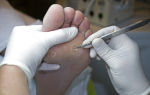
A study using X-rays that produces images of the tissues of the joint and surrounding structures is called a CT scan of the knee joint. Based on the layer-by-layer images of the area under study obtained during scanning, the attending physician is able to assess the condition of the knee joint as a whole.
Indications for use
Computed tomography of the knee joints is recommended if the information content of previous analyzes and studies is insufficient. CT is the main method for visualizing changes in the bone structure of the joint.
Indications for a CT scan of the knee joint may include suspicions of the following conditions and pathologies:
- fracture, cracks or other damage to its musculoskeletal structure;
- arthrosis and arthritis;
- cancerous and cystic neoplasms;
- formed ossifications (ossification of areas caused by calcium deposition in soft tissues);
- inflammatory diseases;
- dislocations and subluxations;
- hematomas and swelling of soft tissues.
A CT scan of the knee may be recommended as a preparation for knee surgery, as well as to assess the effectiveness of treatment.
Contraindications for the study
Since the procedure uses X-rays, the person being examined receives a certain dose of radiation. For this reason, CT scanning of the knee joint will be contraindicated in the following cases:
- during the period of bearing a child;
- in childhood, if the patient is under 14 years of age.
If a contrast agent is used during the study, the list of contraindications expands. CT scanning of the knee will be prohibited:
- in the presence of confirmed pathologies of the endocrine system, in particular with diabetes mellitus;
- in case of renal failure, since the kidneys may not be able to cope with the additional load;
- if you have an allergy to iodine or seafood. In this case, there is a high risk of developing a serious allergic reaction, including anaphylactic shock.
A relative contraindication is the period of breastfeeding. If the examination is necessary for health reasons, then breastfeeding can be resumed 48 hours after completion of the examination.
How is a CT scan of the knee performed?
No specific preparation is required for the procedure. The person must arrive at the appointed time. Clothes should not have metal inserts or decorations.
But if you plan to administer contrast, then you must refuse to eat on the day of the study. This will help reduce the likelihood of side effects when a contrast agent is administered.
How is the procedure itself performed? The person being examined is placed on a pull-out couch and the body, except for the area being examined, is covered with a special apron. It will help protect organs from radiation. The couch is moved into the tomograph and the scan begins.
A CT scan of the knee joint does not last long – literally a few minutes. If contrast is used during the procedure, the duration increases to 15 minutes.
During the entire scanning period, the person must remain completely still. Otherwise, the pictures will turn out to be of poor quality, and diagnosis will be problematic.
The use of contrast will be required if it is necessary to assess the condition of the blood vessels in this area.
What does a CT scan of the knee joint with contrast show? The technique allows you to determine:
- presence of disturbances in the blood circulation process;
- soft tissue hematomas;
- neoplasms;
- existing metastases;
- condition of intra-articular structures and damage that has occurred.
What is this “contrast agent”? Most often, drugs containing gadolinium salts are used as contrast: Magnevist, Gadovist, Omniscan and Dotarem.
The drug is administered intravenously, does not cause tissue damage and is excreted in the urine after a day. Relatively uncommon, contrast can cause an allergic reaction. The signs become:
- attacks of nausea;
- skin itching;
- skin rashes like urticaria;
- slight dizziness.
The development of such symptoms must be reported to medical staff.
MRI or CT – what to choose
Which is better - CT or MRI of the knee joint? It is impossible to give an exact answer, since each of the methods has its own advantages. Thus, MRI of the knee joint is used if there is a need to study the condition of soft tissues. CT is intended to evaluate the bone structures of the knee.
It is worth considering that magnetic resonance imaging can also provide information about the condition of the bones. In particular, a CT scan of the knee joint cannot show the condition of the bone marrow structures. And only MRI can help diagnose intraosseous contusion.
- The radiation exposure is extremely low to cause harm to the human body.
- The images reflect even the slightest changes in the shape of the joint and bone tissue.
- The procedure takes no more than 15 minutes.
The CT scan of the knee can be performed at either a government medical facility or a commercial diagnostic center. In any case, the research is performed for a fee.
Source: https://DiagnostLab.ru/kt/sustavov/kt-kolennogo-sustava.html
Features of CT scanning of joints
Computed tomography (CT) of joints is a modern non-invasive and extremely informative method for studying the bone and soft tissues of the joint area.
CT refers to radiation diagnostic methods, which means the use of X-rays during scanning.
The advantage of CT of joints over MRI is more accurate visualization of bone structures, but MRI provides more accurate information when studying soft tissues.
Why is CT of joints performed and its area of application?
Computed tomography is an x-ray method for studying the internal organs and tissues of the human body. During scanning, a series of images of layer-by-layer sections of the object being studied is taken. The received information is transmitted to the workstation screen, where it is converted into a three-dimensional model.
When scanning, emitters pass X-rays through the patient's body, and special sensors read it.
The principle of obtaining an image is based on the property of X-rays to pass obstacles encountered along the way differently.
Dense structures delay them, soft structures partially, and the rays pass through cavities without hindrance. The final image, like a regular photograph, is negative.
Computed tomography of joints is carried out for the purpose of clear visualization of the bone structures that form the joint, the articular cavity and its constituent structures, articular surfaces, as well as periarticular soft tissues. When performing CT arthrography, cartilaginous structures, joint capsule, ligaments, tendons, menisci, and sesamoid bones are visualized.
This research method is widely used in traumatology, orthopedics, rheumatology, surgery, oncology, dentistry and maxillofacial surgery.
Most often, research is focused on a specific area. The most common CT scans of the following joints (both large and small):
- hip;
- knee;
- ankle;
- shoulder;
- elbow;
- wrist;
- mandibular
Less commonly studied are the hand, clavicle, calcaneocuboid and other joints. A comprehensive examination is also carried out, when neighboring organs and tissues are simultaneously studied.
Hip joint
In this area, CT is most often prescribed to diagnose pathologies such as coxarthrosis, a degenerative-dystrophic disease of the hip joint. The study is also carried out before endoprosthetics.
Knee joint
CT scan of the knee joint in the early stages reveals degenerative-dystrophic processes (gonarthrosis), traumatic injuries, cysts (Baker’s cyst is localized in the popliteal area), arthritis. The method is important for assessing the effectiveness of surgical interventions.
Ankle joint
The ankle joint, like the knee joint, is susceptible to injury, so computed tomography may be needed to diagnose traumatic injuries and their consequences.
Shoulder joint
This anatomical structure is visualized using CT, usually for the diagnosis of osteoarthritis, arthritis, trauma and cancer.
Elbow joint
More often, the elbow joint is examined to identify developmental anomalies, injuries, cysts, and oncological process, before surgical treatment and to evaluate therapy over time.
Wrist joint
Typically, tomographic scanning of this small joint is performed to study the nature of bone injuries, in particular cracks and fractures, as well as ligamentous apparatus, including dislocations, subluxations, and ligament ruptures.
Mandibular joint
CT is used to diagnose osteoarthritis of the mandibular joint, developmental anomalies, and traumatic lesions. In addition, the method is very common in dentistry and maxillofacial surgery when deciding on dental implantation.
Indications and contraindications
Any medical examination, including CT scans of joints, has certain indications and contraindications.
The doctor will write out an appointment for a tomography if the patient has complaints about limited joint mobility, pain in the joint, or if there has been an injury. The goal of diagnosis is to determine the cause of such symptoms. Indications for the study are the following diseases or suspicions of them:
- osteoarthritis;
- developmental anomalies;
- inflammatory processes in the articulation area (arthritis, bursitis, myositis, tendinitis, abscess, phlegmon, osteomyelitis, synovitis);
- osteomyelitis;
- trauma and its consequences;
- tuberculosis damage to structures;
- diffuse connective tissue diseases, for example, ankylosing spondylitis (ankylosing spondylitis), rheumatoid arthritis, acute rheumatic fever (rheumatism);
- articular cysts, including Baker's cysts;
- metabolic diseases (gout, diabetes, rickets);
- tumor process of bone or cartilage tissue, metastatic damage to joints;
- multiple myeloma;
- diagnosis of impingement syndrome (persistent joint pain due to prolonged compression of periarticular soft tissues);
- suspicion of a foreign body in the articular cavity or periarticular tissues;
- planning surgical intervention, monitoring the effectiveness of treatment.
Contraindications:
- pregnancy, suspicion of it;
- discrepancy between the patient’s weight and the technical capabilities of the device (it is designed for a body weight of no more than 150 kilograms).
It is difficult for patients with hyperkinesis to remain still, so they are recommended to take a sedative before the procedure.
Contraindications for contrast-enhanced CT:
- intolerance to contrast agent (iodine);
- diseases of the endocrine system, including decompensated diabetes mellitus;
- decompensated liver, kidney and cardiovascular failure;
- severe general condition of the patient;
- mental illnesses and disorders;
- taking nephrotoxic drugs (non-steroidal anti-inflammatory drugs, Metformin, Dipyridamole);
- relative contraindication – age under 14 years. It is necessary to weigh the risk of complications after the study and its importance for establishing an accurate diagnosis, as well as the inability of other methods to provide a sufficiently informative result.
Preparation and carrying out the procedure
When performing a CT scan of the joint only, the patient does not require special preparation. On the eve of the diagnostic study, the doctor is obliged to ask the patient about concomitant diseases, pregnancy or suspicion of pregnancy, and claustrophobia.
It is necessary to prepare if you plan to conduct a computed tomography scan not only of the joint, but also of the abdominal and pelvic organs.
Visualization of internal organs requires the administration of a staining agent orally, so preparation is required.
To reduce the side effects of the contrast agent, the examination is carried out on an empty stomach, that is, you can eat your last meal no later than 7-8 hours before the examination.
In this case, a special diet is required for 2-3 days before the test to minimize the process of gas formation in the intestines. It is necessary to exclude legumes, sweets, alcohol, cabbage, bread and other foods that provoke intense formation of gases in the gastrointestinal tract.
Examination technique
After preparation (if it was necessary), the patient is sent to the CT room, where he is placed in a supine position on a special mobile CT scanner table.
Then the doctor sets the device to the mode required for the study.
In the case of an examination with contrast, at this stage of diagnosis (a few minutes before turning on the tomograph), a special radiopharmaceutical drug must be administered intravenously or inside the joint.
The patient should remain motionless to obtain high-quality, clear images. A scanning ring will rotate around the area being examined, from which X-rays are emitted. Each revolution of this ring is accompanied by a specific noise, during which time information about one slice is read (one picture is taken). The duration of one revolution is less than 1 second.
During the study, all medical workers leave the room with the tomograph into a separate room, where they can monitor the progress of the scan through the viewing window, and also communicate with the patient via a loudspeaker.
A CT scan is a painless procedure, however, when contrast is administered, there may be a feeling of heat in the body, itching, and slight dizziness, which disappear after the diagnosis is completed.
The duration of the procedure is approximately 10 minutes without the use of contrast, and with the introduction of the drug - about 20 minutes.
After the study, the patient can review the results and pick them up, including on digital media. As a rule, preparing a report takes no more than 30 minutes, in rare cases – several hours, when a detailed study of the 3D model of the joint is necessary.
CT joints with contrast
In some cases, for greater information, it is necessary to conduct a contrast-enhanced CT scan, which involves the introduction of a special staining substance into the vascular bed or into the articular cavity.
The drug enhances the contrast of surrounding tissues by increasing clarity and brightness. Contrast enhancement allows better differentiation of anatomical structures and, as a result, a more accurate conclusion.
Most often, iodine-containing contrast agents are used, among which the most widely used are Ioversol, Iohexol, Iodixanol, Omnipaque, Ultravist, Tomohexol and Scanlux. In addition to these drugs, there are also products based on a mixture of gases.
For research, contrast can be administered in two ways: intravenously through the cubital vein or into the cavity of the joint being examined. The choice of route of administration depends on the purpose of the study.
Thus, the intravenous route is necessary for a thorough examination of the soft tissues adjacent to the joint (ligaments, tendons, muscles, vessels), which are likely affected, for example, by a tumor process, cyst, myositis, ligament injuries, osteomyelitis or bursitis.
Intra-articular injection of the drug, or CT arthrography, may be required to improve the quality of the image of soft tissue structures located in the joint cavity (synovial membranes, menisci, intra-articular ligaments, labrum). It is worth noting that both iodine-containing drugs and a mixture of gases can be injected into the joint cavity.
CT scan of joints - what the decoding of the data obtained shows
On a computed tomogram of the joint, one can clearly differentiate the bone structures: bones that form the articular cavity, bony outgrowths, sesamoid bones that strengthen the articulation. Accordingly, a CT image can visualize any pathology of bone tissue - cracks, fractures, subluxations, dislocations, osteomyelitis, arthrosis, developmental anomalies, osteoporosis, tumors.
CT helps to visualize soft tissues in or around the joint cavity - tendons, ligaments, joint capsules, muscles, cartilage, aponeuroses, joint capsules.
This makes it possible to determine pathological disorders - calcifications, compactions, hematomas, tumors, ruptures, cysts, vascular aneurysms, the presence of fluid in the joint cavity.
Contrast-enhanced CT significantly improves image information.
Computed tomography makes it possible to determine the shape, size, extent of prevalence, nature of the pathology, as well as the presence of changes in the surrounding tissues. The advantage of the method is a qualitative assessment of the effectiveness of the therapy, monitoring the condition of the joint over time in case of chronic pathology or in the postoperative period.
Different anatomical structures have different appearance on the tomogram. For example, air is characterized by a black color, bone tissue is visualized as white, and internal organs and muscles are gray. When contrast is used, soft tissue structures become almost white.
Frequency of the procedure
To minimize undesirable effects, CT scans should be performed once a quarter, but no more than three times a year. If indicated, the attending physician may prescribe a repeat study at a shorter interval, but not earlier than one month after the previous session.
Possibility of research in children
Computed tomography of joints is not recommended for children under 14 years of age. However, in case of insufficient information content of other diagnostic methods, CT is resorted to. In such situations, it is necessary to individually weigh the risks of the consequences of a late diagnosis and the benefits of computed tomography.
Conducting research in children has its own peculiarities. Children under 5 years of age are usually stressed by the procedure and cannot remain still, which may require anesthesia. When scanning with contrast, it is necessary to individually calculate the dosage of the drug, taking into account the body weight in kilograms and the age of the child.
Where can I get tested?
Computed tomography of the joints can be performed both in a government institution and in a private clinic.
To undergo a free examination, you must, in addition to a referral, have a passport, a compulsory health insurance policy (CHI policy) and an individual personal account insurance number (SNILS). However, there are often long queues at government offices. If you don’t have time to wait, you can get examined for a fee in a private clinic.
What is the price
The price of a CT scan is determined by the level of the clinic, the equipment, the size of the joint being examined, the need to create a three-dimensional image model, deciphering the examination results, additional consultation with specialists, and recording on disk. The cost increases when computed tomography of not only joints, but also soft tissues is required, for which a contrast agent is additionally introduced.
The price of a CT scan, depending on the joint being examined, ranges from 1,200 rubles when examining the temporomandibular joint to 4,000 rubles when scanning large joints (knee, shoulder, hip). If it is necessary to conduct a CT scan with contrast enhancement, the price increases to 9,000 rubles.
Source: https://iDiagnost.ru/kt/osobennosti-provedeniya-kt-sustavov
CT scan of the ankle joint: how computed tomography is done, indications for it, who should not do it - Diagnostics
A CT scan of the ankle can be called a more detailed version of radiography - it allows you to examine the condition of the bones, joint and ligaments in various projections. It is prescribed in situations where a radiograph in two projections does not provide the necessary information.
CT scan of the ankle can detect a wide variety of injuries
What is a CT scan of joints?
The technique is based on the use of X-ray radiation. Unlike a conventional X-ray, the radiation source and receiver rotate around the patient. This allows you to obtain many images in different projections (while conventional radiography can only show two). You can even see small defects located on the edges of the bones facing each other.
A CT scan of the foot allows you to see defects in the bone, articular cartilage, and joint cavity. The condition of muscles and ligaments can be judged indirectly by changes in the distance between bones or disruption of the shape of the joint. Hematomas, tumors and other formations also become visible.
In what cases is examination required?
The main indication for CT scanning is various joint injuries. Since the ankle joint has a rather complex structure, its CT scan provides more information than simple radiography. A CT scan confirms the presence of fractures, sprains and dislocations of the joint.
CT scans are done for various injuries
Other diseases that an ankle CT scan reveals:
- chronic skeletal diseases affecting the joint: bone tuberculosis, chondropathy, arthritis;
- malignant and benign tumors of the joint;
- preparation for operations and assessment of its effectiveness;
- clarification of the results of other diagnostic procedures;
- assessing the effectiveness of trauma treatment.
Since the radiation dose is quite small (less than with conventional radiography), the procedure can be performed several times during the treatment to determine the intensity of the recovery processes and the effectiveness of the treatment.
Do you need preparation for a CT scan?
The procedure does not require special preparation in advance. A computed tomography scan of the ankle joint can be performed urgently if there is an injury or suspicion of injury, preparation for emergency surgery, as well as planned for chronic diseases and preparation for surgery or tumors.
Phones are prohibited during the procedure
Before undergoing a CT scan, it is advisable to take a shower if the patient’s condition allows it. Since the process uses X-ray radiation, it is necessary to remove all metal jewelry from yourself, remove your phone, plastic cards and metal money from your pockets - this can affect the information content of the procedure and the operation of the equipment.
Procedure for performing the examination
A CT scan of the ankle takes a few minutes. All this time, the patient needs to lie horizontally, during which time the device will move around the desired part of the limb, gradually moving along it. Medical staff are located in the next room, and the patient can notify specialists if any difficulties arise.
The procedure does not cause discomfort, does not cause harm and, in the absence of contraindications, is completely safe.
During the procedure, the patient must not move, and especially not move the leg on which the examination is being performed. This may result in poor image clarity and an incorrect diagnosis. You are also not allowed to eat or drink during the procedure, and you need to go to the toilet beforehand.
You must lie still during the examination.
What does a CT scan reveal?
- Since the operating principle of a CT scan of the foot is based on X-rays, the resulting image depends on the X-ray density of the tissue.
- Bone tissue has the greatest ability to absorb rays, cartilage has a little less, and soft tissues transmit X-rays without stopping them.
- Therefore, the study is of greatest diagnostic value for determining bone damage.
- A CT scan of the ankle shows the following pathologies:
- fractures, cracks and changes in the shape of bones, including those invisible to radiography;
- changes in the structure of cartilage tissue, changes in the size of the joint space, the shape of the articular surfaces of the bones;
- formations with altered X-ray density - hematomas, pathological choroid plexuses, tumors, including small ones;
- artificial structures in the joint and bones.
For a more accurate examination, MSCT of the foot is used - this type of study involves not one emitter and receiver, but several, which allows you to obtain a three-dimensional image of the affected area and get the most accurate idea of the condition of the joint. CT does not show the condition of soft tissues, and if it is important to evaluate this particular parameter, another type of study is used - MRI.
In this video you will see what a leg image looks like on a CT scan:
Who should not perform the procedure
Since the method is an X-ray examination, a CT scan of the foot involves a dose of radiation. It is significantly smaller than with radiography, which allows the examination to be carried out several times during treatment.
The permissible radiation dose for patients is 1 mSv/year, for radiologists – 20–50 mSv/year. Radiation sickness occurs at 3 Sv/year. The table shows radiation doses when examining the ankle joint using different methods.
ProcedureX-ray in two projectionsComputed tomographyMultispiral computed tomography
| Radiation dose per session | 0.01 mSv | 0.05 mSv | 0.1 mSv |
Thus, the radiation dose is 0.05 of the permissible dose per year. This should be taken into account in the dose load sheet - a document that is necessary for ordering x-ray examinations.
A single CT scan does not exclude the possibility of fluorography, radiography and other studies.
Other contraindications to CT and MSCT of the ankle joint:
- pregnancy;
- age under 3 years;
Pregnant women are not given CT scans.
- nervous and mental illnesses – claustrophobia, hyperkinesis;
- radiation sickness;
- when using contrast – kidney disease, thyroid disease, allergy to contrast.
CT scans can be performed on breastfeeding women, but lactation must be interrupted, especially if contrast was used. In patients who have exceeded the permissible dose of radiation for a year, CT can be performed only for health reasons. A pacemaker is not a contraindication, but you need to warn your doctor about it.
Did the article help? Rate it
Source: https://ndc-sergiev-posad.ru/prochee/kt-golenostopnogo-sustava-kak-delaetsya-kompyuternaya-tomografiya-pokazaniya-k-provedeniyu-komu-nelzya-delat.html
Computed tomography (CT) of joints: what it shows, performance, preparation, interpretation and prices in Moscow
Computed tomography of joints (CT joints) is the most modern and fastest method of studying this part of the human body.
This diagnostic method is used at the Yusupov Hospital to determine or exclude destructive, tumor and inflammatory processes in the joints.
The modern tomograph of the new generation, which the clinic is equipped with, allows you to quickly obtain the clearest images, which are interpreted in detail by experienced, highly qualified specialists.
Compared to conventional radiography, CT of the joints provides more accurate information, so computed tomography is one of the most informative diagnostic methods.
Is CT scanning of joints dangerous?
CT scans of joints are based on X-rays. For many patients, this fact is the basis for worries about the possible harm to health that an x-ray can cause.
However, these fears are absolutely unjustified, since the radiation exposure provided by modern equipment and software and hardware systems is biologically determined and is considered very low.
Computed tomography does not harm human health; it can even be performed several times throughout the year.
How to prepare for a CT scan of the joints?
Due to the fact that computed tomography of the joints is performed without the use of a contrast agent, the patient does not require any special preparation.
The radiologist may need the following documents that the patient needs to take with him:
- referral from the attending physician;
- outpatient card or extract from medical history;
- results of previous studies with images and descriptions (both CT and others);
- other documents that relate to the subject’s disease.
How is a joint CT scan performed?
To conduct the study, the patient enters the equipment room, takes off clothes that may interfere with the procedure, metal jewelry, glasses, watches, leaves unnecessary items there, and a mobile phone. After this, it is placed on a mobile table.
During a computed tomography scan, the tomograph ring rotates around the table on which the person being examined lies. One of the conditions for obtaining high-quality images is complete immobility of the patient, for which special belts can be used.
The duration of the procedure is, on average, about five minutes. As a rule, it takes experienced specialists at the Yusupov Hospital no more than one hour to draw up a conclusion. No preliminary preparation is required.
CT of the joints is an absolutely safe procedure, with virtually no contraindications, except for those standard for any tomography.
Computed tomography of joints is not recommended:
- children under 14 years of age (except in cases of emergency);
- pregnant women (due to possible negative effects on the fetus);
- patients with excessive body weight - more than 150-160 kg (due to the limited size of the tomograph).
Indications for CT scan of elbow joints
A computed tomography scan of the elbow joints is prescribed if the following pathologies are suspected:
- dislocations;
- arthritis and arthrosis;
- epicondylitis;
- osteomyelitis;
- neoplasms;
- developmental anomalies;
- osteochondropathy;
- foreign bodies in joints;
- as a preoperative preparation measure.
Indications for CT scan of the shoulder joints
A CT scan of the shoulder joints may be prescribed to patients to identify the following diseases and conditions:
- shoulder dislocation;
- fractures of the scapula or humerus;
- arthritis and arthrosis;
- neoplasms of bones or cartilage;
- humeroscapular periarthritis;
- shoulder pain;
- osteochondropathy;
- changes in the shoulder joint.
In addition, computed tomography of the shoulder joints is used before surgery and to clarify the results of radiography.
Indications for CT scan of the wrist joint
CT scan of the wrist joint is performed for patients with the following diseases and conditions:
- arthritis and arthrosis of the joint;
- dislocations;
- tumors;
- osteomyelitis;
- developmental anomalies;
- consequences of previous injuries;
- when planning surgery.
Indications for CT scan of the hip joint
CT scan of the hip joint is necessary for the following pathologies:
- arthritis of any etiology (infectious, traumatic, rheumatic);
- destructive changes in the joint (due to tuberculosis, sepsis, tumor, etc.);
- tumors in cartilage, synovial capsule or surrounding tissues;
- deforming osteoarthritis;
- osteochondropathy of the hip joint;
- accumulation of pus, blood, inflammatory fluid in the hip joint;
- fractures of the pelvic bones or femur, incl. intra-articular, etc.
Indications for CT scan of the ankle joint
CT scanning of the ankle joint is part of a comprehensive diagnosis of the following diseases:
- various ankle injuries;
- degenerative-dystrophic diseases of the ankle joint;
- inflammatory diseases of the ankle;
- joint dislocation;
- infectious diseases of the joint (osteomyelitis);
- benign and malignant tumors of the ankle joint, soft tissues, bones.
Computed tomography of the joints also allows you to verify the diagnosis if other research methods do not allow this. In addition, CT scans of joints are used in preparation for surgical treatment and to monitor the results of surgery.
Indications for CT scan of knee joints
CT scan of joints is prescribed for the following purposes:
- determination of the bone structure of the proximal tibia, distal femur and patella;
- obtaining information about the structure of the soft tissues of the joint;
- assessment of the integrity of bone and other structures in case of injury;
- identification of various patella and knee fractures;
- diagnosis of Baker's cyst, Osgood-Schlatter disease;
- assessment of the anatomical features of the knee joint when planning surgical intervention;
- assessment of surgical results.
MRI or CT of joints: which is better?
Several methods are used to diagnose joint diseases at the Yusupov Hospital: ultrasound, X-ray, magnetic resonance and computed tomography. The most modern and informative of them are MRI and CT. When prescribing one or another diagnostic method, the attending physician takes into account the task of examining the joints and the individual characteristics of each patient.
For example, magnetic resonance imaging is not used for patients who have various implants, pacemakers, pins, etc. installed in their bodies, when the above factors are not contraindications for computed tomography.
Bruises and various injuries, as a rule, require the use of MRI, since such injuries damage soft tissues, the visualization of which is better provided by magnetic resonance imaging. For diseases affecting bone structures (various fractures, tumors, osteomyelitis, etc.), the optimal diagnostic method is computed tomography.
You can get a CT scan of joints in Moscow at an affordable price in one of the most modern multidisciplinary medical centers - the Yusupov Hospital.
Experienced, highly qualified clinic specialists conduct research using the latest spiral computed tomograph, which guarantees the most accurate results.
Thanks to the high information content of CT, doctors select an effective treatment regimen individually for each patient.
Neurologist, Doctor of Medical Sciences
- ICD-10 (International Classification of Diseases)
- Yusupov Hospital
- "Diagnostics". — Brief Medical Encyclopedia. - M.: Soviet Encyclopedia, 1989.
- “Clinical assessment of laboratory test results”//G. I. Nazarenko, A. A. Kishkun. Moscow, 2005
- Clinical laboratory analytics. Fundamentals of clinical laboratory analysis V.V. Menshikov, 2002.
*The information on the site is for informational purposes only. All materials and prices posted on the site are not a public offer, defined by the provisions of Art. 437 Civil Code of the Russian Federation. For accurate information, please contact the clinic staff or visit our clinic.
Download price list for services
Source: https://yusupovs.com/articles/neurology/kt-sustavov-chto-pokazyvaet-issledovanie-pokazaniya-k-provedeniyu/
CT scan of bones and joints
Medical center » Diagnostics » Computed tomography (CT) » CT scan of bones and joints
Duration: 30-60 seconds Contrast: no Preparation: no (no contrast). Contraindications: yes. Limitations: weight no more than 200 kg. Preparation of the conclusion: within 1.5 hours. Cost: from 3750 rub. Equipment: Siemens SOMATOM Perspective 64 slices , Siemens SOMATOM Scope Power 16 slices
CT (computed tomography) of bones and joints is a modern method of hardware diagnostics, which allows one to obtain x-ray images of dense bone formations, articular surfaces, muscles, and subcutaneous fat. The CT method is characterized by maximum data accuracy and the absence of special contraindications. CT is the only possible alternative to MRI for patients with implanted metal structures in the body.
Tomographic studies using modern computer technologies at SM-Clinic make it possible to most fully visualize the condition of the joints on an x-ray. An accurate result is achieved by obtaining layer-by-layer images of various areas of the skeletal system.
Advantages of CT scan of bones and joints "SM-Clinic"
At SM-Clinic, computed tomography of bones and joints can be performed any day of the week, including weekends. Doctors and diagnosticians at SM-Clinic have the appropriate certificates for working with X-ray computer diagnostic equipment and extensive experience.
Thanks to the modern diagnostic complex CT scans of bones and joints at the SM-Clinic:
- allows you to display “cut-through” images of bones and joints in any plane
- take CT images of high resolution and maximum accuracy
- takes no more than 1-2 minutes in total
- is safe for people with implanted metal structures
Computed tomography of joints , as a rule, is prescribed by the attending physician (traumatologist, rheumatologist, surgeon) to establish or clarify the diagnosis. At SM-Clinic you can also undergo this procedure without a referral. CT scans show more than just joint changes. Pathological processes in the articular surfaces are in most cases accompanied by changes in the layer of bone located under the cartilage. Only computed tomography can assess his condition. The SM-Clinic's arsenal of diagnostic equipment includes a spiral tomography apparatus. Images taken with this technology show swelling, cysts and soft tissue lumps. Therefore, CT is indispensable for diagnosing anomalies of the osteoarticular apparatus and, consequently, determining the strategy and monitoring the treatment of injuries, arthrosis and arthritis. At SM-Clinic, computed tomography reveals:
- the amount of joint fluid and blood accumulations;
- benign and malignant neoplasms in joints;
- arthritis and arthrosis;
- diffuse, focal, dystrophic changes and developmental abnormalities of joints and adjacent tissues
- osteomyelitis, osteophytes, etc.
- destructive processes in bone structures and joints;
- foreign bodies in joints.
CT scan of the joints can be prescribed as a primary study or to check and clarify the data obtained from radiography. Computed tomography is necessary for:
- establishing a potentially serious diagnosis (pain of unknown nature, suspicion of cancer, etc.);
- planning surgical intervention in emergency situations;
- monitoring treatment results
The procedure for performing computed tomography of joints is contraindicated: during pregnancy; Computed tomography of joints also has limitations for overweight patients:
- for a clinic on the street. Clara Zetkin - maximum weight 120 kg;
- for the clinic on Yaroslavskaya street - maximum patient weight 200 kg;
- for the clinic on Volgogradsky Prospekt - maximum weight 130 kg;
- for a clinic on the street. Marshal Timoshenko - maximum weight 180 kg.
CT scans of bones and joints at SM-Clinic are performed on an outpatient basis and do not require special preparation. Examinations of large joints, such as a CT scan of the hip , are performed in the supine position.
Carrying out a CT scan of the joints of the feet and hands provides various options for patient placement, in which only the limb being examined is placed in the scanner tunnel. With a CT scan of the knee joints, images are taken in increments of 1-3 millimeters.
After the doctor has positioned the patient, he leaves the room, but continues to maintain audio communication with him. The mobile table of the tomograph, on which the patient is located, smoothly slides into the scanner frame, where a large number of images are taken in transverse planes. The main limitation, which the SM-Clinic specialist will remind you of again before the procedure, is to remain motionless during the entire session. Compliance with this condition will not cause any inconvenience, since the exploration of the area of interest will take only a few seconds. The result of a CT scan will be visualization of the condition of the tissues of the joints and adjacent areas (bones, muscles, subcutaneous fat) in the form of cross-sectional images. After completing a computed tomography scan at the SM-Clinic, within 60 minutes you will receive a conclusion based on diagnostic information about the condition of the joints and bones obtained during the scan. You can find out the information you are interested in about performing CT scans of joints at SM-Clinic, as well as sign up for an examination by calling the 24-hour multi-channel number: +7 (495) 292-39-72.
| Name of service | Price, rub.)* |
| CT scan of the temporomandibular joint | 4,800 rub. |
We accept bank cards VISA, MASTERCARD, MAESTRO for payment
The full price list can be found at the reception desk or you can ask a question by calling +7 (495) 292-39-72.
* The administration of the clinic takes all measures to timely update the price list posted on the website, however, in order to avoid possible misunderstandings, we advise you to check the cost of services at the reception or in the contact center by phone +7 (495) 292-39-72.
The posted price is not an offer. Medical services are provided on the basis of a contract.
Source: https://www.smclinic.ru/diagnosis/kompyuternaya-tomografiya-kt/kt-sustavov/
CT scan of joints, indications, contraindications, what shows
We use highly effective medical devices that have proven themselves in clinics in Germany, Italy and the USA.
We'll call you back and make an appointment within 20 minutes
Computed tomography of the joints examines the size, structure, and condition of the joints. Used in the diagnosis of multiple myeloma, osteomyelitis, tuberculosis and many other pathologies and diseases.
Indications for CT scan of joints
- Pain, stiffness, crunching in joints and other symptoms of joint diseases
- Arthritis and osteoarthrosis
- Diffuse connective tissue diseases
- Tumors of cartilage and bone tissue - malignant and benign
- Injuries, operations and any consequences of injuries and surgical interventions
- Myeloma
- Tuberculosis and osteomyelitis
- Foreign bodies in the joint area
What does a CT scan of joints show?
Computed tomography of the joints shows the anatomical features of the joint itself and the condition of the tissues surrounding it. The device performs layer-by-layer scanning of the area under study in several planes, then converts the obtained information into a three-dimensional image using a special program
Preparation for CT scan of joints
There is no need for special preparation for the study - it can be carried out at any time and in almost any condition. If a CT scan is performed with contrast, you should not eat or drink at least 6 hours before the test. Therefore, it is often carried out in the morning, on an empty stomach.
Immediately before the procedure, you need to remove clothing with metal elements and any metal jewelry.
How is a joint CT scan performed and how long does it take?
The study is carried out in a specially equipped room, in a round installation. The patient is given a special robe if he has taken off all his clothes and is placed on the table. The table is pushed into a special installation in which the scanner is located. It reads the information and transmits it to a computer, and the doctor controls the process via a remote control in another room.
The study is carried out in 15-20 minutes. If contrast injection is required, the procedure time is increased by 5-10 minutes.
Equipment for CT joints
Spiral. In it, the table and equipment move simultaneously, so the radiation exposure is reduced compared to previous generation devices. One tomographic slice is obtained per revolution.
Multispiral. It produces more slices per revolution - for example, 64. The equipment is characterized by high research speed and low radiation exposure.
Contraindications to CT of joints
- Absolute - pregnancy, mental disorders and metal implants in the body
- Relative - weight more than 120 kilograms, children under 12 years of age, lactation period
- With caution - the patient’s serious condition, intolerance to iodine-containing drugs during CT scan with contrast
Where and how to sign up
The network of CMRT clinics offers online registration for CT scans of joints. The specialist will carefully examine you, briefly tell you about the problems and offer a consultation with a specialized doctor.
Accurate diagnosis allows you to correctly identify the disease in the early stages and prescribe appropriate treatment. We describe modern high-precision methods that are safe for the body.
We treat diseases of neurology and orthopedics comprehensively. We prescribe an individual course of treatment based on diagnostic results using modern equipment. We relieve pain without surgery.
Diagnostic accuracy and quality service are the main priorities of our work. We value every review our patients leave us.
Panina Valentina Viktorovna
Actress, Honored Artist of the RSFSR
I found out about you on the Internet - I urgently need an MRI.
And after the performance I’m with you. I really liked your staff. Thank you for your attention, kindness and accuracy.
May everything be as good in your soul as I am now, despite all the problems...
Be!!! We're happy! Your Panina V.V.
Open review scan
Array
( [ID] => 107 [~ID] => 107 [CODE] => [~CODE] => [XML_ID] => 107 [~XML_ID] => 107 [NAME] => Panina Valentina Viktorovna [~NAME ] => Panina Valentina Viktorovna [TAGS] => [~TAGS] => [SORT] => 100 [~SORT] => 100 [PREVIEW_TEXT] =>
I found out about you on the Internet - I urgently need an MRI.
And after the performance I’m with you. I really liked your staff. Thank you for your attention, kindness and accuracy.
May everything be as good in your soul as I am now, despite all the problems...
Be!!! We're happy! Your Panina V.V.
[~PREVIEW_TEXT] =>
I found out about you on the Internet - I urgently need an MRI.
And after the performance I’m with you. I really liked your staff. Thank you for your attention, kindness and accuracy.
May everything be as good in your soul as I am now, despite all the problems...
Be!!! We're happy! Your Panina V.V.
Source: https://cmrt.ru/diagnostika/kt-kompyuternaya-tomografiya/kt-sustavov/