Modern methods of examining patients with suspected pathological health abnormalities are mainly based on the use of special devices and instruments, their constant improvement, and expanding the ability to study the necessary areas of the human body. A CT scan of the larynx is performed to identify various types of chronic diseases or tumors in a given anatomical area of the human body.
CT scan of the larynx is one of the modern research methods
What are the features of the study?
Computed tomography is the leading method of hardware research, allowing an experienced specialist to study in detail the structure of the objects being examined without damaging the integrity of the patient’s skin and muscle tissue.
The use of this type of diagnostics has a number of advantages over other examination methods:
- high performance;
- no harm to health;
- speed of the procedure.
A KG of the throat and larynx shows the anatomical structure of the ENT organs in 3D mode, as a result of which a qualified doctor determines the source of the development of negative processes and establishes the correct diagnosis for further effective treatment.
What are the indications for diagnostics?
Modernized computed tomographs make it possible to perform diagnostics with the greatest safety for the health of the person being examined.
In this case, X-ray radiation is characterized by its focus on the object of study, as well as its weak impact, which ensures the absence of ionization destruction of the patient’s body.
Despite this purpose, the need to perform a CT scan of the throat is strictly performed by a specialist. In addition, the doctor selects the scanning method and indicates the approximate parameters of the examination.
CT scan is performed if laryngeal cancer is suspected
The main signals requiring research include:
- various injuries in the neck area;
- inflammation in the area of the ENT organs;
- tumor formations in a given area of the body;
- narrowing of the lumen, swelling of the larynx;
- problems with the thyroid gland;
- the need to check the clinical picture of the neck vessels.
What preparation is required
Preparing a patient for a CT scan does not involve performing special actions for the examination. The only thing that can interfere with the correct diagnostic process is the presence on the patient’s body of any objects with the presence of metals, including jewelry. Therefore, before the procedure, it is recommended to get rid of existing similar things.
How to perform the procedure
The examination session involves the patient lying down on a mobile table. The patient's head should be slightly elevated with the help of a pillow and secured with special straps. The person must remain motionless until the end of the procedure.
In this position, it is sent inside the tomographic ring, while the X-ray unit moves evenly in a spiral around the larynx, scanning and sending the corresponding image to the device monitor. The test usually lasts approximately 15 minutes.
The procedure itself is very simple and does not take much time.
Features of diagnostics with contrast
In some cases, when there is a need to accurately isolate normal tissue structures and structures with existing deviations, computed tomography of the ENT organs is performed using contrast.
For this purpose, a special preparation is used, the main component of which is iodine. The substance is administered to the patient intravenously before the procedure.
Next, it penetrates into the vascular system, and this allows you to view a specific shade on the screen, after which the product is deposited in the tissues, contributing to the expression of abnormal formations.
CT scan of the larynx with contrast is prescribed in current clinical situations when diagnostics are required to identify tumors, as well as study the movement of blood flow in the vessels of the neck.
What does the study provide?
Using computed tomography diagnostics, we study:
- Upper respiratory canals.
- Soft and hard fabrics.
- Region of the thyroid and parathyroid glands.
- Blood and lymphatic vessels.
CT scan can examine the condition of the thyroid and parathyroid glands
Performing a CT scan of the trachea and larynx helps to identify various disorders and abnormalities in this area of the human body. Among them:
- neoplasms and formation of metastases;
- cysts, adenomas;
- presence of inflammation;
- polyps;
- presence of foreign bodies;
- change in blood flow in the vessels of the neck.
In addition, a CT scan of the larynx shows the existence of the following diseases:
- lymphadenitis;
- bone formations in the nuchal ligament;
- destruction of cartilage tissue;
- atherosclerosis;
- stenosis;
- abscess;
- diverticula of the larynx, as well as areas of the esophagus;
- thyroid diseases;
- laryngitis;
- narrowing of the larynx.
In this video you can see how a CT scan of the larynx and trachea is performed:
Are there any risks during the examination?
The use of MSCT of the larynx (multispiral computed tomography) should be abandoned if certain factors exist:
- pregnancy;
- children under fourteen years of age;
- people with excess body weight over 150 kg;
- lactation period;
- acute diseases of the urinary system;
- serious heart disease.
The category of people being studied who are contraindicated for the use of this type of diagnosis includes patients suffering from claustrophobia.
Such patients are prescribed examinations at medical research centers with open-type devices. Then the risk of an attack or anxiety is significantly minimized.
If a person suffers from claustrophobia, then this procedure is not recommended for him.
In the modern world, with tremendous achievements in the field of medicine, CT of the throat is one of the most productive diagnostic methods of examination, in which the anatomy of the larynx is reliably studied.
As a result of the examination, the patient is given pictures, as well as disks with images in 3D mode.
After this, the doctor takes measures to prescribe effective treatment for the pathology in accordance with the data obtained.
Did the article help? Rate it (No Ratings Yet) Loading…
Source: https://infouzi.ru/kt/golova-i-sheja-3/gortan.html
CT scan of the throat and larynx - what it shows, how and why it is done
A CT scan of the neck and larynx may be required if a chronic or tumor disease of this anatomical area is suspected. This diagnostic method will provide comprehensive information about the health of the initial parts of the respiratory tract and a number of other systems.
CT scan of soft tissues of the larynx and neck - what kind of technique
CT of the larynx is understood as a radiographic method for assessing the morphological changes that occur in a specified area of the body. The method is based on the property of X-rays to penetrate soft tissue, linger in dense structures and allow them to be visualized on medical photographs. Computed tomography of the larynx is the most accurate instrumental diagnostic method, giving an idea of absolutely any type of pathology:
- Dystrophic
- Inflammatory
- Degenerative
- Tumor
- Destructive
During the procedure, the tomograph takes many layer-by-layer images with a section size from 1 mm to 0.5 cm, depending on the type of device.
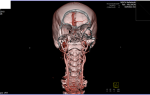
CT images will be presented in the form of a three-dimensional image, and the scanning is carried out in several projections.
This approach makes tomography data very accurate, and it will be almost impossible to miss even the slightest changes in the condition of the tissues. Even more detailed information is provided by MSCT of the larynx, where the number of sections is greater.
A CT scan of the larynx will show not only the upper respiratory tract. The tomography observation area includes:
- Thyroid
- Epithelial body
- Regional lymph nodes
- Blood vessels
- Upper esophagus
Contrast enhancement in CT and diagnosis of laryngeal cancer
Research plays a huge role in the diagnosis of cancer. Conventional CT is undoubtedly very accurate, but contrast-enhanced CT may be recommended for non-invasive differentiation of cancerous tumors from benign tumors. The procedure will help identify:
Source: https://DiagnostLab.ru/kt/golova-sheya/kt-gorla-i-gortani-chto-pokazyvaet-2.html
CT scan of the larynx, neck and throat: what does a study with and without contrast show?
At times, it can be difficult for a doctor to make a diagnosis. If a patient complains of discomfort in the neck area, it is not entirely clear what the reason is. Perhaps the lymph nodes are affected, the salivary glands are out of order, there is a pathology of the thyroid gland, soft tissue hurts, damaged blood vessels, the larynx, esophagus or trachea.
Separate examination of these organs requires a lot of time. Some patients cannot afford a complex of ultrasound, radiography and endoscopic examinations. In such a situation, the doctor prescribes computed tomography (CT) - a modern diagnostic method that provides information about the condition of all organs in the throat area.
Within 60 minutes, the patient learns the cause of his illness.
In what cases is a CT scan of the throat, larynx, neck and nasopharynx prescribed?
In the presence of a number of diseases, the initiative to prescribe a CT scan of the throat and larynx or MRI to the patient comes from the doctor. Indications for diagnosis are:
- oncological formations - tumor lesions of the larynx, esophagus, thyroid gland, metastases of a malignant tumor in the spine and lymph nodes;
- stones in the salivary glands;
- enlarged goiter;
- neck injury;
- entry of foreign bodies into the soft tissues of the neck, into the hollow structures of the throat;
- purulent diseases of the neck organs;
- pathological conditions of blood vessels;
- deformation of the lining of the esophagus with further bulging of its layers into the mediastinum;
- cancer of the lymphatic system;
- spinal injuries in the cervical region;
- injury to the nasopharynx and paranasal sinuses;
- inflammation in the retropharyngeal region;
- determining the location of the tumor;
- monitoring the patient’s condition after surgical treatment of the neck organs;
- checking the results of radiation therapy and chemotherapy.
When the patient is not feeling well, the doctor will also recommend a CT scan of the larynx or an MRI. Symptoms for which a doctor prescribes a study based on patient complaints are:
- feeling of squeezing of the larynx;
- pain in the neck;
- purulent discharge in the mouth;
- temperature increase;
- swelling of the larynx;
- persistent laryngitis;
- increase in goiter, bone formation in this area.
In case of nasopharyngeal disease, computed tomography is rarely prescribed, since an experienced doctor can make a diagnosis after a visual examination. Oncological diseases of the nasopharyngeal system are rare. The cause of constant laryngitis and the appearance of tumors in the nasopharynx is poor ecology, gastrointestinal diseases, and bad habits.
What does a CT scan show?
Computer diagnostics of the neck is a fairly informative way to determine the disease.
She can show the doctor the presence of a tumor of the neck organs, the appearance of metastases, pathology in the development of the throat, atherosclerosis of the vessels of the neck, protrusion of the esophageal wall, the formation of nodules in the thyroid gland, destruction of the cartilage of the larynx, the presence of foreign bodies in the organs, the appearance of cysts, pathological growth of lymph nodes, inflammatory processes, deformation of the cervical vertebrae and intervertebral discs.
The methodology and scope of research is determined by a specialist. Depending on the prerequisites for CT, different diagnostic methods are used. For example, to determine the source of headaches, a CT scan of the neck vessels is performed. When diagnosing diseases of the esophagus, a contrast agent is used.
Differences between CT scans with and without contrast
If the task is to determine the condition of the cervical spine, to identify protrusions and hernias, contrast in CT is not used. When diagnosing diseases of the neck and soft tissue organs, it is better to use the method of introducing a contrast substance into the blood, since without it it is difficult to distinguish organs and formations from each other.
Iodine-containing drugs Ultravist or Omnipaque are administered to the patient intravenously by injection or drip. Sometimes the patient is asked to drink a contrast agent.
When examining the condition of blood vessels, contrast is administered dropwise throughout the procedure.
The consequence of the administration of the drug for the patient may be a slight feeling of nausea, a feeling of warmth in the body, and a metallic taste in the mouth.
Before using contrast, it is necessary to check whether the patient is allergic to the substance. It can be expressed in swelling of the face and ears, difficulty breathing, sore throat, skin rash, bronchospasm, and a drop in blood pressure below normal. If these signs appear, you must report them to the doctor, who will interrupt the procedure.
When is the best time to make a diagnosis?
Before starting the procedure, the specialist will ask the patient to remove all metal objects - earrings, hairpins, chains, beads. When using contrast, the patient will be asked to arrive on an empty stomach. You can drink water. You are allowed to eat no later than 6 hours before the examination.
Contraindications
If diagnostics are carried out without a contrast agent, contraindications to tomography are pregnancy at any stage and the patient being overweight. The design capability of the tomograph allows examination of people weighing no more than 120 kg.
There are categories of citizens for whom the doctor prescribes a CT scan of the larynx with caution - if absolutely necessary. These include nursing mothers, children under 12 years of age, and patients suffering from renal failure or myeloma.
When using tomography with contrast, the study is contraindicated for nursing and pregnant women, patients with liver or kidney failure, diabetes mellitus, and people with intolerance to iodine-containing substances. They are more often prescribed an MRI.
The procedure for carrying out the procedure and interpretation of the results
The tomograph looks like a massive pipe. For examination, the patient lies down on a table. His head is fixed with holders to ensure immobility.
When using a contrast agent, a catheter connected to an automatic dispenser with a thin tube is inserted into the patient’s vein in the elbow area. The patient is then moved inside the tomograph and the examination begins.
In order to obtain an accurate result, the doctor from time to time asks the patient to hold his breath for a few seconds.
To provide an opportunity to report poor health, a rubber bulb is placed in the patient’s hand, which makes a sharp sound when pressed. If the patient gives a signal, the examination is interrupted and the patient moves out of the tomograph.
The study is deciphered by a specialist. Based on the images taken, it produces a result with identified pathologies.
The patient receives photographs and files with images of diseased organs on digital media, as well as a specialist’s report.
The decoding is not a diagnosis; it serves as source material for a specialist - it shows problems in the body. Deciphering takes from half an hour to an hour.
Which is better, CT or MRI? With a CT scan, the body is exposed to x-rays, while with an MRI, it is exposed only to a magnetic field. Magnetic resonance imaging is considered safer, but computer diagnostics is 1.5-2 times cheaper and faster. The choice is up to the doctor.
Source: https://vedmed-expert.ru/kt/golova-sheya/kt-gortani.html
What will a CT scan of the neck show and how to prepare for the examination - indications for computed tomography of organs and tissues of the neck
This type of diagnosis provides accurate visualization of the bone structures and soft tissues of the neck, mucous membranes of the esophagus and larynx. In its information content, computed tomography is superior to X-rays, ultrasound diagnostics, FGDS, and in some situations, when it is necessary to study the condition of the bone tissues of the neck, even MRI.
However, during this examination the patient receives a certain dose of radiation, so it is not recommended to carry out the procedure more than once a year.
This procedure cannot be performed on all patients.
In addition, each device has its own weight restrictions. For some devices this is 130 kg, for others 200 kg - it all depends on the model.
This manipulation does not require any specific preparation. However, in the case of contrast, the patient must refrain from eating and drinking for several hours before the procedure. Otherwise, the gag reflex may be triggered.
- Among other things, the use of a contrast agent has a number of contraindications, so you need to donate blood in advance to determine the level of creatinine and urea.
- In order not to distort the results of the study, all jewelry must be removed.
- Parts of clothing can also interfere with the scanning process, so it is best to bring a hospital gown with you.
- Device for scanning the neck and cervical spine
- When contrast is performed, a dye is injected into the patient a few minutes before the scan begins.
- If the object of study is the digestive tract, contrast is administered orally .
- For all other cases, bolus contrast - a small amount of iodine-containing X-ray contrast agent enters the body through an installed catheter in a vein throughout the study.
- In general, at the time of scanning, the patient is in a separate room, and communication with medical staff is maintained thanks to a special intercom.
At the very beginning of the procedure, the nurse helps the patient lie down on a retractable table, which, after connecting the system, automatically enters the tomograph, where layer-by-layer sections of the area under study are made. At the time of scanning, the person should not move or even swallow saliva.
Computed tomography of the cervical spine takes, on average, 5-10 minutes . The scanning process itself lasts no more than a minute , which is very convenient for examining people who are in serious condition.
After the diagnosis is completed, the patient is sent home.
He is notified in advance that the first day after the procedure, urine and feces may change color. This phenomenon is a consequence of the removal of the contrast agent.
Source: https://www.operabelno.ru/chto-pokazhet-kt-shei-pokazaniya-dlya-kompyuternoj-tomografii-organov-i-tkanej-shei/
CT (computed tomography) of the neck and larynx with contrast
A computer scan of the neck, head, and larynx is prescribed by an ENT doctor, gastroenterologist, or oncologist. The study shows the structure of the vertebrae, the condition of the blood vessels, and neighboring organs.
The quality of tomography is significantly superior to radiography, although it is based on X-ray radiation. Making a series of sections of the area under study every few millimeters, software creation of a spatial model - these features increase the sensitivity and specificity of the study.
Native computed tomography of the thyroid gland and neck shows bone structures, enlarged lymph nodes, and reveals neoplasms.
Contrast scanning (CT with contrast) allows you to track vessels, study the contours of anatomical formations, verify tumors growing in and out of walls (endo, exophytic growth).
CT scan of the larynx with contrast - how it is done, what it shows
The cancerous focus is nourished by its own vascular network. Without contrast, it is impossible to detect abnormal microcirculation. Intravenous injection of a drug based on gadolinium salts enhances the visibility of areas that are not visible on native scanning.
A CT scan of the neck vessels with contrast is performed after a provocative allergy test. Before manipulation, a small concentration of the product is injected. Tomography can be continued in the absence of local and general allergic reactions.
The high quality of the procedure is guaranteed when the person maintains a stationary position on the diagnostic table. The head is secured with straps. The position is selected according to the assigned diagnostic tasks.
Computed tomography of the cervical region (multispiral) is a high-quality method with high speed of section production and improved image resolution. MSCT of the neck is done for people with muscle twitching after taking a sedative.
A comprehensive CT scan of the larynx and neck shows:
- Condition of the thymus gland, thyroid gland;
- Lymphatic and arterial vessels;
- The wall of the larynx, pharynx;
- Cervical vertebrae.
Scanning is used before planning surgical elimination of displacements of the cervical bones, soft tissues after severe injuries, and choosing tactics for radiation therapy of tumors. In order to clarify the diagnosis, an examination is prescribed only after alternative studies that are not accompanied by radiation have been performed.
The three-dimensional modeling mode of multislice tomography shows the osteochondral skeleton of the trachea and larynx.
Traumatic injuries can be accompanied by transverse ruptures, separation of the hyoid bone, cricoid cartilage. Self-healing of the defect leads to wall atrophy. Timely detection of pathology prevents complications.
CT scan of the larynx with contrast - progress of the study
If the native examination shows oncology, a CT scan of the larynx with contrast is performed to exclude metastatic foci. Injecting the drug into the cubital vein leads to entry into the vessels of the neck within a few seconds. The bolus amplification method has an advantage, when the injection of the drug is carried out by an automatic dispenser, and tomography is done as the compound spreads.
The extensive microcirculation requires sequential scanning to study the vasculature at different levels.
The patient is placed on the table. The diagnostic system contains a tunnel - a section of the device with an internal circular cutout and peripheral expansion.
The X-ray source recording the sensor rotates in a circle. The patient automatically moves inside the system as the examination progresses.
In older installations, doctors ask the patient to hold their breath for several seconds to prevent image blur.
Modern multispiral devices make sections quickly. There is no need to stop breathing for a short time. The use of MSCT of the neck allows you to make tomograms in a few minutes. It takes 30-40 minutes to analyze the images, describe them, and formulate a conclusion.
The procedure is not performed on pregnant women, children under 14 years of age, or those with intolerance to iodine contrasts. Enhancing drugs are eliminated in 2 days, so you need to stop breastfeeding during this period.
Contrasting through the mouth (oral) is rational if there is a suspicion of narrowing of the esophagus, large growths of the mucous membrane with difficulty in passing food.
Contrast examination of the neck and head (CT angiography) - how it is done
After intravenous enhancement with Omnipaque and Ultravist, the cervical vessels are contrasted. The contrast passes from the ulnar vein to the vessels of the upper shoulder girdle in a few seconds.
CT scan of the soft tissue lymph nodes of the neck visualizes the jugular vein, vertebral artery, internal and external carotid arteries. An informative image is obtained after three-dimensional modeling (3D mode).
The software application creates a spatial display that clearly shows aneurysms (dissections of the vascular wall), blood clots inside the lumen, external compression of the artery or vein, inflammatory and destructive changes.
Main indications for contrast CT of the neck:
- Diagnosis of venous and arterial anomalies;
- Studying the characteristics of injuries to the neck and larynx after injuries for vascular compression;
- Fatty deposits inside the artery wall, blood clots;
- Identification of the causes of progressive headaches, loss of concentration, visual acuity, hearing;
- Inflammatory changes in the vascular wall, assessment of healing after surgery, radiation exposure of the tumor.
The advantage of CT scan of the neck is visualization of the spinal cord canal. With neoplasms and displacement of the cervical vertebrae, infringement occurs, leading to disability and stroke. Concomitant examination of the larynx and other anatomical formations of the cervical region helps determine the number of tumors, metastases, and the degree of involvement of surrounding organs.
Computer scan of the neck organs: indications
X-ray diagnostic methods are performed on the recommendation of a doctor. Compliance with the indications is necessary in order to avoid harm from ionizing radiation. Before prescribing the method, the specialist evaluates the benefits of tomography and the harm of the procedure for the patient.
The use of computed tomography for suspected cancer takes precedence over imaginary complications from ionizing radiation. Malignant tumors are deadly. Early detection and proper treatment help save a person’s life.
Verification involves the use of an integrated approach - CT, MRI, radiography, laboratory tests. The task is to find all malignant foci for early destruction.
Main indications for tomography:
- Clarification of the localization of the foreign body;
- Determination of the nature of blood flow, the degree of blood supply to the brain;
- Detection of purulent, destructive changes, inflammatory processes;
- Diagnosis of cervical diverticula and cysts;
- Verification of lymphatic tissue diseases;
- Detection of anomalies, aneurysms.
Scanning the neck with contrast has contraindications - allergy to iodine, renal failure, severe diabetes. The relative limit is weight more than 120 kilograms.
MSCT anatomy of the cervical part
The radial anatomy of the cervical region is well visualized on tomograms with contrast. In the images, specialists monitor the condition of the brachiocephalic system, the vessels of the circle of Willis, the main brain stems, and collateral circulation.
The neck consists of a complex of formations of different sizes, density, and length. When using multislice computed tomography, visualization is improved due to the use of several X-ray emitters and a system of sensors installed at a certain distance.
The anatomy of the neck is not visible on radiography. The smallest tumors are detected only after CT and MRI, so the harm from radiation fades into the background. Early detection of malignant neoplasms of the larynx and other structures is the initial task.
The advantage of computer scanning is the determination of tissue density (densitometry). The mode allows you to distinguish papillomas from scars, cysts, and inflammatory foci.
Enhancing contrast shows narrowing of the artery lumen, abnormalities, wall expansion, atherosclerotic deposits. The procedure is carried out before surgery.
About the safety of CT scans of the neck
The effectiveness of computer examination is beyond doubt, since a CT scan of the head shows neoplastic processes and neoplasms. Scanning with contrast allows you to study the features of soft tissues. Bolus enhancement (intravenous administration with an automatic syringe) visualizes organs as the substance moves through the vessels.
Cancer that grows along the organ wall (endophytic) has no structure. Only through densitometric measurement can a neoplasm be detected.
Scar formation is recorded using densitometry. The mode shows areas of narrowing of the larynx and trachea.
Other diagnostic methods cannot obtain the information described.
Let us draw a conclusion about the advantages of CT scan of the neck and larynx:
- Detection of space-occupying formations, traumatic defects, tumors;
- Identification of scars, degree of stenosis of the trachea, larynx;
- A painless, gentle method for examining patients with difficulty breathing;
- Evaluates criteria for the effectiveness of surgical treatment (three-dimensional reconstruction, virtual bronchoscopy)
Early detection of stenosis, scar changes, and pathology of paratracheal tissue prevents dangerous complications.
CT scan of the larynx, throat, pharynx - what does computed tomography without contrast show?
The close location of the larynx, pharynx, and throat makes it possible to examine organs on CT in one procedure. Computer scanning is based on recording the reflection of X-rays by tissues during circular rotation of the source. Tomography is carried out through a specified number of millimeters. The step of the procedure is determined by the estimated diagnostic results.
Native computed tomography of the pharynx and larynx has less information content when compared with the contrast variety. After intravenous administration of iodine preparations, the visibility of the contours of blood vessels increases. Tracking the microcirculation of the cervical organs makes it possible to study the prevalence of the pathological process, identify formations, and determine the depth of tissue damage.
Improves visibility and allows for multi-plane examination. Once the tomograms are obtained, software applications create a three-dimensional image. The slightest damage is visible on the spatial model. Remodeling is used before planning surgical interventions to remove cancer and large formations.
CT scan of the throat and larynx – what will it show?
After performing a CT scan of the pharynx and larynx, specialists have the opportunity to clarify the diagnosis, identify a new nosological form, and dynamically assess the healing of the defect after surgery.
What a neck tomography will show:
- Traumatic injuries to the upper body;
- Inflammation – phlegmon, infiltrate, abscess;
- Tumors of the parathyroid gland, thyroid gland, soft tissues;
- Additional outgrowths of the wall of the esophagus, larynx, pharynx - diverticula;
- Calcification of lymph nodes;
- Anomalies in the development of the thyroid gland;
- Swelling of the larynx, suffocation;
- Foreign bodies in the throat;
- Ossification of tracheal cartilage;
- Pathology of the lymphatic and vascular system.
Determining the disease requires a highly qualified radiologist. A diagnosis is possible after a layer-by-layer analysis of several hundred sections obtained, spatial models, and contrast photos. The examination of the larynx and pharynx is carried out carefully so as not to miss small oncological formations.
After detecting a defect, the dimensions are measured and the thickness of the walls surrounding the tissue is assessed. Scanning with contrast is prescribed to detect or rule out metastases.
Computed tomography (CT) of the throat and larynx determines the condition of the cricoid zone, where important anatomical structures are localized - lymph nodes, jugular vein, vascular bundle of the neck.
Examination of the cervical region reveals destruction of tracheal cartilage, oncological invasion into the wall with a spread of more than 3 mm, tumor damage to soft tissues, and the preepiglottic fissure.
CT scan of the pharynx and larynx: indications, contraindications
Classic CT of the larynx is indicated for:
- Cystic cavities, adenomatous foci (benign tumor);
- Serious injuries to the neck area;
- Vascular pathologies (thrombus, stenosis, wall dissection);
- Bulging of the wall (diverticula);
- Thyroid abnormalities;
- Phlegmon, abscesses;
- Inflammation of the larynx (laryngitis);
- Foreign bodies of the respiratory tract, digestive organs;
- Pathological changes in lymphatic and blood vessels.
Does not contain dense structures of the larynx and pharynx, enhances visualization with iodine.
You cannot do a CT scan of the throat or larynx with enhancement for thyroid diseases. The substance is excreted by the kidneys, so if the organ is insufficient, a contrast procedure is excluded. The procedure cannot be performed if there is a significant decrease in liver function.
Computed tomography of the neck and larynx: how to do CT, MSCT
The larynx and pharynx are qualitatively visualized after multislice tomography. The research is performed faster than its classic counterpart. Several combined source-receiver complexes make it possible to make as many scans in one rotation as there are installed heads.
Classic CT scan of the throat is less informative in displaying the internal structure of the hollow organ. The lack of dense tissue structures limits visibility. The soft tissue component is studied after strengthening followed by 3D reposition.
An alternative to research is MRI and ultrasound, but each method has advantages, disadvantages, and solves its own diagnostic problems.
Virtual endoscopy of the neck and larynx
To study hollow organs, scanning may be performed after filling with air and contrast (double enhancement). The virtual endoscopy method differs from the classic CT scan of the pharynx, larynx, and trachea.
Source: https://mrt-kt-golovnogo-mozga.ru/article/kt-shei-gortani
Preparation and performance of computed tomography of the neck
A CT scan of the soft tissues and organs of the neck helps visualize the muscles of the neck, the condition of the thyroid gland, and the lymph nodes in the area.
It is also used to examine hollow organs of the neck, such as the nasopharynx, trachea, and esophagus. Computed tomography is one of the most informative methods for studying the cervical region.
CT examination helps to detect malignant formations, foci of inflammation, cysts, organ injuries, hematomas, the presence and location of a foreign body in the neck, as well as lymph node hyperplasia.
The area that is examined also includes the cervical spine.
A soft tissue CT scan of the neck is a test that takes very thin slices (3.5-5 mm) of the neck, starting just above the level of the ears and ending just below the collarbone. This allows for more accurate diagnosis of conditions related to areas such as the nasal passages, mouth, throat, thyroid and parotid glands.
A computed tomography (CT) scan uses a special X-ray machine to view organs and tissues inside the body in detail.
CT scanning of the soft tissues and organs of the neck provides more detailed information about neck injuries, tumors and other diseases than other types of X-rays. A CT scan can also show bones, soft tissue, and blood vessels in the same images.
A CT scan scans the inside of the body. The images are more detailed than typical x-rays. During a CT scan of the neck, pictures are taken of cross-sections, or slices, of structures in the body.
When contrast is used during a CT scan, the structures are highlighted even more. CT scan can help determine the diagnosis at an early stage. The doctor uses this information to determine the best treatment for the patient.
Application of CT of soft tissues and organs of the neck
Computed tomography is very informative for diagnosing diseases of the soft tissues and organs of the neck. In particular, this study is able to document tumor size, location and relationship to adjacent structures, and CT can demonstrate the path of tumor spread.
By using intravenous contrast agent during the examination, the best visualization of the vessels is ensured and thereby improves the identification of key vascular structures.
CT does not provide a histological diagnosis and does not allow differentiation between benign and malignant processes.
CT is very useful in the evaluation of parotid tumors and is likely to replace traditional sialography for the evaluation of neoplastic pathology of the parotid salivary gland. However, sialography remains the procedure of choice for the evaluation of inflammatory diseases of the salivary glands.
CT is extremely valuable in the evaluation and diagnosis of laryngeal carcinoma. Computed tomography provides better imaging accuracy for assessing cartilage damage and tumor growth in the peripharyngeal spaces.
Current limitations of laryngeal CT are motion artifacts, lack of dynamic information, inability to differentiate edema from tumor infiltration, and lack of adequate characterization of mucosal surfaces.
CT of the soft tissues and organs of the neck is most useful in detecting metastatic adenopathy, especially in obese patients.
Reactive nodes, however, cannot be distinguished from metastatic nodes. Normal lymph nodes are usually less than 5 mm in diameter and reactive nodes are usually less than 15 mm in diameter. Large nodes with central necrosis usually indicate the presence of metastatic disease.
The role of CT in the evaluation of thyroid nodules is limited due to the accuracy of radionuclide and ultrasound diagnostic methods.
Indications for the study:
- the presence of abnormalities of the neck organs;
- damage to the neck organs caused by trauma;
- malignant and benign neoplasms;
- metastasis to lymph nodes;
- foreign bodies in the neck for subsequent planning of their surgical removal;
- diagnosis of neck cyst;
- damage to the cervical spine;
- infectious diseases of neck tissue (abscess, infiltrate, phlegmon);
- diverticula;
- damage to the blood vessels of the neck.
A contraindication for computed tomography is pregnancy at any stage due to the negative effect on the fetus.
Contraindications to the administration of a contrast agent are:
- history of severe reaction to contrast;
- the presence of allergic diseases, bronchial asthma;
- presence of hyperthyroidism;
- severe liver or kidney failure;
- period of pregnancy and lactation.
CT scans use X-ray technology and advanced computer analysis to create detailed images of the body.
Preparing for CT
You are allowed to eat a light meal before a CT scan of the neck. You should drink plenty of fluids on the day of your scan. You are allowed to take medications as usual.
You should definitely tell your doctor or CT specialist if there is a possibility of pregnancy.
Some items may interfere with CT images, so your doctor may ask you to remove the following items:
- any clothing with zippers or snaps;
- hairpins and jewelry;
- glasses;
- Hearing Aids;
- removable dental prostheses.
The patient will be asked to lie on his back, place his arms at his sides, and be very quiet. The table on which the patient is located will slide into the scanner. The scanner will be covered only at the level of the head and neck. The scanner is open at the back and front, allowing the patient to see. This procedure usually takes 15 to 30 minutes.
After this procedure there are no restrictions for the patient. You can eat and drive as usual. If the test was done with contrast material, you should drink six to eight glasses of water to flush it out of your body.
If a CT scan of soft tissues and organs of the neck with contrast is planned, before administering a contrast agent, it is necessary to clarify whether there is an allergy to any medications or iodine, which is its basis. If the patient has had an allergic reaction to contrast dye in the past, doctors will give medication to prevent a reaction to the contrast dye before the scan.
If you have underlying medical conditions such as asthma, diabetes, heart problems, multiple myeloma, or kidney disease, you may increase your risk of a contrast reaction. For example, kidney disease makes it difficult to remove contrast material from the blood.
Because a CT scan uses x-rays, no other people are allowed in the room during the test.
How to scan
The specialist helps the patient take the correct position on the CT scanner table. Soft straps or tape may be used to help keep the neck in the correct position.
During the scan, the patient will be inside the CT scanner. X-rays will pass through the patient's body as the X-ray tube rotates around him. The device will take pictures from many angles, creating cross-sectional images (slices) of the neck.
The x-ray technician leaves the room and during the scan the patient will be alone in the room, but can always see, hear and talk to him through a special feedback device.
The patient must remain still while the table is moved to the center of the scanner.
If a contrast study is necessary, patients will receive contrast (x-ray dye) during the scan. The contrast makes tissues and blood vessels more visible to the doctor's view in CT scans.
How does the patient feel during the scan?
A CT scan is painless, but the patient may experience some discomfort while standing on the scanning table.
If contrast is used during injection, the patient may notice a warm, fuzzy feeling. He may also experience a metallic taste in his mouth. These reactions are normal. They should go away within 1-2 minutes.
From time to time, the patient experiences itching for several hours after contrast injection. You need to inform your doctor about this so that he can prescribe appropriate treatment.
You should also immediately tell your doctor if you experience dizziness, shortness of breath, or shortness of breath after a CT scan with contrast, as this may be an allergic reaction. A doctor or nurse will be nearby during the scan to assist the patient if needed.
Who interprets the results and how to get them
A radiologist experienced in CT reading will review and interpret the CT images and send a detailed report to the physician who referred the patient for testing. And the doctor will talk with the patient about the results of a computed tomography scan of the soft tissues and organs of the neck.
More fresh and relevant information about health on our Telegram channel. Subscribe: https://t.me/foodandhealthru
Specialty: therapist, radiologist.
Total experience: 20 years.
Place of work: SL Medical Group LLC, Maykop.
Education: 1990-1996, North Ossetian State Medical Academy.
Training:
1. In 2016, at the Russian Medical Academy of Postgraduate Education, she underwent advanced training in the additional professional program “Therapy” and was admitted to carry out medical or pharmaceutical activities in the specialty of therapy.
2. In 2017, by the decision of the examination commission at the private institution of additional professional education “Institute for Advanced Training of Medical Personnel”, she was admitted to carry out medical or pharmaceutical activities in the specialty of radiology.
Work experience: therapist – 18 years, radiologist – 2 years.
CT scan of the cervical spine
CT scan of the cervical spine is a modern, informative, non-invasive method for examining the vertebrae, intervertebral discs, spinal canal and spinal cord located at the level of the neck.
Typically, the examination is performed covering the area of the base of the skull above and the first thoracic vertebrae below in order not to miss pathological changes that may be located in these transitional areas.
Computed tomography of the cervical spine allows you to obtain images of layer-by-layer sections of the human body.
The dark and light areas of the images are a reflection of various organs and tissues, which differ from each other in their ability to retain X-rays.
For example, bone tissue appears light, almost white in photographs due to its high ability to absorb X-rays.
The thickness of the sections ranges from 1 to 3 mm. Large slice thickness significantly speeds up the procedure, but will reduce the information content of the examination. Modern models of tomographs allow, in addition to images, to obtain three-dimensional models of the organs and anatomical structures being studied.
The doctor can examine such a model from all sides, rotate it, enlarge it, and zoom it in.
All this makes it possible to obtain more information about the structure of tissues and organs of the area under study, as well as with a greater degree of probability to identify violations in the structure of individual elements and their relative position relative to each other. This is especially useful in cases of cervical injuries.
What does a CT scan of the cervical spine show?
The following types of disorders can be identified in the images obtained during the examination::
- congenital features of the development of the spine;
- cervical injuries: fractures and displacement of the vertebrae, hematomas;
- narrowing of the spinal canal;
- tumors;
- osteochondrosis, disc herniation;
- changes in vertebral bodies, their deformations;
- inflammatory processes localized in the vertebrae, spinal cord, soft tissues surrounding the spine;
- changes in the spine, characteristic of a number of diseases, such as rheumatoid arthritis or ankylosing spondylitis;
- consequences of injuries and surgical operations.
CT scan of the cervical spine with contrast
The examination can be performed either with or without the use of a contrast agent. Bone tissue is clearly visible in images without contrast enhancement. But vessels, benign and malignant formations, foci of inflammation can be practically indistinguishable on native images.
The decision about the need to administer a contrast agent is made by the radiology doctor who conducts the examination. The need to administer contrast may arise during an examination in cases where the doctor sees changes in the images, but cannot identify them due to the lack of details important for diagnosis.
Examination of the cervical spine is carried out using an iodine-based contrast agent intended for intravenous administration..
Most often, the drug is administered once before or during the procedure. Bolus (long-term, dosed) administration of contrast is indicated in cases where it is necessary to obtain information about the condition of the vessels of the neck and the characteristics of the blood supply to the tissues.
The following symptoms may occur in response to the administration of a contrast agent::
- sour or metallic taste in the mouth;
- pain at the injection site;
- flushes of heat in the body;
These reactions are not harmful to the patient's health.
Contrast drug intolerance is manifested by the following symptoms::
- swelling of the face, ears;
- difficulty breathing due to swelling of the larynx;
- sore throat;
- nausea and vomiting;
- itchy skin;
- hives
- bronchospasm;
- drop in blood pressure.
Source: https://doctorsustav.ru/diagnostika/podgotovka-i-provedenie-kompyuternoj-tomografii-shei