The cervical spine is made up of 7 bones called vertebrae. Thanks to their presence, a person can freely move his head and neck. This is the most mobile part, which is subject to heavy loads, and at the same time the most vulnerable. A CT scan of the cervical spine helps to see and identify abnormalities in the functioning of this structure.
Indications for use
The main indication for scanning is pain. It can appear gradually, with increasing force, or begin abruptly in the shoulder area. The pain is often accompanied by numbness in the fingers. Previously, before prescribing a CT scan, other examinations, X-rays, and MRIs are performed. If these research methods provide insufficient information, computed tomography is prescribed.
Reasons for examination:
- constant headaches, frequent dizziness;
- fainting;
- darkening of the eyes;
- injuries, damage to the head, neck;
- pain in the hands;
- spasms, shooting.
The most common indications for CT scanning are:
- To confirm malignant and benign neoplasms. To search for metastases and determine their boundaries.
- In case of injury to the head or neck.
- If a foreign body gets into the soft tissues of the larynx or neck.
- To determine the level of blood flow in the head and neck area.
- To identify inflammatory processes such as infiltrates, phlegmon, soft tissue abscesses.
- When diagnosing inflammation of the lymph nodes.
- For the diagnosis of diverticula and soft tissue cysts of the larynx and neck.
- With a detailed examination in the case of spinal osteochondrosis.
- In case of detection of intervertebral hernias, sprains in the cervical spine.
- To identify vascular disorders in cases of suspected atherosclerosis and cerebral aneurysms.
Pain in the cervical spine has different origins. Poor circulation, headaches, and dizziness signal a pathology such as vertebrobasilar insufficiency.
Neck shooting, or cervicago, often occurs. The disease occurs as a result of sudden compression of the muscles. The main reason is osteochondrosis or intervertebral hernia.
Spinal curvatures often cause pain in the cervical vertebrae.
Tomography helps to examine in detail all parts of the spine and vertebrae. The photographs show minute disturbances in the structure.
How to prepare for the examination
No special preparation is required before scanning the spine, cervical spine, or vertebrae. But it is better to refrain from eating 2 hours before the procedure. During preparation, the patient is asked to leave all metal-containing objects outside the office. For the procedure, the patient brings comfortable clothing himself or is provided with it on the spot.
Sometimes contrast enhancement is used to better view an organ or soft tissue. Contrast is injected into a vein or spinal canal. Some time is necessary for the substance to spread throughout the body through the bloodstream and accumulate in places where the inflammatory process occurs. A CT scan takes 15 to 30 minutes.
The dye helps to clearly see tissues and organs in photographs. It is also used to check blood flow, look for tumors, areas of inflammation and nerve damage.
Before the procedure using contrast, it is recommended to refrain from eating for 6-8 hours. After the examination, you must drink water. The dye leaves the body with fluid within 24 hours.
Many doctors recommend following a gentle diet after the scan. It is advisable to include the following products in your diet:
- nuts;
- parsley;
- beans;
- seafood;
- milk;
- dry red wine (within reasonable limits);
- grape juice.
The main danger is the radiation received during the procedure. Therefore, a long time must pass between CT, MRI, and x-rays, if they are necessary. After the procedure, you can take antihistamines as prescribed by your doctor.
How is the diagnosis carried out?
The scan is performed by a radiologist, then the images are analyzed by a radiologist.
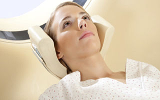
During the examination, the patient lies on a movable table attached to the scanner. A CT scan sends x-rays into the body of the person being examined.
Each revolution takes no more than a second and provides an image of a layer-by-layer section of the organ under study. Part of the scanner can tilt to view the curves of the spine.
All pictures are transferred to a computer and can also be printed. You must lie still during the examination.
During the scanning process, the technologist observes the patient through the window, maintaining two-way communication with him.
The entire examination process takes from 30 to 60 minutes. Most of the time is spent on preparatory activities. The scanning process itself takes a few seconds.
Contraindications for tomography:
- allergic reactions to contrast agent (iodine);
- taking Metformin, whose reaction to the dye can cause problems;
- the occurrence of nausea and vomiting after contrast administration;
- pregnancy, breastfeeding;
- inability to remain still during the test;
- the presence of metal dentures (when scanning the head area).
It is not advisable for children under 14 years of age to have a CT scan. For people with thyroid disease or kidney failure, examination with a contrast agent is not performed.
What does the procedure determine?
- After the examination and printing of the images, a conclusion is written about the condition of the internal tissues and organs.
- The condition of the cervical spine, vertebrae and spine is considered normal if:
- The dorsal bones are natural in shape, number and alignment.
- The discs and joints that support the spine are normal.
- The spinal canal is consistent in size and shape.
- If the contrast material used spreads evenly along the spinal canal, there is no narrowing or blockage of the latter.
- None of the spinal cord nerves are pinched or pinched, and there is no bulge growth.
Deviations from the norm:
- Vertebrae are damaged, missing or misaligned.
- One or more discs are damaged, herniations are found in the intervertebral discs.
- The flow of contrast through the spinal canal is restricted or blocked, indicating spinal stenosis.
- The vertebrae show signs of arthritis and bone problems caused by osteoporosis.
- An abscess or spinal tumor is detected.
- CT scans can help identify abnormalities such as diffuse disc bulging and herniation.
- Doctors often refer for examination to confirm the diagnosis of diseases that require urgent intervention:
- Osteochondrosis, dystrophic damage to the 6th and 7th intervertebral discs.
- Protrusion is the stretching of the fibrous ring beyond the boundaries of the vertebrae at the early stage of intervertebral hernia.
- Extrusion, or severe disruption of the integrity of the fibrous ring, often accompanied by its rupture.
- A herniated disc caused by a rupture of the fibrous ring.
A timely scan for a cervical fracture guarantees a favorable prognosis for treatment.
Conclusion
CT is an informative examination method with minimal risk of negative effects of x-ray radiation on the body. The resulting high-quality three-dimensional images of diseased organs help to choose the optimal treatment option. Using the procedure, doctors can not only correctly diagnose the disease at an early stage, but also check the effectiveness of the prescribed treatment.
Source: https://OrtoCure.ru/diagnostika/kt-shejnogo-otdela.html
CT scan of the cervical spine: objectives of the study, preparation for the procedure
One of the most vulnerable points of the human body is the neck. Here, in a small area, numerous blood vessels, arteries, tracheas, and nerves converge. It is quite difficult to identify disease in this area. Especially for such cases, there is a CT scan, or computed tomography. Thanks to it, you can find out exactly which organ needs treatment.
CT scan of the cervical spine
In this article we will understand how a CT scan of the cervical spine is performed, how to prepare for such a procedure and what results it gives.
What is computed tomography (CT)?
Tomography is an examination method in which individual areas of the body are examined using a lumen in different planes. Initially, tomography was performed using x-rays. However, this method did not become very popular.
With it, researchers did not receive the necessary information about the area of interest to them. Therefore, scientists began to think about improving this method. As technological progress developed, a new method was created through the use of a computer. It was called computed tomography, or CT for short.
Using this method, doctors can study the organ being examined in great detail.
Using this technique, the spine in the neck area is examined. Based on the data obtained, three-dimensional and two-dimensional models of the cervical vertebrae are created, which facilitates the study of this section. CT is a high-precision x-ray examination that is widely used in the study of bone lesions and soft tissue pathologies.
Using a CT scan of the cervical spine, various pathologies in this area can be identified.
It is based on X-ray technology with computer processing and analysis, so the output is detailed and clear images.
With the help of computed tomography, humanity has the opportunity to see the whole picture, which is especially useful when examining internal organs and the structure of many tissues.
The peculiarity of this approach lies in taking numerous images using a tomograph, which, as it were, “spins” around the selected examination area. This is followed by processing on a computer, thanks to which the images acquire their clarity.
The images allow you to peer into the smallest details; the size of the smallest detail can reach up to 0.5 mm. In addition, all photographs are stored digitally. They can be viewed at any time, rotated at the desired angle and in the required plane.
This method most accurately allows you to determine possible damage to the cervical spine.
The negative impact of the procedure is minimal. X-ray exposure is low and examination time is shorter than conventional CT.
During the procedure, pathologies and damage can be found in the vertebrae of the neck, larynx, pharynx, muscles, blood vessels, lymph nodes, thyroid gland, etc.
Computed tomography is usually used for:
- confirming the diagnosis;
- drawing up a treatment plan;
- operations planning;
A complex type of study of the neck using CT is an image of all the “layers” of the neck, where all the tissues representing this area are clearly visible. Also here you can notice hollow organs, such as the esophagus and trachea, bone structures and at the same time all kinds of changes in them.
CT scan is prescribed for many diseases of the neck and organs in this part
Thanks to CT it is possible to examine:
- developed abnormalities in organs;
- tumors and their metastases in the soft tissues and bones of the neck;
- violations of the walls of blood vessels associated with the manifestation of atherosclerosis;
- aneurysms and compression of blood vessels;
- damage to the esophagus (its expansion and protrusion);
- nodules in the thyroid gland;
- deterioration of cartilage in the larynx;
- stones in the salivary ducts;
- injuries;
- tumors and other foreign bodies;
- cysts;
- changes in lymph nodes, their enlargement;
- inflammation, abscesses, phlegmon;
- cervical deformity.
Safety of the technique
Since the inception of CT scanning, various studies have been conducted regarding the radiation dose a person receives during these procedures. One of the last such studies was conducted in Belarus in 2008. Scientists tested the impact of CT scanners from different manufacturers. Results vary depending on the time spent under the scanner and the area of the body examined.
The average radiation exposure during neck examination was 2.6±1.0 mSv. The acceptable radiation dose for humans per year is 3-4 mSv.
Although CT does not expose the body to much radiation, it is not recommended for pregnant women.
CT scan is a relatively safe procedure
The examination of children should also be approached with caution. Children are sent for CT scans only in cases of urgent need to obtain information.
Who should do this research?
Examination of the cervical spine, its soft and bone tissues, as well as internal organs is most effectively done using computed tomography. Thanks to it, all structures belonging to this department are studied one by one. The bones, spine, blood vessels and lymph nodes are examined one after another.
When is a CT scan of the neck recommended?
- A CT scan is indicated for any injury to the head or neck, mainly for severe subsequent manifestations.
- It is prescribed to confirm the diagnosis, the presence of malignant or benign neoplasms, detect metastases, and determine their growth rate.
- To check nearby organs and tissues for tumor lesions.
- It is prescribed to determine the location of tumors, to identify their number and structural features.
- CT scan helps to assess blood circulation. Determine how normal the nutrition of organs and tissues of the cervical spine is.
- Computed tomography is performed when inflammatory processes are detected. This includes abscesses, phlegmons, infiltrates, etc. It is also prescribed for previously diagnosed inflammations for which ineffective treatment was prescribed.
- Used to identify cysts and cervical diverticula and soft tissue of the larynx.
- Recommended for diagnosing spinal osteochondrosis.
- Used to detect all kinds of abnormalities in the blood vessels and lymph nodes located in the neck. For example, aneurysms, atherosclerosis and abnormalities in the functioning of the lymph nodes.
Contraindications
Computed tomography is based on x-ray technology. In essence, it is similar to an x-ray examination. The only difference is that during a CT scan the beams travel in a fan-shaped direction.
As a result, they converge at one point. In X-rays, the rays travel in parallel directions. Despite all the differences, both of these procedures have a negative effect on the human body in one way or another.
Although computed tomography is less harmful, it is not completely harmless.
This procedure has some contraindications
Based on this, this procedure is contraindicated for the following people:
- pregnant women at any stage;
- CT scanning is not suitable for breastfeeding mothers. If the procedure has been carried out, feeding the baby is not allowed for two days;
- Computed tomography is not recommended for children under 14 years of age. Allowed only in emergency cases, in case of severe pathologies;
- due to the narrow arch of the tomograph, the maximum weight of the examined patient should not exceed 200 kilograms;
- the patient undergoing a tomographic examination using contrast should not be allergic to iodine and seafood;
- contrast staining is contraindicated in people with kidney disease, renal failure, heart disease and thyroid disease;
- CT scanning is prohibited for people with metal dental implants and various prostheses in the neck;
- the procedure is not performed on patients with severe stages of diabetes;
- CT scanning is unacceptable for people with mental disabilities, poor coordination and those in a coma.
Which specialist can prescribe a CT examination of the cervical region?
Diseases of the neck area are extremely diverse. Thus, a referral for a CT examination can be prescribed by various specialists. They can be an orthopedist, surgeon, oncologist, traumatologist, etc.
The main symptom used for CT diagnostics is pain in the cervical region.
After the process is completed, the patient leaves with an image in his hands, a disk with information about the examination performed and a text conclusion.
Any specialist can prescribe a CT scan of the neck, since in this area there may be disorders associated with both the spine and internal organs
Which procedure is better for diagnosing diseases of the spinal column in the cervical section - MRI or CT?
One of the most frequently asked questions that arises from people undergoing treatment is what is most effective in identifying diseases of the cervical spine? Computed tomography or magnetic resonance examination?
MRI is primarily aimed at identifying pathologies in the bones of the spine. Thanks to the MRI procedure, the doctor has access to both the external and internal structures of the spinal column in all details.
Important! Modern tomographs are able to determine the strength of bone structures, their integrity and thickness. And with the availability of special computer applications, it became possible to find out densitometric data, that is, bone density.
Thanks to CT examination, specialists can learn about the presence of spinal diseases and various pathologies in a patient, for example, abnormal development of the spinal column, demineralization processes, sclerotic changes, spinal hemorrhages, tumors, etc.
The results of MRI and CT may be similar, but the operating principles of the devices differ significantly
CT is a more accurate examination of the cervical spine and all its structures. No other diagnostic examination will provide such detailed and accurate information. However, despite this accuracy, if abnormalities in the intervertebral structures of the cervical spine are suspected, preference is given to magnetic resonance imaging.
With the help of a CT scan, the doctor is able not only to detect the disease itself, but to predict its further course.
For example, if there is a strong blow to the back of the head, fractures of the processes and bodies of the cervical vertebrae may occur, as well as their unstable behavior.
With this information, the doctor can assume that without proper treatment, compression of the nerve endings may occur in the future. This in turn will lead to paralysis of some organs and systems of the body, for which the damaged nerve processes were responsible.
Considering all the advantages of CT, magnetic resonance imaging has fewer contraindications. This is due to the absence of X-rays, which are used in CT scans. Thanks to the above, magnetic resonance examination can be prescribed even for a newborn baby and a pregnant woman. Under equal conditions, CT scans are not prescribed to such patients.
Thyroid examination
The thyroid gland is the largest in the human body. It is located in the throat area. The gland consists of two lobes, which are connected by an isthmus. In rare cases, its absence is revealed.
The gland is protected by fibrous and outer membranes. The space between them is filled with adipose tissue. It is permeated by a network of blood vessels and nerves.
The thyroid gland is involved in the formation of hormones necessary for the body - thyroxine, calcitonin, triiodothyronine and thyrocalcitonin.
In the diagnosis of diseases and pathological neoplasms, CT examination is not inferior in information content to procedures such as MRI and ultrasound. Using computed tomography, the doctor can promptly detect the presence of cystic formations, adenomas and other pathological nodes.
CT scan can examine the thyroid gland
Examination of the trachea and larynx
The larynx is connected by nine movable hyaline cartilages. They are connected by ligaments, membranes and joints. The inside of the larynx is covered with mucous tissue. The larynx is made up of three parts - upper, middle and lower.
Source: https://spina-expert.ru/diagnostika/kt-shejnogo-otdela-pozvonochnika/
Computed tomography (CT) of the neck: what the doctor examines and what pathologies are detected
When prescribing a CT scan of the cervical region for a patient, the doctor expects to receive information about the soft tissues in the throat, blood vessels in the neck and head. In some cases, a CT scan of the neck with contrast is performed. This is necessary for a more accurate determination of the state of the vascular system.
A CT scan of the cervical spine helps doctors determine possible damage in this area.
What are the goals of the procedure?
CT is the most complete and informative diagnostic method.
During the examination, it is possible to determine both problems in the neck and disorders in bone and soft tissue, and visualization of the larynx, spinal cord, thyroid gland, and spinal structure is also carried out.
During the procedure, the doctor determines the location of the inflammatory focus, as well as benign or malignant neoplasms. In addition, the specialist identifies whether there are foreign bodies and determines the presence of lymph node hyperplasia.
During the examination, the diagnostician examines the brachiocephalic region using x-rays. CT scans provide a three-dimensional image of the vascular system from different angles. To improve visualization, the patient is injected with a contrast agent, in this case the examination is called CT angiography.
In what cases is a CT scan of the neck performed?
A CT scan of the soft tissues of the neck is performed in the presence of a herniated disc or to determine whether the ligaments in the neck are stretched. The procedure is also necessary if the doctor suspects that the patient has a vascular pathology or an abnormality of the lymph nodes. This allows you to determine the presence of atherosclerosis or aneurysm.
Doctors often prescribe MSCT of the cervical spine
The patient is referred for MSCT of the neck:
- with severe neck and head trauma;
- to determine the level of blood supply to the brain;
- to determine and confirm the formation of a tumor, its location, nature and anatomy are determined;
- in the presence of a cyst;
- to determine the presence of a foreign body;
- to confirm osteochondrosis of the spinal column;
- with purulent lesions (abscesses, phlegmon, infiltrates);
- if therapy for inflammation in the head, neck and throat does not bring results;
- in the presence of diverticula in the throat.
With the help of CT angiography, the condition of the cervical vessels is determined with greater accuracy. During the examination, the specialist identifies blockages, abnormal tortuosity in the carotid arteries, damage and aneurysm, scans arterial and venous blood clots, and determines the anatomy of neoplasms where the vascular system is involved.
If the doctor has doubts about the diagnosis and suspects that the patient has a stroke, MSCT of the neck vessels is mandatory.
How is the procedure performed?
Computed tomography is performed only on the recommendation of a doctor. A contrast agent is often used for diagnosis, so you should avoid eating for 5 hours before the procedure. Doctors advise not to even drink water before the examination.
If you have jewelry, you should remove it all before the examination.
Before a computer examination is carried out, the patient must free himself from jewelry and clothing with metal elements. Sometimes in the CT room the patient is asked to change into specially prepared clothes.
The patient must have with him a surgical report (in case of previous surgery), an otolaryngological report (if the problem is in the larynx or nasopharynx) or an endocrinological report (if the thyroid gland is to be examined).
Carrying out the procedure
When scanning without contrast injection, the patient is positioned on a movable table. Typically, the person should lie on their back. Sometimes the patient is secured with leather straps and a bolster. This is necessary to ensure that the position of the body does not change.
When the tomograph is set up and the positions are set, the doctor turns it on and goes into the next room. The patient needs to remain in a motionless position for several minutes.
During the procedure, the table moves in a spiral, and a tube in which beams and detectors are mounted rotates around the patient. The patient should not be afraid, since the diagnosis is completely painless.
During the procedure, the patient may hear a noise that appears as the ring of the device moves.
The procedure is often performed with the injection of a contrast agent.
If the diagnosis is carried out with contrast, then a special substance is injected into the patient intravenously and scanning continues.
After the contrast is administered, the patient may feel hot or cold, a metallic taste in the mouth, and slight dizziness.
If a person does not have an allergic reaction to iodine or consumption of seafood, the negative reaction will be minimal and will go away on its own after the examination is completed. In another situation, the doctor prescribes antihistamines.
The duration of the usual procedure is 15 minutes, and with contrast - 30-50 minutes.
Within an hour, the diagnostician collects the necessary images, records the information on a disk and enters the examination results into the documentation. Then the patient receives the document with the results and gives it to the attending physician for decoding.
This video shows what a CT scan of the cervical spine looks like:
What can be revealed on a CT scan of the neck?
CT scan of the soft tissues of the neck shows:
- abnormal development of cervical organs;
- the presence of benign and malignant tumors in the bones and soft tissues of the neck, metastases;
- the presence of atherosclerotic changes in the walls of large cervical vessels, compression of the vascular system by a large neoplasm, aneurysm, etc.;
- whether there are expansions and formations of a diverticulum (protrusion of the walls) in the esophagus;
- the presence of pathological thyroid nodules;
- the presence of destructive (destroyed) foci in the cartilaginous tissue of the throat;
- the presence of stones in the salivary glands;
- whether the neck tissues are damaged due to injury, whether there are foreign bodies;
- is there a cyst in the neck;
- are the lymph nodes enlarged?
- is there inflammation in the cervical tissue (phlegmon, abscess);
- whether the cervical region of the spinal column is changed, whether the vertebrae are deformed.
This method is used to diagnose cysts in the neck area
What exactly will be examined on a CT scan of the neck - hollow organs or bone structure - depends on the goal of the diagnosis. In accordance with the appointment, the diagnostician determines the scope of the upcoming procedure and the need for contrast.
Did the article help? Rate it ( 2 votes, average: 5.00
Source: https://infouzi.ru/kt/golova-i-sheja-3/tomografija-shejnogo-otdela.html
Diagnostic CT scan of the neck
Computed tomography has long gained popularity in medicine and has won the title of the most informative diagnosis. A CT scan of the neck shows what the condition of the vessels of the head and upper spine is. The study also reveals differential and destructive changes in the soft tissues of the larynx and neck. Scanning is especially important before or after surgery.
General characteristics of the examination
The diagnostic method is based on the exposure, transmission and reflection of x-rays. The examination of the organic structures of the neck is performed layer by layer. X-rays expose thin sections of tissue. The method eliminates blurring of images located near slices.
In terms of projection capabilities and image contrast, CT is superior to all known x-ray methods.
There are several generations of CT scanners. The design has a rotating X-ray tube, which delivers a beam to the area required for diagnosis. The beam beam is wide, it covers the entire desired area in diameter. Using the control, the diagnostician can change the width of the beam beam.
The beam reaches the detector and emits an electrical signal. It is detected by the device's sensors. The received signals are transmitted to the computer. The CT detector is several times more sensitive than conventional x-rays. The tomograph generates an X-ray beam of the required width. Cumulated beams of rays pass only through the required area of the human body. The remaining areas of the figure are not exposed to radiation.
Conducting a CT scan of soft tissues and bones of the neck is possible with 50-100 tomographic images. A clear image can easily differentiate what a CT scan of the neck shows.
Types of examination
For a full CT scan of the entire neck, either a standard procedure or a session with contrast is chosen. The staining agent is injected immediately before the neck scan begins.
The drug makes the color of the image more intense and helps to distinguish the nature of the tumor and the direction of blood flow.
There are several types of procedure:
- standard conventional tomography;
- spiral tomography;
- multislice tomography with many images and high tube rotation speed;
- study with the introduction of a contrast agent;
- angiography of cervical vessels.
The standard CT procedure perfectly shows all cervical regions. The anatomy of the neck is clearly visible on CT. The organ includes the larynx, thyroid gland, and the beginning of the esophagus. A CT scan of the neck with contrast can detect circulatory disorders in the neck and proliferation of thyroid tissue. To obtain complete information, the patient is often prescribed MSCT (CT) of the neck.
Indications for use
Computed tomography of the neck is prescribed in various clinical situations:
- diagnosis of neck pathologies;
- examination of the ligaments, cartilages and vertebrae of the neck;
- detection and treatment of disc herniation;
- postoperative observation;
- diverticula of the larynx and esophagus;
- cystic formations;
- purulent inflammation of the throat;
- presence of a foreign body;
- detection of cancer and metastases.
A complete examination of the upper spine is necessary for osteochondrosis, osteoporosis, arthritis and arthrosis. A CT scan of the neck can more accurately determine the cause of pain and shooting in the neck than a routine medical examination in the office. A CT scan of the neck is also necessary to examine the cerebral vessels.
Anomalies and curvature of the veins may occur in the vascular branch of the neck. This disrupts blood circulation and blood supply to the brain, disrupts microcirculation of blood masses and the delivery of nutrition to the head. Examination of the vessels of the head is necessary in the pre-stroke and post-stroke state.
The study is required to diagnose thyroid diseases. The gland is exposed to many negative factors and requires regular inspection. CT scan of the neck shows degenerative processes and features of diseases of the soft tissues of the larynx and trachea.
Diagnosis of the cervical spine CT allows you to see disc protrusion and displacement of the vertebrae. If a patient suffers from frequent headaches, CT scans can help identify their causes.
Indications for examination include hypertension, thyroid and throat diseases.
Contraindications to the procedure
Neck scanning is not always permitted by doctors. The procedure is especially contraindicated during early pregnancy. Doctors prohibit exposing the body to radiation during the period when all the internal organs of a person are developing.
The fetus is more sensitive to ionizing radiation than the maternal adult organism. After radiation exposure, the embryo may develop anomalies and defects in internal organs. Pregnant women are only allowed to undergo ultrasound diagnostics.
Among the restrictions:
- fear of closed spaces;
- epilepsy;
- obesity of the third degree;
- pregnancy;
- childhood;
- kidney pathologies.
The contrast examination method is not used for kidney diseases. The introduction of a staining solution entails a huge load on the kidney. An unhealthy organ is unable to cope and remove the quickly staining drug from the body.
Brain scans are not performed if you have permanent dental implants or dentures with metal pins. Metal can distort CT scans of the neck and head.
Preparation for the procedure
Tomography is sometimes performed on patients with a contrast agent. Staining neck tissue allows you to obtain the necessary information about the vessels or tumor process.
During the study, contrast is introduced in advance. An allergy test is carried out. After the procedure, the contrast agent is independently excreted from the body in the urine.
Contrast CT places a heavy burden on the kidneys, so this organ must be healthy.
On the day of neck scanning, it is advisable to avoid eating in the morning. The night before you should have a light dinner. Stop drinking alcohol ten days before the expected CT procedure. The neck scanning procedure does not require special preparation.
How is the examination carried out?
A CT scanner has complex devices, sensors and mechanisms. A separate room is allocated for conducting examinations on the device. The diagnostician and the monitor are in the next room during the examination. This way, the specialist does not receive a dose of x-ray radiation and can observe the patient during the study.
The tomograph capsule is long - the tube has a number of detectors and an X-ray tube. The equipment rotates around the area of the body being examined. With the spiral diagnostic method, the tomograph beams and table constantly move in a spiral with specified speed parameters. The tomograph allows you to obtain several panoramic images of the neck in different projections.
The research time does not take more than ten minutes. The patient is placed in the tomograph capsule. A prerequisite for obtaining an accurate result of a CT scan of the neck is to be in an immobilized position. It is forbidden to turn your head, turn, or raise your limbs while the X-ray tube is working. If a person has increased physical activity, the procedure will not be performed.
The patient can communicate with the doctor. He is in the next room and monitors the person being examined through a transparent glass window. The tomograph apparatus has a special communication device. The patient can always ask the diagnostician a question or report the onset of an attack or discomfort. Through the device, the doctor gives the patient specific commands - to hold his breath or turn his head. Don't be afraid to explore. It is painless and not dangerous if radiation exposure standards are observed.
After the CT scan, you must wait for the results and the issuance of a diagnostic report. The obtained tomographic images show all the details of organic structures. The image and diagnostic report are sent to the attending physician who referred for examination.
New growths of various benign and malignant nature, cysts, pathology of the lymphatic system, and disc displacement can be detected. If a pathology is detected, the doctor examines the obtained images in detail and prescribes therapy depending on the data obtained from scanning the neck.
The method itself does not carry a large radiation dose, but scanning can only be done once every 3-4 months. If you do tomography more often, you can exceed the permissible x-ray exposure per year. Tomography is not performed on severely weakened people, so as not to expose them to radiation.
The examination can be done at any diagnostic center. However, it is carried out only according to indications and with a referral from the treating doctor. Usually, registration for the procedure is open, which makes it possible to avoid the queue and undergo the necessary examination in a timely manner.
Source: https://mrtdom.ru/diagnostika-kt/kt-shei/provedenie-diagnostiki-kt-shei
CT diagnostics of the neck and its soft tissues
CT and MRI are completely different diagnostic tests. They differ in the preparation of the patient, the procedure, the basic principles of scanning, and images, but they have a common goal - identifying pathological changes in internal organs, in particular the neck area.
CT diagnostics for some parts of the human body and certain diseases is considered more informative compared to magnetic resonance imaging. Thanks to the information obtained using a CT scan, the doctor will be able to make an accurate diagnosis and prescribe the necessary treatment for the patient.
CT and MRI: differences
CT and MRI procedures have both similarities and differences
Computed tomography began to be used in medicine earlier than MRI. Preparation for the procedure is simple and does not require much effort. The principle of CT scanning of the neck is carried out using X-rays, which help to recognize even small changes in tissue. They affect the entire thickness of the throat area being examined, but layer by layer. The device constantly changes the energy of the directed beam, which collides with tissues of different density (structure). Therefore, images of the neck organs are obtained layer by layer. In the photographs, the internal organs of the throat have blurred boundaries, and the spine is clear, which makes it possible to better see pathological changes in the bones. This occurs due to X-rays, which visualize denser tissue structures.
MRI is considered no less informative than CT, but still these are two different diagnostics. A neck MRI does not require any extensive preparation. An MRI machine scans the throat organs using nuclear magnetic resonance.
Pulsating magnetic waves, located in a constant high-intensity magnetic field, are repelled by hydrogen atoms, which makes it possible to study the chemical structure of soft tissues. The photographs show clear boundaries of the internal organs of the neck. MRI is not informative for determining pathological changes in bones.
For example, total or partial fusion of the vertebrae as a result of spondylosis. MRI diagnostics are best used to examine soft tissues.
Important! MRI and computed tomography are diagnostics that differ from each other in many factors. Therefore, for a specific disease of the organs of the neck area, one of these examinations is prescribed.
Thyroid examination
The thyroid gland is a large gland in the human body. Location in the anterior-lateral part of the neck, in the throat area. The thyroid gland has two lateral lobes connected by an isthmus (sometimes it is absent).
The membranes are in the form of a capsule - fibrous, outer. Between them there is a slit-like space consisting of loose adipose tissue. The glandular tissue is penetrated by vessels, nerves, and follicles are located in its loops.
Responsible for the production of thyroxine, calcitonin, triiodothyronine, thyrocalcitonin.
The thyroid gland is one of the most important glands in the body, and therefore it is necessary to monitor its health
Computed tomography helps evaluate the structure of the gland and also shows the presence of nodes of pathological origin. If necessary, allows for targeted biopsy of a pathological node.
Often, the above examination is prescribed to patients with a history of various types of cysts, adenomas, colloidal proliferating goiter and others.
In general, diagnostic data are not inferior in information content to MRI or ultrasound.
Examination of the larynx and trachea
The larynx consists of nine hyaline cartilages, movably connected to each other. They are connected by ligaments, joints, and membranes. The larynx is covered on the inside with mucous tissue. The organ has three sections: upper, middle, lower.
Source: https://diagnostinfo.ru/kt/golova-i-shea/kt-organov-shei.html
What will a CT scan of the neck show and how to prepare for the examination - indications for computed tomography of organs and tissues of the neck
This type of diagnosis provides accurate visualization of the bone structures and soft tissues of the neck, mucous membranes of the esophagus and larynx. In its information content, computed tomography is superior to X-rays, ultrasound diagnostics, FGDS, and in some situations, when it is necessary to study the condition of the bone tissues of the neck, even MRI.
However, during this examination the patient receives a certain dose of radiation, so it is not recommended to carry out the procedure more than once a year.
This procedure cannot be performed on all patients.
In addition, each device has its own weight restrictions. For some devices this is 130 kg, for others 200 kg - it all depends on the model.
This manipulation does not require any specific preparation. However, in the case of contrast, the patient must refrain from eating and drinking for several hours before the procedure. Otherwise, the gag reflex may be triggered.
- Among other things, the use of a contrast agent has a number of contraindications, so you need to donate blood in advance to determine the level of creatinine and urea.
- In order not to distort the results of the study, all jewelry must be removed.
- Parts of clothing can also interfere with the scanning process, so it is best to bring a hospital gown with you.
- Device for scanning the neck and cervical spine
- When contrast is performed, a dye is injected into the patient a few minutes before the scan begins.
- If the object of study is the digestive tract, contrast is administered orally .
- For all other cases, bolus contrast - a small amount of iodine-containing X-ray contrast agent enters the body through an installed catheter in a vein throughout the study.
- In general, at the time of scanning, the patient is in a separate room, and communication with medical staff is maintained thanks to a special intercom.
At the very beginning of the procedure, the nurse helps the patient lie down on a retractable table, which, after connecting the system, automatically enters the tomograph, where layer-by-layer sections of the area under study are made. At the time of scanning, the person should not move or even swallow saliva.
Computed tomography of the cervical spine takes, on average, 5-10 minutes . The scanning process itself lasts no more than a minute , which is very convenient for examining people who are in serious condition.
After the diagnosis is completed, the patient is sent home.
He is notified in advance that the first day after the procedure, urine and feces may change color. This phenomenon is a consequence of the removal of the contrast agent.
Source: https://www.operabelno.ru/chto-pokazhet-kt-shei-pokazaniya-dlya-kompyuternoj-tomografii-organov-i-tkanej-shei/