There are times when a doctor prescribes an MRI of the liver to complete the picture. This is a completely safe diagnostic method that provides high-quality images that can identify pathology even at the very beginning of its manifestation.
Magnetic resonance imaging is a fairly informative method, which significantly differs from other types of research. This article talks about what an MRI of the liver shows and how this study is carried out.
How the research is carried out
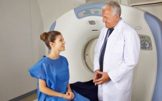
MRI, which allows you to evaluate liver function, can be performed on both an open and closed type tomograph. Most often, diagnosis is made on the last type of examination, represented by a tunnel into which a table with a patient enters. However, it has a drawback, which is associated with the long-term presence of the subject in a closed space. This often causes discomfort.
An open examination takes place in a room that looks like an X-ray room. During diagnosis, it is possible to approach the patient and talk, which is important for studying elderly people and children. The duration of the procedure varies from 30 minutes to 1 hour. Under some circumstances, the time may take up to 1.5 hours.
An important condition that improves the quality of the study is the exclusion of mobility throughout the entire examination. Because even minimal movement reduces the quality of pictures.
To make the patient comfortable in a closed-type device, there is built-in ventilation and lighting. Usually the diagnosis does not cause any discomfort. However, it happens that the patient feels slight stiffness as a result of prolonged lack of movement, tingling in the catheter area. This discomfort usually goes away quickly after the procedure.
It will take about an hour for the attending physician or specialist conducting the examination to process the results and draw up a conclusion. If there are any difficult issues, then other specialists are involved in decoding, and the processing time increases accordingly. In this case, the results are delivered to the patient every other day or sent to an email address.
Indications for the study
Magnetic resonance imaging techniques will allow us to study the structure of tissues, identify affected lesions, their exact location, size, and character. The doctor also receives information about the condition of the biliary tract.
This survey allows you to obtain information:
- about the structure of the liver and its size;
- the presence of inflammation, dystrophic changes, tumors, abscesses;
- narrowed bile ducts;
- whether healthy tissue degenerates into adipose tissue;
- are there stones, polyps, cysts in the organ;
- how damaged the liver is;
- is there an anomalous structure;
- is the treatment effective?
- whether the liver is suitable as a transplant.
The doctor who issued the referral is responsible for interpreting the diagnostic results.
MRI of the liver with contrast is recommended for patients who suspect the presence of neoplasms of a malignant or benign nature. This is usually a hemangioma formed from blood vessels.
When a contrast agent is introduced into the blood, tumor processes become most noticeable.
Diagnostics with contrast can detect not only tumors, but also metastases, detect impaired vascular function and narrowed bile ducts.
This study shows:
- with constant pain in the right hypochondrium;
- the incomprehensible nature of the febrile state;
- any malignant neoplasms to assess the presence of metastases;
- enlarged liver;
- an increase in “liver” tests in the blood;
- if there is a diagnosis of liver cirrhosis.
Advice: it is better to examine the pancreas at the same time as the liver, since these organs are closely related to each other and are simultaneously affected by pathological processes.
Diagnostics with contrast
The dye is injected into a vein and distributed throughout the body. It soon reaches the organ being examined. Today, there are different forms of contrast administration during MRI. In the first option, a single intravenous injection occurs immediately before the diagnosis itself. The coloring preparation is calculated from the following proportion: 0.2 mg to 1 kg of patient weight.
How does ultrasound differ from MRI?
In the second option, a drop of contrast is introduced, with the help of which you can control the level of the dye directly during the study period.
Most often, this method is used when conducting dynamic studies. The dye allows you to more accurately determine the size of the tumor, its structure, and outline. As a result of its introduction, the liver segments are more clearly detailed.
The contrast does not pose a danger to the patient and is eliminated from the body through the kidneys within 2 days. Based on this, this procedure is not performed on people with kidney failure.
Allergic reactions to the introduction of a dye are quite rare.
However, if there is a suspicion of a predisposition to hypersensitivity, then it is better not to perform this type of examination.
A study with Primovist allows you to identify pathologies in the initial stages
In the modern world, there is a growing trend in the success of surgical interventions to eliminate liver tumors. This happens due to its early recognition. A significant role in this was given to innovation in colorants: “Primovist”. After injection into a vein, the drug quickly reaches the liver and makes it possible to examine not only this organ, but also the bile ducts.
An examination with Primovist allows determining the presence of tumors, whether they are malignant or benign, detecting metastases, distinguishing a primary disease from a secondary one. This examination is the most preferable and takes the quality of diagnosis to a new level.
Contraindications
There are cases when a magnetic tomography examination of the liver is not performed due to the presence of complete contraindications. Which include:
- pregnancy up to 12 weeks;
- installed pacemaker, insulin pump, prostheses;
- tattoos made using metal-containing paint;
- It is impossible to conduct an examination with a traditional tomograph if the patient weighs more than 120 kg.
MRI in the 2nd and 3rd trimester of pregnancy does not pose a risk to the fetus.
Examination with contrast is not carried out in case of pregnancy or if there is suspicion of pregnancy, hypersensitivity to the components of the administered drug, or while the child is breastfeeding.
In the latter case, it is recommended not to breastfeed the baby for 2 days after the study. Absolute prohibitions include renal failure and the need for hemodialysis.
Contraindications are determined individually
Relative contraindications for liver tomography include:
- psychical deviations;
- hyper-reactivity;
- severe stage of claustrophobia;
- poor health of the patient.
If there are relative contraindications to the study, you should talk with a doctor who will help stabilize the patient’s condition and conduct a diagnosis. Usually, in cases of mental illness, the patient is prescribed sedatives.
How to prepare for research
Before undergoing the examination, it is important to prepare for it. Preparation for liver diagnostics begins 3 days before the MRI. Therefore, before the procedure, you should visit a doctor and strictly follow all his recommendations, only in this way is it possible to obtain accurate results.
Important! Before an MRI, you must exclude any sweets, baked goods, vegetables, fruits, and cereals for 3 days. It is not allowed to consume fermented milk products or fiber in any form 1 day before the event.
Before the test, you should not eat food for 6 hours and water for 4 hours.
To avoid an excess of gases, it is recommended to take activated carbon on the day of the examination at the rate of 1 tablet per 5 kg of the subject’s weight.
Before carrying out diagnostics using a dye, you should drink an antispasmodic. If liver tomography is prescribed for women, then it is important to come to the procedure without decorative cosmetics or hairspray.
Before an MRI, you should completely avoid eating fatty and floury foods.
What test is an alternative to MRI?
Alternative methods for examining the liver include ultrasound or CT. However, these methods do not provide complete information about the examined organ and do not allow identifying pathology at the very beginning of its manifestation.
In addition, when performing a computed tomography scan, the patient is exposed to x-rays. Of course, the dose of radiation exposure is lower than with standard radiographic examination.
If the patient has a neoplasm of an oncological nature, then this effect will be extremely undesirable.
Therefore, you do not need to prescribe a diagnostic method for yourself; it is important to follow all the recommendations of the attending physician, who, before referring you for the procedure, weighs the advantages of a particular method and its risks, and gives preference to the safest diagnosis. MRI of the biliary tract and liver has a number of advantages, which include:
- high accuracy of the result, allowing you to make a diagnosis;
- painlessness;
- no exposure to x-rays.
The only disadvantage of this scan is its high price. However, the high efficiency of this method completely justifies this drawback. In addition, the price factor is influenced by the location of the study.
For example, the usual price for an MRI in Moscow is about 7-10 thousand rubles. In the capital, MRI, carried out on Miklouho-Maclay Street, is used not only by state-run clinics, but also by private ones.
MRI of the biliary tract and liver is the most effective technique that allows you to identify pathology at the initial stage and begin treatment in a timely manner.
Source: https://apkhleb.ru/mrt/pecheni
MRI of the liver
- Magnetic resonance imaging (MRI) of the liver is a method of non-invasive examination of the gland and its ducts, which is based on a change in the polarity of hydrogen atoms upon contact with radio waves and a constant magnetic field.
- When scanning, accurate data is obtained, since photographs of given sections with a minimum step make it possible to construct a three-dimensional model of the organ, but due to the high cost of the study, MRI is prescribed after simpler diagnostic procedures (ultrasound), when there is a need to supplement and clarify the data.
- During an ultrasound examination, a specialist receives a low-resolution image, but on it one can still see the inflammatory process and neoplasms, and it is possible to assess the general condition of the organ, its size and morphology.
MRI allows one to judge the type of formation and structure of the parenchyma; with its help, one can distinguish between oncology, hemangioma, fatty degeneration, and find out what is inside the cyst. The main advantages of ultrasound are its availability and the safety of the procedure when carrying a child, and the use of MRI of the liver during pregnancy is limited.
When is an MRI of the liver prescribed?
A hepatologist, gastroenterologist, or oncologist can refer you for a diagnostic procedure. MRI is performed only after ultrasound of the liver and laboratory tests to identify or confirm pathology. A specialist may suspect diseases of the hepatobiliary system if the patient complains of pain in the right hypochondrium, dyspeptic and asthenovegetative syndrome.
During the initial examination, the doctor discovers an enlarged and thickened liver, yellowing of the skin and sclera, and spider veins on the body. Patients often wonder what an MRI of the liver shows and how the procedure goes. The indication for the procedure is diffuse damage to the organ.
Scans are performed to determine the properties of intrahepatic formations, namely, to find out whether the cells are malignant and benign tumors that are inside the cystic cavities. MRI can also identify focal lesions. In addition to the parenchyma of the gland, vessels are visible on the images and it is possible to assess their patency.
Magnetic resonance imaging of the liver is used in the diagnosis of many diseases of the hepatobiliary system
The study is prescribed if it is necessary to clarify the results of other diagnostic procedures, as well as after surgical removal of the tumor. The scan will show congenital abnormalities in the development of the liver and its ducts. In the pictures you can see hepatosis, congenital cyst, dystopia, duplication and other malformations.
Tumors that have formed in other organs often metastasize to the liver, so MRI is prescribed to identify or exclude them. Due to acquired or congenital disorders, iron can accumulate in the gland, which negatively affects hepatocytes. Elevated levels of the trace element can be detected during MRI of the liver.
An examination is necessary if a gland transplant is being prepared, since there is a need for a complete assessment of its condition and structure.
The examination is prescribed to examine the biliary tract and diagnose diseases associated with a decrease in their diameter up to its complete occlusion. Scans in a specific situation help determine the cause of a patient's symptoms.
In the images, you can distinguish choledocholithiasis, or a tumor in the bile ducts, which causes motility disorders. MRI is used to detect congenital anomalies and inflammation of the bile ducts. Cholangiography allows you to refute or confirm the presence of a malignant formation in the pancreas, pseudocysts and other malformations.
Tomography is performed after liver surgery to exclude iatrogenic changes. A scan of the liver and gallbladder is often required at the same time because the structures are anatomically related.
Thus, MRI of the gland is prescribed:
- to determine congenital disorders of the hepatobiliary system;
- to detect neoplasms, cysts, stones, iron accumulations;
- to assess the condition of the organ parenchyma, how the structure differs from the norm;
- in the postoperative period to monitor the course of the disease and detect iatrogenic changes;
- to search for metastases when an extrahepatic tumor is detected (if there is cancer, then there is a high probability that it will metastasize to the liver);
- to assess the condition of the bile ducts.
MRI has a number of advantages over other diagnostic procedures, although it is not prescribed first.
In the photographs, a specialist will be able to distinguish:
What tests do I need to take to check my liver?
- hemangiomas (benign formations). Unlike malignant tumors, the contrast agent is washed out from the periphery of the hemangioma;
- nodular focal hyperplasia (benign tumor). In the picture it looks like a formation with clear and smooth edges, a pronounced capsule;
- hemosiderosis (iron accumulation). In the image, the organ is hypointense, since iron is ferromagnetic;
- changes characteristic of cirrhosis. Shows altered size of the gland, diffuse hypointense foci, congestion and increased pressure in the portal vein;
- abscesses. They look like rounded formations with fuzzy edges, the intensity of coloring depends on the stage of the process;
- cysts. In the image, the formation has clear edges, echogenicity depends on the type.
Most often, MRI of the liver with contrast is prescribed, since the use of a contrast agent makes it possible to differentiate between benign and malignant formations. Gadolinium passes through the vessels and, under the influence of a magnetic field, even creates capillaries.
The intensity of blood supply is increased in tumor tissue and decreases in areas of parenchyma death (cirrhosis, cyst, abscess). For MRI of the liver, contrasting with Primovist (a drug containing gadolinium) can be performed, which allows one to obtain additional data on the structure and classification of focal lesions of the gland, thereby increasing the accuracy of diagnosis.
Contraindications for carrying out
Magnetic resonance imaging is contraindicated in patients who have any metal structures in their body. A person with a pacemaker, a hearing aid, or metal fragments that remained after an injury should not be in a magnetic field. These items can fail or become dislodged, causing a number of difficulties.
The presence of dental implants is not a contraindication to MRI examination of the liver. Even if a person has prostheses in his body, they can be MR-compatible (you need to re-read the implant passport), since they consist of titanium, so the doctor determines contraindications on an individual basis.
Complex electronic devices, such as hearing aids, may malfunction in the tomograph.
Scanning is not recommended during pregnancy, especially during the first trimester, but if the risks to the woman’s health exceed the risks to the fetus, then an MRI is performed. The examination is not prescribed if the patient is in a general serious condition, since during the procedure it is not possible to monitor the patient’s vital signs.
Contrast-enhanced tomography is contraindicated in some patients. Such limitations include renal failure, since the contrast is excreted from the body by the kidneys. Gadolinium and its compounds should not be administered if the patient has a confirmed allergic reaction to the substance.
MRI with contrast is prescribed with caution to people who have a history of severe allergic manifestations; in this case, pre-medicated hyposensitization is carried out. Breastfeeding is a relative contraindication, since the contrast is completely eliminated from the body within two days.
If the patient has epilepsy, scanning is possible under general anesthesia. Under sedatives, people with mental disorders, hyperreactivity, and young children who are not able to remain in one position for a long time can be examined.
How to prepare for a tomography
Before magnetic resonance imaging is performed, it is determined whether the patient has an allergic reaction to the contrast agent, and it is checked whether the excretory function is normal, since the vast majority of the study is performed with contrast.
Preparation for the examination includes dietary recommendations. Two days before the procedure, the patient must remove from the diet any foods that cause increased gas formation. This includes black bread, legumes, cabbage, and dairy products.
Preparation for MRI of the liver includes collecting anamnesis, determining tolerance of the contrast agent, studying kidney function
The study should be carried out on an empty stomach, at least 4-6 hours after the last meal. 1-2 hours before the scheduled procedure, it is recommended to drink a remedy that eliminates smooth muscle spasm (No-Shpu, Drotaverine) to improve visualization of the biliary tract.
Before going into the tomograph, the patient must get rid of any metal objects (rings, earrings, chains, piercings), so it is better to prepare in advance and leave all valuables at home.
Scanning technique
Since MRI of the liver is usually done with contrast, before the patient is placed in the tomograph, a catheter is installed for intravenous administration of contrast. To prevent thrombosis, saline solution is injected dripwise. The tomograph looks like a large tube into which the person being examined is placed on a special gurney.
The tomograph can be of open or closed type. An open camera produces images that are inferior in quality, but it is better suited for people with large body weights or those who cannot stand confined spaces. The examination lasts about half an hour, but can be extended to 90 minutes, and it is often psychologically difficult for people to remain in a closed tomograph for so long.
A closed device is suitable for people whose body weight is less than 150 kg and whose abdominal volume does not exceed 150 cm. During operation, the device makes loud sounds, which can cause some discomfort, so patients are given earplugs or headphones.
When a contrast agent is administered, the patient may feel hot in the body, dry mouth, and dizziness. These symptoms do not require medical attention and go away on their own within a few minutes.
During the examination, communication between the doctor and patients is carried out using two-way communication.
The subject can inform the doctor about changes in his condition, and the doctor informs when it is necessary to hold his breath in order to obtain the best quality images.
Most of the time it is necessary to lie still and not breathe, which is the most difficult thing for patients.
Often during the procedure, tingling occurs in the catheter area or limbs swell due to uncomfortable body position.
The patient receives the results of the examination a couple of hours after the procedure, he is given printed images and a radiologist’s report, and if necessary, the data is copied to an electronic medium.
Complications that may arise during diagnosis are usually associated with an allergic reaction to the contrast agent, chronic renal failure, or the presence of metal objects in the patient’s body.
Magnetic resonance imaging is one of the most informative diagnostic methods. MRI of the liver is used to obtain data on the homogeneity and density of the liver parenchyma, to examine the vessels and bile ducts. Using this study, it is possible to detect space-occupying formations of various natures, inflammation, atrophy, and structural changes.
In difficult cases, a consultation can be assembled to decipher the results, then the conclusion is issued the next day or sent by email
MRI is used to confirm developmental abnormalities, the presence of stones in the gland and its ducts, as well as to assess the general condition of the organ. Due to the high accuracy, MRI of the liver is indicated in the postoperative period, since it allows one to determine the effectiveness of therapy, identify possible consequences associated with the surgical technique, and detect metastases.
Unlike ultrasound, scanning can detect even small tumors and differentiate between benign and malignant tumors. MRI has an advantage over CT, since the images are more detailed and there is no radiation exposure. The scan is completely harmless, so it can be done on a child.
Source: https://vrbiz.ru/diagnostika/mrt-pecheni
Why MRI is rarely used to study the liver and what you need to know if you are referred to it
The liver is an unpaired organ of the abdominal cavity with high functional activity and the ability to regenerate. Disorders of the liver and the closely related biliary tract system often require in-depth examination in the form of magnetic resonance imaging (MRI).
This technique is based on the ability of the nuclei of hydrogen atoms to change their configuration under the influence of a magnetic field. The energy released during this reaction serves as the source of the image on the MRI machine screen.
However, despite the safety and high information content of the technique, it is not always used to diagnose liver pathology.
Briefly about the structure and functions of the liver
The liver is an exocrine gland that produces bile. Bile flows through the ducts into the gallbladder, from which it is released into the small intestine and is involved in the digestion of food.
The liver consists of the right and left lobes and is conventionally divided into 8 segments with its own system of arteries, veins, and nerves.
The elementary structure of the gland is the hepatic lobule, which includes functionally active cells, vessels, and bile capillaries.
The functions of the liver in the body are multifaceted:
- digestive – production of bile;
- detoxification – neutralization of aggressive substances;
- energy – glucose formation, glycogen storage;
- metabolic – storage of vitamins and minerals, regulation of the metabolism of fats, carbohydrates, proteins;
- protein synthetic – the formation of proteins involved in blood clotting and activation of biochemical reactions;
- hormonal – synthesis of insulin-like biologically active substances.
In addition, the liver is a depot of blood, which in emergency situations enters the general vascular bed and prevents complications after massive blood loss.
Indications and contraindications for MRI
MRI of the liver shows changes in its structure with high accuracy, which makes it possible to identify almost any pathology of the organ. However, in many cases, the use of this technique is considered inappropriate, since ultrasound is not much inferior to MRI without contrast.
Indications for liver MRI:
- neoplasms of the gland;
- cysts;
- abscesses;
- anomalies in the structure of the organ;
- changes in the bile ducts;
- planned liver surgery;
- impossibility of performing computed tomography with contrast for cirrhosis, abscesses, cysts or other liver pathologies;
- non-tumor diseases of the liver or gall bladder.
The latter indication is considered relative, since ultrasound can successfully detect diseases such as hepatitis, cirrhosis, echino- or alveococcosis, cholecystitis, and cholelithiasis.
Contraindications to magnetic resonance examination are mainly related to the peculiarities of the operation of tomographs. An absolute ban on performing MRI is imposed if the body has:
- pacemaker;
- ferromagnetic prostheses, implants, fragments;
- clipped vessels to stop bleeding;
- metal Ilizarov apparatus;
- transplanted kidney.
MRI of the liver with contrast injection is contraindicated in:
- severe renal failure;
- pregnancy;
- individual intolerance to contrast;
- hemolytic anemia.
There are a number of other conditions that impose restrictions on the performance of magnetic resonance imaging. These are relative contraindications:
- non-ferromagnetic implants and prostheses;
- insulin pumps;
- I trimester of pregnancy;
- breast-feeding;
- severe heart failure;
- claustrophobia;
- mental or motor agitation (this is especially true for examining children);
- episyndrome;
- tattoos with metal particles in the dye;
- overweight (more than 120–200 kg).
The presence of non-ferromagnetic objects in the body is not a strict contraindication for the procedure, but can significantly reduce its accuracy.
Which doctor should I contact for a prescription?
Magnetic resonance imaging of the biliary tract and liver is usually prescribed by therapists or surgeons, sometimes by doctors of narrower specialties: gastroenterologists, hepatologists, infectious disease specialists, oncologists.
It all depends on what problem the patient comes to the doctor with, what disease the specialist suspects.
For example, an oncologist prescribes an examination to identify a tumor formation and clarify its nature, and an infectious disease specialist may recommend an MRI in case of severe hepatitis or liver parasitosis, when ultrasound does not provide complete information about the disease.
How to properly prepare for research
A planned MRI of the liver requires special preparation for the examination:
- For 3 days before diagnosis, a diet is recommended that excludes dishes that increase gas formation in the digestive tract: white cabbage, legumes, fresh fruits and vegetables, baked goods, spices, carbonated drinks. If you cannot cope with flatulence with diet alone, then doctors prescribe medications: Espumisan, Smecta, Pancreatin and others. This is necessary so that intestinal loops distended with gas do not interfere with visualization.
- 5-6 hours before the MRI, you should not eat anything or drink large amounts of liquid. It is allowed to drink only still water in small quantities. Fasting is required to give the gallbladder time to fill with bile, then its examination will be more informative.
- Before the examination, you are not allowed to smoke or drink alcohol, drugs or stimulants.
- If the patient suffers from claustrophobia, then doctors advise one day before the MRI to start taking light sedatives: Novo-passit, Persen, Tenoten, valerian infusion. Only a doctor can prescribe more serious medications. It is also important to prepare mentally: keep in mind that the procedure is harmless, the device has a ventilation system, and you can contact the doctor at any time and ask to stop the procedure.
- When going for an MRI, be sure to take with you a referral, an outpatient card with the results of the studies, and a medical insurance policy. Wear comfortable clothing without metal elements and a minimum of metal accessories. Decorative cosmetics containing metal particles will need to be washed off before the procedure. If you have implanted implants and other structures, then take with you a passport for them or a discharge summary from the hospital, which indicates the type of implant and its composition.
- Immediately before the examination, the patient removes all jewelry, removable dentures and other metal objects, and also leaves magnetic media in a special container: cell phones, plastic cards with magnetic tape, mp3 players.
Preparation for an MRI of the liver with contrast is practically no different from the standard one. The only important point: if you have chronic kidney disease, you will first need to take a blood test to determine their function and take the result with you.
What will happen in the office - tomography technique
From a special room in which preparations are carried out and all unnecessary things are left, the patient goes directly to the room with the MRI machine.
Here the nurse helps him lie down correctly on the tomograph bed, explains how he should behave during the procedure and shows how to contact the doctor.
Usually for this purpose there is a special intercom or remote control with a “panic button”, which the patient presses if he feels unwell.
It is important to make yourself comfortable in the tomograph, as you will have to lie still for at least 20 minutes.
The nurse secures the patient with bolsters, then goes to the doctor in the next office and closes the door. The doctor can observe the patient through the window and communicate using an intercom. At the right moment, he will give the command to “breathe and not breathe.” Then you will need to pause breathing for half a minute.
After turning on the tomograph, the couch with the patient moves inside a closed tube in which the shooting takes place. Keep in mind that the MRI machine makes loud, unpleasant noises during operation, so sometimes the patient is asked to wear headphones or earplugs. There should be no other uncomfortable sensations: only a feeling of slight heating of the area being examined.
You will have to lie in the tomograph for 30 to 60 minutes. If an MRI examination of the liver and biliary tract is carried out with contrast, then after taking regular pictures, the process is interrupted, the doctor injects contrast into the patient’s vein and resumes the procedure. After completing the examination, the couch automatically moves out of the pipe. This means that you can get up and put yourself in order.
In some cases (early childhood, unconsciousness, psychomotor agitation), MRI is performed under general anesthesia. Such interventions require special preparation and are discussed in advance. The report is given to the patient 2 hours after the MRI or the next day.
Application of contrast agent
MRI of the bile ducts and liver with contrast is prescribed in the presence of space-occupying formations or obstructions to the outflow of bile to clarify their nature. The contrast used during MRI diagnostics contains gadolinium. Several gadolinium-based substances have been developed. Primovist is best suited for liver imaging, as it accumulates in larger volumes in liver tissue.
The drug is administered intravenously during the examination. If there is a risk of developing an allergy, then an allergy test is first performed by first introducing a small volume of contrast to assess the body’s reaction to a foreign substance.
The drug is excreted by the intestines and kidneys, so renal failure is a contraindication for contrast.
It is also not given to pregnant women, since there have not been enough clinical studies on the effects of gadolinium on the fetus.
MRI images show well-perfused areas that accumulate contrast, as well as areas with destroyed liver cells. This allows us to make a conclusion about the characteristics of the neoplasm (malignant or benign tumor, metastasis).
Interpretation of the liver tomography results, what the study will show
The conclusion of an MRI study of the liver and biliary tract contains the following information:
- scanning mode (T1-, T2-weighted, FLAIR, DWI);
- projections, sections in photographs;
- presence of contrast, name and volume of contrast agent;
- size, shape of organs;
- tissue structure;
- the presence of pathological formations, their localization;
- features of the vessels and bile ducts of the liver.
Normally, on MRI images, the liver looks like an organ with a homogeneous structure without additional inclusions, normal size and shape, without dilatation of the bile ducts. The gallbladder has homogeneous contents without stones, its walls are not thickened, and the size is average. All visible bile and blood vessels are without pathological dilations or narrowings, the lymph nodes in the porta hepatis are not enlarged.
If there are diseases of the liver and biliary tract, MRI will show abnormalities in their structure.
With cirrhosis, the organ is enlarged or reduced in size, with tuberous contours, dilated vessels of the portal vein system.
The cyst looks like a round formation of relatively regular shape, filled with liquid that does not absorb contrast. Parasitic cysts additionally contain septa and chambers in the cavity.
Tumors typically accumulate contrast material and have different intensities in different imaging modalities. Metastases appear as multiple irregularly shaped foci that do not absorb contrast. With fatty hepatosis, the liver tissue becomes too dense, and foci of growth of adipose tissue are visible.
Alternative diagnostic methods
The main “rivals” of MRI are ultrasound and computed tomography (CT). Ultrasound benefits from high information content and greater accessibility.
As a rule, it allows you to obtain a similar result with less financial and time costs. That is why ultrasound is prescribed for the primary diagnosis of the disease or simply as a preventive examination.
Based on the results obtained, the question of whether an MRI or CT with or without contrast enhancement is necessary is decided.
If ultrasound reveals signs of cirrhosis, hepatitis, fatty hepatosis, cysts, or stones in the gallbladder cavity, then additional diagnostics, as a rule, are not required. Of course, CT and MRI provide a more accurate picture of the disease, but this does not affect the tactics of treatment and further management of the patient.
If an ultrasound reveals a space-occupying lesion of an unspecified nature, further examination is prescribed in the form of an MRI or CT scan with contrast. They will help the doctor understand whether the tumor is benign or malignant. The information content of these methods in this situation is equivalent.
If an ultrasound of the liver indicates the presence of fluid formations (cysts, abscesses) or possible metastases, then a CT scan of the liver with contrast enhancement will be more informative.
It is important to understand that instrumental techniques are not always able to provide complete information about the condition of the biliary tract and hepatic structures. In addition to them, a biochemical blood test is required to assess the function of the organ.
Summarizing
If liver and biliary tract disease is suspected, diagnosis using MRI is usually carried out only after ultrasound examination. MRI with contrast injection is in great demand.
This technique allows you to obtain the most complete information about pathological formations identified in the liver.
MRI is considered a safer technique compared to CT, so it is often prescribed when the latter is not possible.
Source: https://diagme.ru/mrt/bryushnaya-i-grudnaya-polost/pecheni
MRI of the liver
Magnetic resonance imaging plays a special role in the diagnosis of liver diseases. The sensitivity of MRI in detecting focal lesions is currently not inferior to computed tomography.
The undoubted advantage of the method is its very high specificity.
Even without the introduction of a contrast agent, good diagnostic results are achieved in identifying abscesses, cysts, hemangiomas and nodular hyperplasia.
The liver is an organ that ensures metabolic processes in the body. With its participation, the transformation of some substances into others occurs. Its other important function is barrier, which is to neutralize toxic substances that enter the body.
Due to its barrier function, the liver is subjected to great stress, which often leads to various liver diseases. The most commonly diagnosed:
- organ abscesses;
- cirrhotic changes;
- neoplasms of benign and malignant nature;
- dystrophic changes;
- hepatitis of viral and other nature;
- fibrosis - replacement of healthy cells with connective tissue and others.
Indications for the study
A referral for a liver scan using MRI is usually given by a hepatologist or gastroenterologist. The reasons for referral for tomography may be symptoms of organ pathology: complaints of pain in the right hypochondrium, loss of appetite, yellowing of the whites of the eyes, darkening of urine, etc.
Also, a study may be prescribed in the following cases:
- if a malignant neoplasm is suspected in the tissues of the liver or organs closest to it;
- hepatomegaly (an increase in the size of an organ) is diagnosed, the cause of which is illness or if the cause is unknown;
- to clarify data from other diagnostic methods;
- there is reason to suspect that stones or salt deposits have formed in the organ, interfering with the flow of normal processes;
- to clarify the etiology of hepatitis and the consequences of this disease for the organ;
- liver cancer has been previously diagnosed and there is a threat of metastatic damage to nearby organs;
- assessing the effectiveness of treatment, for example, after chemotherapy or surgery;
- assessment of the condition of the liver in cirrhosis, etc.
MRI not only diagnoses diseases, but also monitors the progress of treatment.
What does an MRI of the liver show?
Magnetic resonance imaging is a study that evaluates not only the structure of the liver, but also the bloodstream and bile ducts. A comprehensive assessment allows doctors to most accurately diagnose the disease and draw conclusions about its causes in order to fully provide treatment.
An MRI will show diseases such as:
- hemangiomas ; These are small benign neoplasms that appear as hypointense lesions on imaging. When contrasting, the contrast agent will be washed out from the periphery, this will allow differentiation from malignant neoplasms.
- nodular focal hyperplasia ; The second most common benign tumor of the liver. In the photographs it looks like a single or multiple formation, having a pronounced capsule with clear and even contours.
- hemosiderosis ; The disease is associated with excessive accumulation of iron in the liver tissues. In the images, the organ will be hypointense, since iron is ferromagnetic.
- cirrhotic changes ; Cirrhosis is a fatal, incurable disease that can be detected in the early stages using MRI and thereby significantly prolong life expectancy. Magnetic resonance imaging will show that the lobe of the liver on the right is reduced compared to normal, and the caudate lobe and lateral segment, on the contrary, are enlarged. Diffuse hypointense foci will be visible in the structure of the parenchyma. MRI for cirrhosis also allows you to assess the condition of the portal vein, which is also affected in this disease. From the photographs one can judge the presence of stagnation in it and an increase in pressure.
- abscesses ; An abscess on MRI will look like a round formation, the echogenicity (intensity of staining on the image) of which may vary depending on the stage of the process. The outlines are usually unclear.
- cysts and others; Cysts on MRI of the liver are visible as formations with clear contours. Echogenicity may also vary depending on the type of cyst.
When is an MRI of the liver with contrast performed?
MRI with contrast is prescribed to patients who have a tumor in the liver, but for some reason it is not possible to accurately determine its nature. Using contrast, it is possible to differentiate malignant from benign tumors with almost 100% accuracy.
Another indication for MRI of the liver with contrast is the presence of any symptoms of organ damage in combination with the lack of objective data from other previously performed studies. Since the method is highly accurate, it can be used to detect even very small tumors or minor changes in the organ.
Contrasting allows you to establish the most accurate diagnosis, and the effectiveness of treatment depends on this!
Preparation
An MRI of the liver is a procedure that usually does not require complex preparation. 24 hours before the test, gas-forming products are excluded from the patient’s diet. It is not recommended to eat food at all for 2-3 hours immediately before the test.
Order of conduct
The patient, upon entering the office, removes all metal objects and lies down on the tomography table, which is placed inside the device.
During the study, a person does not experience any discomfort while lying quietly in the device for half an hour. In some cases, an attack of claustrophobia may occur, but you can combat it by talking to a doctor using a special microphone built into the device.
Sometimes the study is extended to 1-1.5 hours if it is necessary to obtain more accurate information. It is important to remain still during this entire time so that the pictures are of high quality.
If MRI is performed with contrast, then before the procedure a contrast agent is injected into the patient’s vein, which is one of the stages of preparation for the study.
Advantages of the method
The main advantage of MRI of the liver is that the procedure is completely safe for the human body and is highly informative. The study is prescribed not only to children at any age, but also to pregnant women, which indicates the high safety of magnetic resonance imaging. During the diagnosis, the patient is not exposed to X-rays or other radiation harmful to the body.
Which is better MRI or CT scan of the liver?
One of the alternative diagnostic methods is computed tomography. With CT, the patient’s body is exposed to X-rays, which is why its use is not always possible in the early stages of the disease, so as not to provoke its progression.
CT is also a less informative diagnostic method, despite the fact that it is often performed with contrast. Computed tomography provides the most accurate data for large lesions of the organ. It is prescribed for the following diseases:
- a strong increase in organ volume and mass;
- massive damage to liver tissue due to cirrhotic changes;
- neoplasms were discovered;
- there is a constant and prolonged feeling of discomfort or pain in the liver area.
Which is better: MRI of the liver or ultrasound?
Ultrasound is one of the routine examinations that are prescribed to all patients with abdominal pathologies. If the data obtained after an ultrasound is sufficient for the doctor to make a diagnosis, he will limit himself only to this diagnostic study. If there is too little information to make a diagnosis, CT and MRI may be additionally performed to assess the condition of the liver.
It is not practical to compare the effectiveness of ultrasound and MRI, since MRI is never prescribed as a first-level study, unlike ultrasound.
Ultrasound diagnostics will help the doctor, saving the patient’s money, to diagnose simple diseases or, if the patient suffers from claustrophobia, to obtain at least some data for making a diagnosis. MRI, in turn, will make it possible to clarify the diagnosis in doubtful cases and determine the presence of a neoplasm and its nature.
MRI of the liver is an effective, but still quite expensive, examination method, prescribed to patients in controversial cases when the diagnosis is not entirely clear. The technique has a high resolution ability, which allows it to be used for diagnosing oncology in the early stages.
This article contains general information only, is not scientific material and should not be construed as a substitute for medical advice.
Source: https://mrt-kt.info/vidy-mrt/bryushnoy-polosti/pecheni/
MRI of the liver: pros and cons
The World Health Organization has ranked liver cancer as the eighth leading cause of death. The insidiousness of serious liver diseases, including cancer and cirrhosis, is that they often develop asymptomatically. A person may find out about his illness by accident or when it is too late.
What to do to prevent liver pain? What are the symptoms of liver disease? How is liver disease diagnosed and what role does MRI play in it? We addressed these and other questions to the executive director and chief physician of MRT Expert Lipetsk LLC, Oksana Egorovna Volkova.
— How do liver diseases begin?
As a rule, it all starts with an inflammatory process in the liver, the trigger for which can be a variety of reasons, including hepatitis C or B, alcohol abuse, congenital diseases, poor diet and a number of other factors.
Changes in the liver occur in several stages. It all starts with steatosis, in which an increased content of fat cells accumulates in the liver. Steatosis and can lead to hepatitis (inflammation of liver tissue). Then follows the stage of fibrosis - the liver is “overgrown” with connective tissue. If treatment is not applied at this stage, the patient develops cirrhosis of the liver, an irreversible disease.
— What symptoms does the liver signal about its troubles?
The liver is a very large and extremely responsible organ of the digestive system; it is not for nothing that ancient healers called it “the seat of the human soul.” About 20 million chemical reactions occur in the liver every minute.
It has a huge number of different functions, up to five hundred. Among the latter are the synthesis of protein and bile acid, the accumulation and breakdown of glucose, and the neutralization of toxins dangerous to humans.
Therefore, when the liver is damaged, the entire body suffers.
One of the signs of liver disease is suddenly appearing bruises on the patient’s body. Moreover, the origin of these bruises is not related to external damaging factors.
Other symptoms include bitterness in the mouth in the morning, yellowing of the tongue, sclera of the eyes and/or skin, red palms (palmar erythema), bad breath, itching, digestive problems (decreased appetite, diarrhea or constipation, nausea, discoloration of stool, dark urine).
Since the liver itself cannot hurt due to the absence of pain receptors in this organ, discomfort and pain in the right hypochondrium may also indicate liver disease. Such a pain syndrome most likely indicates that the liver is enlarged, which entails stretching of the capsule in which it is located.
— Oksana Egorovna, tell us about the methods for diagnosing the liver
With an ultrasound, you can’t find out everything about the liver, but a lot – size, tissue structure, condition of blood vessels, i.e. everything that concerns physiological changes in the organ. A biochemical blood test will tell you about functional liver disorders.
Other liver diagnostic methods include positron emission tomography (PET) and magnetic resonance imaging.
The PET method is advanced in nuclear medicine. It allows you to identify various neoplasms in organs with a high degree of probability. Before diagnosis, the patient is given an intravenous radiopharmaceutical that is quickly distributed throughout the body. During the scan, cancer cells will reveal themselves with a bright glow.
As for MRI of the liver, this diagnosis allows you to confirm what was revealed during an ultrasound examination, as well as to find liver pathology that was not detected during an ultrasound examination.
I would like to note that recently patients who are prescribed magnetic resonance imaging of the liver as an independent diagnosis, as an alternative to ultrasound, are increasingly coming to the MRI Expert centers.
And such doctor’s prescriptions are justified.
So, not so long ago, at the MRI Expert Lipetsk center, a case occurred when a patient, who was diagnosed with a liver tumor during an ultrasound examination, left us absolutely healthy and happy.
MRI diagnostics allowed us to refute the diagnosis. Why did it happen? One of the intestinal loops was mistaken by the ultrasound doctor for a tumor in the patient's liver.
This happened because ultrasound, unlike MRI, does not allow you to “decompose” organs into layers.
— What diseases can MRI of the liver detect?
During MRI of the liver, we see an extremely wide range of pathologies: benign and malignant formations, even the smallest (from 2-3 mm), hemangiomas, liver adenomas, hyperplasia, congestion in portal hypertension, traumatic and cirrhotic changes, parasitic diseases (among including echinococcosis, alveococcosis), hemochromatosis (pigmentary cirrhosis), as well as diseases of the gallbladder and its ducts. MRI also visualizes secondary metastatic changes in the liver well.
MRI can reveal traces of fluid around the liver, potentially indicating inflammatory diseases such as cholangitis or hepatitis. If fluid is visible throughout the abdominal cavity, then a conclusion is drawn about the presence of ascites, indicating primary liver cancer or its secondary changes in the form of metastases from other organs. Ascites also occurs against the background of cardiovascular pathology.
As a rule, having seen traces of fluid during an MRI of the liver, the radiologist will send the patient for the necessary additional examinations.
— Is MRI of the liver performed with or without contrast?
An MRI of the liver without contrast is not very informative. Any liver formations require confirmation, for which dynamic contrast is performed. Each of the formations accumulates contrast specifically, which makes it possible to differentiate hemangioma, adenoma, focal nodular hyperplasia, metastases, cysts and other pathologies.
— Is special preparation required before performing an MRI of the liver?
Yes, preparation is necessary. You should not eat food 4 hours before the test.
Since rare patients come for diagnostics only with an MRI of the liver, and not of the entire abdominal cavity, in order to reduce intestinal motility, it is recommended to take a couple of No-Spa tablets and 2-3 Espumisan tablets an hour before the examination.
— Is MRI of the liver a separate study or part of a comprehensive diagnosis of MRI of the abdominal cavity?
Sometimes an MRI of the liver is performed as a separate diagnosis. But since the liver affects the functioning of other organs, it is highly advisable to conduct an examination of the entire abdominal cavity. Thus, in medical practice, cholecystogenic pancreatitis often occurs against the background of disruption of the bile-excretory system.
— If liver disease has already been identified, how often is it necessary to undergo an MRI for dynamic monitoring?
This depends on the identified pathology, therefore, in the conclusion, MRI Expert diagnostic doctors write recommendations, including the date for repeating the examination. As a rule, dynamics are necessary after 3 or 6 months, less often - after a year.
— How are liver diseases treated?
Until recently, patients with serious liver disease had two options: either die or wait for a transplant. Now everything has changed. There are new generation drugs.
But surgical treatment methods are also widely used. The liver is a very regenerative organ, capable of restoring the volume of resected tissue 1.5-2 months after resection.
Functionally, it takes a little longer to recover.
But it is still important to remember that if changes in the liver are detected in time, at least at the stage of fibrosis, then the chances of returning to health are high. However, even with cirrhosis you can live a long time.
Take care of your liver and be healthy!
Source: https://www.mrtexpert.ru/articles/384