A tumor of the inner layer of the uterus of a malignant type has unexpressed symptoms, so a comprehensive diagnosis is carried out to determine the diagnosis.
Modern ultrasound examination makes it possible to obtain data on the characteristics of the development of a neoplasm in order to select the most effective treatment regimen.
Signs of endometrial cancer on ultrasound have characteristic features that, unfortunately, not every specialist can notice. To determine the correct diagnosis, a lengthy examination by an experienced diagnostician is necessary.
Diagnosing uterine tumors can be difficult
How is the procedure performed?
Endometrial cancer is detected by ultrasound by two methods of gynecological examination: transabdominal or transvaginal. It is possible to combine both diagnostic methods.
The procedure is performed according to the standard method:
- with transvaginal ultrasound: a hardware sensor with a medical condom and lubricated with a special gel is inserted into the vagina;
- with transabdominal ultrasound: the lower abdomen is lubricated with gel and a sensor is moved over the skin.
At the moment the sensor contacts the surface of the tissue, an image of the genital organs being examined appears on the computer screen. The duration of the entire procedure is on average about 15 minutes. In complex cases, diagnosis may take a little longer.
In private clinics you can undergo 3D ultrasound of the uterine cavity. This method is the most accurate and allows you to obtain the most accurate information about all the characteristics of the tumor.
Small pathological formations are difficult to detect during ultrasound, but a large tumor in the body of the uterus is clearly displayed on the screen. If endometrial cancer is suspected, a comprehensive diagnosis is recommended, thus increasing the chances of identifying even small tumors.
One of the main diagnostic methods is ultrasound
When should you get tested?
Ultrasound diagnostics is prescribed by a gynecologist when a patient detects signs of a cancerous tumor of the endometrial layers in the uterus. The procedure can be carried out both in municipal and private medical institutions according to indications.
Ultrasound of the uterus and nearby organs is recommended for patients with signs of cancer:
- uterine bleeding;
- severe sharp pain in the genital area;
- significant disturbances during the menstrual cycle;
- purulent or mucous vaginal discharge;
- sharp pain in the lumbar region;
- pain during sexual intercourse.
The likelihood of diagnosing uterine cancer on ultrasound at the first and second stages is low.
The difficulty in making a diagnosis lies in the fact that, despite the detection of small tumors, it is difficult to determine exactly the nature of the formation, since no special signs appear on ultrasound in the first and second stages of cancer.
Examination is necessary for abdominal pain
Who is at risk
Regular ultrasound examinations are necessary for women with chronic pathologies of the reproductive system and for patients undergoing treatment for gynecological diseases.
The risk group mainly includes women of childbearing age:
- diagnosed with infertility;
- have had one or more abortions;
- those who have not given birth (over 25 years of age);
- having hormonally active neoplasms on the ovaries;
- those suffering from thyroid dysfunction;
- people with diabetes;
- have undergone hormonal therapy;
- having cystic formations of the genital organs.
Diabetes requires stricter control
Risk factors also include difficult childbirth, prolonged absence of intimacy, early or, conversely, late onset of menopause.
According to statistics, endometrial cancers develop in postmenopausal women in the age group of 50–60 years. But diseases at a younger age also occur.
What are the first signs of cancer
An ultrasound scan is necessary to obtain information about the depth of penetration of cancer cells, the site of formation and size of the tumor, and the possible presence of metastases in the lymph nodes and neighboring organs.
The very first sign of uterine endometrial cancer on ultrasound is a change in the structure and thickness of the surface layer in the organ cavity. Normally, in women after menopause, the thickness of the endometrium is up to 6 mm, and before - from 16 mm. The thinner the endometrial layer in young women, the higher the likelihood of cancer.
A doctor can accurately diagnose cancer using ultrasound data.
Another symptom is tissue hyperplasia. Endometrial hyperplasia on ultrasound is characterized by the display of a layer of uniform thickness with a pronounced clear contour, as well as ruggedness of the displayed contours and heterogeneity of echogenicity.
What does an ultrasound show?
Only an experienced doctor can accurately determine an oncological tumor affecting the endometrium during an ultrasound examination. Symptoms are often so imprecise that differential diagnosis is required to confirm the diagnosis.
In case of uterine cancer, the following picture will be displayed on the monitor screen when using an ultrasound machine for examination:
- presence of a tumor;
- changes in the structure of endometrial tissue;
- location of the tumor;
- characteristic features of the tumor.
If a cancerous process in the endometrium is suspected, other internal genital organs, as well as lymph nodes, must be examined.
- Often, ultrasound reveals cervical cancer, multiple papillomas, cysts and other formations that provoke cancer.
- What you should pay attention to in order not to miss uterine cancer, you will learn about this from this video:
Recommendations from experts
Do not panic when receiving preliminary results of an ultrasound. Ultrasound does not always correctly determine the nature of the developing tumor.
For this reason, even if the doctor makes a preliminary diagnosis of endometrial cancer, the patient is prescribed a number of additional tests, the main of which is a biopsy. Only by confirming the activity of cancer cells in all examination results can a correct diagnosis be determined.
Women over the age of 20, even if there are no complaints about the functionality of the genital system, should undergo an ultrasound examination at least twice a year. Preventive examinations will help not only to identify the disease at an early stage, but also to promptly detect pathologies that provoke endometrial cancer of the uterus.
Did the article help? Rate it (No Ratings Yet) Loading…
Source: https://infouzi.ru/ultrazvukovoe-issledovanie/malyj-taz/rak-jendometrija.html
Endometrial cancer of the uterus
Endometrial cancer (lat. Carcinoma corporis uteri) or, as it is also called, uterine precancer is an oncological disease that affects only women, since the disease originates from endometrial cells - the inner layer of the uterus. The impetus for the formation of this type of pathology is an increase in the level of the hormone estrogen.
According to statistics, about 9% of all diagnosed cancers among women are endometrial cancer. The majority of patients are women who have crossed the 50-year-old threshold. This figure is more than 60%. But even among young women, cases of uterine precancer formation are not uncommon.
Endometrial cancer: what is it?
Estrogen is a hormone produced by the ovaries in the first phase of the menstrual cycle. Thanks to this hormone, the endometrium thickens. This is necessary so that the fertilized egg can attach to the wall of the uterus.
But due to certain factors, estrogens begin to be produced more than normal, which leads to excessive growth of the endometrium and rapid proliferation of endometrial gland cells. This process is called endometrial hyperplasia.
This phenomenon becomes a favorable environment for tumor formation.
In addition to hyperplasia, the presence of polyps in the uterus can lead to endometrial cancer. Polyps most often occur in older women. Endometrial polyps are formed from the lining of the uterus through its proliferation.
Causes
There are a number of factors that can trigger the disease. These include:
- the presence of gynecological diseases that were not detected and treated in time;
- disruptions in the menstrual cycle, as well as its cessation (menopause, postmenopause);
- taking medications containing large amounts of estrogen;
- the disease is hereditary. If anyone in the family suffered from this disease, then there is a high probability that one of the relatives will subsequently suffer from uterine precancer;
- prolonged absence of sexual intercourse;
- having more than one sexual partner and changing them frequently;
- repeated artificial termination of pregnancy;
- early onset of the monthly cycle (under 12 years);
- late pregnancy and childbirth. Especially if a woman carries her first child after thirty years;
- the presence of obesity, which subsequently leads to diabetes;
- alcohol abuse and long-term smoking;
- immunosuppression for one reason or another;
- ignoring visits to the gynecologist for preventive purposes;
- frequent stress;
- Strict dietary restrictions, that is, all kinds of diets, can lead to decreased immunity and hormonal imbalances in the body.
There may be several reasons for the formation of the disease, but one thing is clear: endometrial cancer is formed due to an imbalance in the hormonal background of a woman.
Kinds
Experts distinguish the following types of endometrial cancer:
- Estrogen-dependent cancer has the property of slow growth and metastasis. This is due to the fact that cancer cells are highly differentiated. Responds well to therapy. Occurs in 75% of all diagnosed uterine precancers;
- estrogen-independent cancer - has aggressive properties, as it quickly metastasizes to other tissues and organs. Malignant cells have low differentiation;
- A rare type of endometrial cancer is the pathogenetic type. Formed in combination with colorectal cancer. Experts attribute the formation of this type of cancer to the presence of hereditary factors.
According to histological data, the following types of endometrial cancer are distinguished:
- adenocarcinoma is a glandular cancer that occurs in 65% of cases among all types of endometrial cancer. This type of carcinoma mainly affects women who have crossed the age of 50. Adenoacanthoma is considered a rare type of endometrial adenocarcinoma. It is distinguished by the presence of islands in the tumor, which consist of squamous epithelium.
- clear cell adenocarcinoma is diagnosed in 18% of endometrial cancer cases.
In addition, histology distinguishes squamous, glandular-squamous, serous, and mucinous types.
Stages
- The TNM classification is used for almost all types of cancer in oncology.
- This abbreviation stands for:
- T(tumor) – tumor. Indicates the size of the tumor and its location;
- N (nodus) – node. Indicates the extent of spread to the lymph nodes;
- M (metastasis) – movement. Determines the presence of metastases and their prevalence.
| Stage 0 | TisN0M0 | Cancer cells do not extend beyond the upper layer of the endometrium |
| First stage | T1N0M0 | Oncological process is detected at the borders of the uterus |
| 1A (T1a) | The tumor does not extend beyond the endometrium | |
| 1V (T1v) | Half of the myometrial layer is involved in the oncological process | |
| 1C (T1s) | The neoplasm rushes deep into the myometrium, more than 50% | |
| Second stage | T2N0M0 | Without leaving the uterus, cancer cells spread into the mucosa and stroma. The cervix is affected |
| 2A (T2a) | Atypical cells affecting the endocervix do not extend beyond its boundaries | |
| 2V (T2v) | The oncological process spreads to the stroma of the cervix | |
| Third stage | T3N0-1M0 | The neoplasm can be found in the part of the peritoneum that is in contact with the uterus, ovaries and fallopian tubes |
| 3A (T3aN0M0) | Cancer cells spread to the serosa and ovaries, and can also be found in ascitic fluid | |
| 3V (T3вN0M0) | Atypical cells reach the vagina | |
| 3C (T3N1M0) | Beginning of the metastatic process to regional lymph nodes | |
| Fourth stage | T4N1M0-1 | The oncological process extends beyond the pelvis, affecting the bladder and rectum. Possible distant metastases |
| 4A (T4aN1M0) | The metastatic process involves the bladder and intestines | |
| 4V (T4вN1M1) | Metastases can be detected in distant organs. Most often in the liver, lungs and bones. |
Symptoms
The main sign of endometrial cancer is spontaneous bleeding not associated with menstrual bleeding.
In addition to the presence of uterine bleeding in the early stages, the disease manifests itself as follows:
- menstrual bleeding may become more heavy and prolonged;
- an increase in the number of discharges is observed. They become watery, although there are no inflammatory processes;
- Periodically, mild pain is felt in the lower abdomen.
In later stages of the disease, other more pronounced symptoms appear:
- pain increases. The reason for this is that due to the growth of the tumor, organs close to the uterus are compressed. The pain is usually cramping in nature, spreading to the lumbar region;
- heavy bleeding that is not associated with menstrual bleeding;
- if infections or inflammatory processes occur, then purulent discharge is observed;
- blood impurities can be found in feces and urine;
- if cancer cells have spread to the bladder and intestines, disorders of the excretory system are inevitable. A person is worried about flatulence, constipation;
- at the beginning of the intoxication process, due to an increase in the decay products of cancer cells in the body, rapid fatigue is observed, weight loss occurs, and appetite decreases.
Get an estimate for treatment
*Only upon receipt of data on the patient’s disease, a representative of the clinic will be able to calculate an accurate estimate for treatment.
Diagnostics
Diagnosis of the disease is carried out in stages, starting from a routine examination in a gynecologist’s office, ending with modern instrumental and laboratory diagnostic tools.
- A gynecological examination allows you to determine the size of the uterus, ovaries, and the condition of the vaginal mucosa. During the examination, a smear is usually taken for cytology. The doctor finds out whether any immediate relatives have had cancer. After a preliminary examination, the patient is sent for laboratory and instrumental studies;
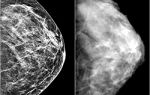
- Ultrasound of the pelvic organs allows you to assess the condition of the gynecological organs. If a tumor is present, using this diagnostic tool, you can determine the size of the tumor, as well as the presence of a metastatic process. In the presence of pathology, during an ultrasound examination, unclear contours of the endometrium are detected, and the echogenicity of some of its parts increases. The boundaries of the uterus are visualized blurry;
- collection of biomaterial for subsequent histological examination (biopsy), allows us to determine the presence or absence of cancer cells. This diagnostic method is mandatory and the most reliable method for diagnosing endometrial cancer. Hysteroscopy allows you to assess the condition of the female organs, as well as collect biomaterial for further research. Hysteroscopy is an endoscopic method that allows using a hysteroscope to examine the layers of the uterus and cervical canal from the inside;
- One of the innovative methods for diagnosing uterine precancer is fluorescence. The method involves introducing special substances that are absorbed by the tumors. Fluorescence allows you to study the tumor in detail at the microscopic level;
- CT and MRI are used to determine the presence of metastases and their extent of spread to other organs;
- A general blood and urine test allows you to assess the general condition of the patient. Today, when diagnosing cancer, they often resort to determining the level of tumor markers. For endometrial cancer, the level of tumor marker CA-125 is most often checked. This indicator is indirect when making a diagnosis.
Treatment
Therapy for endometrial cancer is selected individually depending on the type of tumor and the degree of spread. Age and the presence of chronic diseases are taken into account. In the fight against the disease, complex therapy is mainly used.
Treatment methods include:
- Surgery is the main treatment method for uterine precancer. Complete removal of the uterus and ovaries allows further avoidance of relapses and metastatic processes. In addition to the uterus and appendages, nearby lymph nodes are removed during the operation. If the disease is detected at the initial stages of development, or there are contraindications to this type of surgical operation, then hysteroresectoscopic ablation is used. This procedure involves excision of part of the endometrial and myometrial layer using an endoscopic method. This type of therapy involves strict monitoring of the patient over a long period of time to avoid tumor recurrence;
- Radiation therapy is most often used in combination with other treatment methods. If the tumor has highly differentiated properties, then external radiotherapy is used, since this type of tumor responds well to treatment. If the formation has low differentiation, then most often it quickly grows into the lower part of the uterus. In this case, external radiotherapy is effective in combination with intracavitary radiotherapy. Radiation therapy is most often used after surgery to eliminate any remaining cancer cells;
- Chemotherapy is also used more in combination with other therapies, since the use of chemotherapy drugs alone is often ineffective. This is due to the fact that endometrial tumors are insensitive to some cytostatic drugs. The ATS regimen has proven itself well, providing for the administration of drugs - Cyclophosphamide, Doxorubicin, Cisplatin;
- In the early stages of endometrial cancer, hormone therapy gives good treatment results. This therapy takes place in two stages. The first stage involves taking gestagens and antiestrogens. The patient is under strict supervision of specialists. The effectiveness of treatment is checked by hysteroscopy and biopsy every two months. The first stage lasts a year. After the positive effect of this stage, specialists try to restore the woman’s fertile function if she is of reproductive age. First and second generation estrogen-gestagen drugs are prescribed that can restore ovulation and endometrial function. The duration of this stage is about six months.
Forecast
The earlier the pathology is detected, the better the prognosis for life. If the pathology is detected at the first stage, the five-year survival rate is 95%. At the second stage, the indicators drop to 70%. In later stages of the disease, namely in the third, the survival rate after five years is 30%, in the fourth stage it is only 5%.
In the first three years, endometrial cancer may recur in 75% of cases. Subsequently, this figure decreases.
After therapeutic measures and their positive application, the patient is under the supervision of a doctor. Most often, in the first year after treatment, a preventive examination is carried out 4 times a year, in the second year – once every six months. Subsequently, it is enough to visit a specialist once a year to prevent relapses.
Video on the topic:
Prevention
To avoid endometrial cancer, every woman must follow the following preventive measures:
- try to maintain a certain body weight, avoiding obesity or excessive weight loss;
- for any changes in the menstrual cycle or other gynecological changes, promptly contact a gynecologist;
- to refuse from bad habits;
- Before planning a pregnancy, undergo a full examination. It is advisable to plan your first pregnancy no older than 30 years. Early pregnancy is also undesirable;
- have regular sex life, preferably with one partner. At the same time, in order to avoid an unwanted pregnancy, you need to contact a gynecologist and choose a means of contraception. Frequent abortions lead to bad consequences for a woman’s body;
- It is necessary to visit a gynecologist at least once a year for preventive purposes.
Source: https://pro-rak.com/reproduktivnaya-sistema/rak-endometriya-matki/
Endometrial cancer
Endometrial cancer of the uterus is the most common malignant tumor disease. Pathology more often manifests itself in women who have crossed the fifty-year mark. The main cause of endometrial cancer of the uterus is considered to be changes that occur during menopause.
Signs of a precancerous condition
Malignant formations in most cases are formed against the background of hyperplastic symptoms. The abnormal condition is associated with local or total proliferation of the endometrium.
In a healthy state, women shed the inner layer of the uterus every month, which then comes out with menstrual blood.
Sometimes endometrial rejection does not occur, and against this background, atypical structures are formed that have the ability to turn into cancer cells.
Atypical hyperplasia in medicine is considered a precancerous condition.
Women who have been diagnosed with hyperplasia and who have concomitant pathologies of metabolism and endocrine balance need to undergo a gynecological examination 2 times a year. Endometrial cancer detected in the early stages of development has the best prognosis for complete cure.
Symptoms by stage
Symptoms of uterine cancer appear depending on the stage of development of the pathology. In the initial stages of cancer, the signs are weakly expressed, but in the last stages they manifest themselves clearly.
Stage 1
At stage 1 of uterine endometrial cancer, a woman’s body experiences a hormonal imbalance and an increase in the production of the hormone estrogen. All this causes the lack of ovulation and the presence of cysts in the ovaries. There is no specific clear clinical picture for the disease.
Women planning pregnancy experience infertility. It is also characterized by heavy menstruation, the color of the discharge is dirty brown, and acyclic bleeding is possible. You can suspect a problem thanks to a set of laboratory tests. Ultrasound is an unreliable way to determine the initial stage of oncology.
Reliable signs are obtained during hysteroscopy.
Stage 2
The second degree of pathology is characterized by the presence of symptoms and probable signs of endometrial cancer. Endometrial hyperplasia and polyposis, against which malignancy occurs, manifest themselves as follows:
- uterine bleeding;
- bloody discharge during sexual intercourse;
- menstrual discharge the color of meat slop;
- purulent vaginal discharge;
- regular pain in the lower abdomen;
- loss of appetite;
- nausea.
Cancer at stage 2 can be suspected using ultrasound by the thickening of the endometrium, its heterogeneous structure, the presence of inclusions and characteristic changes in the cavity of the reproductive organ (presence of fluid).
The main way to detect pathology in the initial stages of development is hysteroscopy with a biopsy, examination of aspirate from the uterus, and biopsy performed by various methods. Ultrasound is considered only as an auxiliary diagnostic method.
Stage 3
At the third stage of endometrial cancer, precancerous changes in the endometrium of the uterus are detected in the functional or basal layer. In addition to the symptoms of the first stages of cancer, the following are added:
- general deterioration of health;
- a feeling of compression of the abdominal organs;
- vomiting;
- intense pain in the lower abdomen.
During an ultrasound, the gynecologist can observe a formation of significant size in the uterine cavity, which has its own network of vessels. Damage to the cervix and regional lymph nodes is diagnosed; metastases may be present in the bones, liver, lungs, and brain stem.
Stage three endometrial cancer has a poor survival prognosis. On average, 30% of patients have a five-year survival rate.
Another characteristic sign of the disease in its advanced form is metastasis to neighboring reproductive organs and tissues. With stage 3 uterine cancer, the fallopian tubes, periuterine space, and sometimes the ovaries are most often affected.
Stage 4
At the fourth stage, a malignant tumor forms in the uterine cavity with invasion into the underlying layers of the organ. Cancer cells spread throughout a woman's body through the lymph and bloodstream. Symptoms of the disease are pronounced:
- constant weakness;
- chronic fever up to 37.5 degrees;
- vaginal discharge mixed with blood, not associated with menstruation;
- signs of intoxication of the body;
- stomach ache;
- problems with urination.
Metastasis necessarily involves not only the vagina and cervix, but also distant organs - mammary glands, liver, kidneys, bone tissue, and brain. According to ultrasound, the specialist clearly sees a formation with uneven boundaries and a heterogeneous structure.
Ultrasound is complemented by hysteroscopy, laparoscopy, CT and MRI, histological examination to more accurately identify the stage of pathology and the presence of metastases in the body.
Signs by pathogenesis
Symptoms of the disease also appear depending on the cause of the cancer. The causes of uterine cancer are divided into estrogen-dependent and estrogen-independent. In the first case, tumor development is associated with increased estrogen production.
An increase in estrogen in the body is diagnosed in 70% of women with endometrial cancer of the uterus.
A malignant formation formed as a result of hyperestrogenism occurs against the background of the following symptoms:
- weight gain;
- infertility;
- dysfunction of the adrenal cortex (hirsutism, hyperpigmentation, acne);
- uterine bleeding;
- delaying menopause;
- pathological changes in the structure of the ovaries (PCOS).
Estrogen-independent factors in the development of pathology in 30% of cases cause uterine cancer. The tumor formation is formed due to atrophy of the endometrium of the reproductive organ. The disease is characterized by:
- high rate of spread of metastases to neighboring tissues and organs;
- the occurrence of heavy and prolonged bleeding;
- weak differentiation;
- difficulty in choosing the appropriate method of therapy.
Cancer of the uterine body may also have a genetic cause. Women at risk are advised to regularly visit a gynecologist and undergo all necessary types of examinations.
Description of symptoms
The disease has several characteristic signs. Each of them manifests itself depending on the degree of progression of endometrial cancer and the individual characteristics of patients (age, genetic predisposition, body type).
Uterine bleeding
Bleeding from the reproductive organ is the main symptom of uterine cavity cancer. The clinical picture of the disease manifests itself differently in patients of different ages. Scanty or copious discharge is observed in 90% of cases of illness.
Bleeding in women of reproductive age may indicate cervical pathology, failure of hormonal regulation of the menstrual cycle, and endometriosis.
For this reason, it is difficult to make a correct diagnosis based on symptoms alone.
Uterine bleeding during the postmenopausal period is a clear symptom of developing endometrial cancer.
Thanks to this manifestation of pathology, it is possible to diagnose a malignant tumor in the early stages of development.
Beli
Leucorrhoea is the second most common symptom of uterine cancer, which most often manifests itself when the tumor reaches a significant size. In advanced stages of the pathology, leucorrhoea can be profuse. The accumulation of mucus in the uterine cavity causes regular nagging pain in the abdomen, similar to discomfort during menstruation.
If leucorrhoea accumulates in the uterine cavity, then the clinical picture is supplemented by the following signs:
- bursting pain;
- rise in body temperature;
- febrile syndrome;
- general deterioration of condition.
Endometrial cancer is characterized by watery discharge. When a secondary infection occurs, bloody or purulent clots are released from the uterus. As tumor cells decompose, leucorrhoea acquires an unpleasant putrefactive odor. Pain
Pain syndrome often worries women in the last stages of uterine cancer, when atypical cells spread to the pelvic organs. When the tumor compresses the ureter, discomfort appears in the lumbar region, symptoms reminiscent of renal colic. If the tumor is large, it can put pressure on the rectum or ureter, causing:
- pain during bowel movements and urination;
- frequent urge to go to the toilet;
- tensem - the urge to defecate without passing feces.
In the later stages of cancer, the symptoms of the disease become more pronounced. Women note a general deterioration in their health, a rise in temperature and signs of intoxication. The pathological condition is accompanied by pain in the lower abdomen and leucorrhoea.
The presence of a tumor, its location and size can be determined by ultrasound, CT or MRI. The diagnostic method is complemented by laparoscopy and a number of laboratory tests to determine the type of tumor and the possibility of its metastasis to other organs and tissues.
Source: https://ginekola.ru/ginekologiya/shejka-matki/rak/simptomy-i-priznaki-raka-endometriya-matki.html
Endometrial cancer: the main secrets and problems of treatment
One of the most common types of diseases in women is endometrial cancer. This is a disease that is the formation of a malignant tumor on the walls of the uterine body.
The tumor is formed from the cells of the inner layer of the uterus, which in medicine is called the endometrium. Every year the number of women diagnosed with cancer is steadily growing.
Why women are susceptible to such a dangerous disease, what causes the disease, as well as the peculiarities of treatment, we will learn about all this from the material.
Anatomical features of the structure of the uterus: influence on the occurrence of cancer
Endometrial cancer of the uterus is a tumor that forms in the walls of the mucous cavity of this organ. To understand this type of disease in more detail, you need to know the anatomy of the uterus.
The uterus is an unpaired hollow organ, the main purpose of which is the conception of an embryo, gestation of a fetus and the birth of a child. The uterine cavity is the place where the baby is formed. The location of the uterus is the pelvic cavity, and the main components of this organ are:
- Endometrium . The inner layer of the uterus that lines it from the inside. The main purpose of the endometrium is to facilitate the implantation of a fertilized egg during pregnancy. If a malfunction occurs in the endometrial cells, then they become the sources of cancer.
- Myometrium. The middle layer of the uterus, the main feature of which is the ability to increase during pregnancy. The myometrium can also act as a basis for tumor growth.
- Serous membrane . The part of the abdominal cavity that covers the outside of the uterus.
The endometrium functions through a complex relationship between two systems: nervous and endocrine. The hypothalamus, ovaries and pituitary gland are the organs that produce a certain amount of hormones.
It is through these hormones that the growth, development and rejection of the endometrium is regulated if pregnancy does not occur.
From the anatomical structure of the uterus, we can conclude that due to disorders occurring in the nervous and endocrine systems, a disease such as endometrial cancer develops.
Causes of the disease
The causes of mutation in endometrial cells are still not fully understood. However, it is known for sure that the main reason for the development of a malignant process is a hormonal imbalance.
If there is a dysfunction in organs such as the hypothalamus, pituitary gland and ovaries, then an increased amount of estrogen is produced.
High levels of estrogen lead to the development of changes in the endometrium, which leads to malignant formation.
In addition to hormonal imbalance disorders, the following causes are also identified that have a direct impact on the occurrence of malignant endometrial tumors:
- Disruption of the functioning of the endocrine system. Women who have problems with the thyroid gland experience various diseases, including the possibility of endometrial cancer.
- Genetic predisposition. If a woman has ancestors in her family who suffered from genital cancer, then it is possible that the disease will develop at any age.
- Pathological deviations of the menstrual cycle.
- Gynecological diseases. The risk of cancer increases tenfold if a woman is diagnosed with gynecological diseases for which no measures are taken to cure them.
- Late menopause, observed after the age of 60 years.
- If there are abortions. The abortion procedure is one of the most dangerous, so doctors recommend resorting to the procedure only in rare cases. After abortion, almost every woman is subsequently diagnosed with endometrial cancer.
- No pregnancies. The anatomy of the human body is such that all organs perform their tasks. If an organ is not used, such as the uterus for bearing a child, this can cause the development of neoplasms in its cavity.
- Endometrial cancer can occur after surgical treatment.
- When carrying out therapeutic treatment using drugs containing estrogen.
- Immunodeficiency.
- Frequent experiences and stress, which puts a strong strain on the nervous system, causing it to malfunction.
- In case of non-compliance with a healthy lifestyle.
- Long-term sexual abstinence.
- If during sexual intercourse with different partners protection means (condoms) are not used.
From the above reasons it is clear that it is extremely difficult to avoid the development of a uterine tumor. To prevent this disease from causing death, women (absolutely all) should visit a gynecologist twice a year for preventive purposes. Such measures are mandatory for women, since this method will prevent the development of serious complications.
It is important to know! Diseases of the genital organs in women are quite common, the complications of which can only be prevented by timely diagnosis.
How the disease manifests itself: symptoms
Symptoms of endometrial cancer are quite difficult to determine, which is a significant problem for such a pathology. Symptoms and signs of the disease are quite difficult to determine, due to the wide variety of manifestations.
Every woman experiences signs of endometrial cancer differently. At the initial stage, the development of oncology has practically no symptoms.
That is why it is possible to detect the disease at an early stage only in exceptional cases during a gynecological examination.
At the acute stage, the disease manifests itself differently in each woman. However, it is impossible to judge the presence of an endometrial tumor by the symptoms that appear, since these signs may indicate other diseases of the genital organs. Most experienced specialists cannot always decide to make a 100% diagnosis based solely on the symptoms that appear.
The main symptoms that may indicate endometrial cancer are:
- Uterine bleeding and various discharges. If a woman discovers blood on her underwear after her menstrual period ends, then this is a cause for concern. However, uterine bleeding may indicate other diseases of the genital organs.
- Pain in the lower abdomen. This symptom appears mainly in the later stages of cancer. The pain is predominantly spasmodic in nature.
These two signs are the main symptoms of endometrial cancer in women. Vaginal discharge can be of the following types:
- purulent;
- bloody;
- watery;
- whitish.
Uterine bleeding occurs in 90% of cases and indicates disruptions occurring in the reproductive system. If any negative symptoms are detected, you should consult a specialist.
Any vaginal discharge makes it clear that it is necessary to determine the cause of such consequences and eliminate it.
Symptoms of uterine cancer in postmenopausal women over 60 years of age manifest themselves mainly in the form of copious bleeding from the vagina.
It is important to know! Even the fact that blood is found in the urine is a reason to contact a specialist. This may be the first signal of an exacerbation of cancer.
Features of oncology detection: diagnostics
If symptoms and complaints occur, a woman should immediately visit an antenatal clinic. Diagnosis of endometrial cancer includes the following procedures:
- Examination of the uterine cavity by a gynecologist using special mirrors.
- Carrying out a biopsy procedure, as well as curettage of the uterine cavity and cervical canal.
- Conducting an ultrasound of the pelvic organs.
- Patients are also prescribed a chest x-ray.
- Hysteroscopy, as well as CT or MRI.
- Laboratory research: blood test, urine test, coagulogram.
Carrying out such diagnostic procedures makes it possible to determine the degree of histopathological tumor. These techniques make it possible to determine the size of the tumor, the presence of signs of enlargement, as well as the location and nature of damage to neighboring organs. Each of the methods proposed above has its own characteristics and allows you to determine the corresponding deviations.
- Examination of the vagina using special mirrors reveals the absence of lesions of the vagina and cervix.
- A biopsy or curettage makes it possible to collect fragments of tumor tissue for subsequent laboratory testing.
- Ultrasound allows you to identify the location of the tumor, size, contours and other useful information.
One of the most popular methods for diagnosing cancer is the fluorescence test technique. The procedure is accompanied by the introduction of special substances that accumulate in the tumor with their subsequent registration. The procedure has a significant advantage - the ability to detect even microscopic foci of tumors.
Features of endometrial cancer treatment
Treatment for endometrial cancer is based on surgical removal of the carcinoma. In addition to removing the tumor, the oncologist may decide to carry out therapy in the following ways:
- Radiation therapy;
- Chemotherapy;
- Hormone therapy.
However, to effectively combat tumors, surgical intervention is predominantly used. The scope of such assistance and methods of performing the operation are determined depending on the stage of the cancer. The surgical procedure involves complete excision of the uterus and ovaries.
The principle of this operation is to remove the uterus, ovaries and fallopian tubes. This option is possible for women over 40-50 years old. If there are age restrictions for the operation, then hysteroresectoscopic ablation is performed.
This method of treatment requires high precision, so it is carried out mainly in exceptional cases and in the initial stages of cancer development.
Radiation therapy is the basis of complex treatment. The principle of treatment is based on ionizing radiation or radiography. If the tumor has a low degree of differentiation, double-acting radiation therapy is used: external and intracavitary. Radiation therapy is usually given after surgery.
Chemotherapy treatment of pathology is also not used as a monotherapeutic method. After all, endometrial cancer cells are not highly sensitive to all cytostatic drugs. Often, the chemotherapy treatment regimen includes the use of cyclophosphamide, doxorubicin and cisplatin.
Hormone therapy is possible as an independent treatment method. However, it is carried out mainly in the early stages of the development of the disease. Hormone therapy includes 2 stages of treatment.
At the first stage, gestagen and antiestrogen preparations are used, under the control of hysteroscopy and biopsy. One of these procedures is carried out once every 2 months. The second stage involves the use of estrogen-gestagen drugs of 1-2 generations.
The duration of the first stage is 1 year, and the second is about 6 months.
It is important to know! Any treatment method is highly effective if it is used at an early stage of the development of the disease. Self-medication of endometrial cancer is strictly contraindicated and can lead to active tumor growth.
Forecasts and conclusions
Endometrial carcinoma is one of the most common diseases in women of all ages. How long a woman will live depends on the timeliness of detection of pathology.
If the pathology is diagnosed at the first stage, then 95% of patients live more than 5 years. In subsequent stages, life expectancy predictions decrease exponentially.
At stage 3, life expectancy is up to 5 years in only 30% of women, and at stage 4 metastases this value is 5%.
Many factors influence the prognosis of cancer. These factors include:
- The degree of differentiation of cancer cells.
- Morphological structure.
All patients who have suffered from the disease must be monitored by an oncologist. The routes of metastasis depend on the stage of the pathology. If at stage 1 neoplasms form within the boundaries of the uterus, then at stages 3 and 4 the tumor spreads beyond the pelvis. Moreover, the bladder and rectum (the organs closest to the uterus) are also affected.
In the international classification of diseases ICD-10, endometrial cancer has the following coding:
- C54. Malignant neoplasm of the uterine body.
- C54.1 Endometrial cancer.
It ranks first among malignant tumors of the female genital organs. It is almost impossible to prevent the development of the disease, so the only way to avoid its development is to visit an antenatal clinic in a timely and regular manner.
To summarize, it should be noted that disease prevention includes maintaining normal hormonal levels, as well as the ovulatory menstrual cycle.
It is easier to cure any disease at an early stage of development than to fight it at the stages of complications.
(1
Source: https://OncoVed.ru/matka/rak-endometriya-glavnye-tajny-i-problemy-lecheniya
48. Endometrial cancer
Endometrial
cancer is a malignant neoplasm
that occurs in the cervix.
Histologically, there are two
main types: adenocarcinoma
and squamous cell carcinoma.
-
Risk factors for developing endometrial cancer
include : - - endocrine metabolic
disorders (obesity,
diabetes, hypertension); - - hormonal-dependent
dysfunction of the female genital
organs (anovulation, hyperestrogenism,
infertility); - - hormonally active
ovarian tumors (granulosatheca cell
tumor, Brenner tumor); - -genetic
predisposition;
lack of sexual activity, pregnancy, childbirth;
late onset of menarche, menopause (
over 55 years of age);
hormonal therapy (tamoxifen).
Pathogenesis
of Uterine Cancer.
Three hypotheses for endometrial cancer have been proposed .
The first
of them (estrogenic) is characterized by
the manifestation of hyperestrogenism in
combination with endocrine and metabolic
disorders (obesity, diabetes
mellitus, hypertension), which is observed in 70%.
The second
(estrogen-independent) theory implies
the absence of endocrine metabolic disorders
and ovulation disorders, 30%.
The tumor develops against the background of atrophied
endometrium, is characterized by a predominantly
low degree of differentiation and
has greater autonomy in
development, a high potential for
metastasis, and insensitivity
to gestagens.
-
The third
theory is genetic.
The main stages of the development of a malignant
are noted .- The first
stage is functional disorders
(anovulation, hyperestrogenism). - The second
stage is the formation of background morphological
changes (glandular cystic GPE,
polyps). - The third
stage is the formation of precancerous
morphological changes (atypical
hyperplasia with epithelial dysplasia
stage III). - The fourth
stage is the development of malignant
neoplasia: ♦ pre-invasive cancer;
♦cancer with minimal invasion into the myometrium;
♦severe forms of endometrial cancer. -
CLASSIFICATION
OF UTERINE CANCER (TNM)
Since
1971, the International
Classification of Endometrial Cancer has been used.
Stage
0 - histological data with suspected
malignancy of a hyperplastic
process of the endometrium.
Stage I - the tumor
is limited to the body of the uterus ;
It should be especially noted: a) the age and
condition of the patients;
b) the size of the uterine cavity c) the histological shape
of the tumor.
For stage I endometrial cancer,
division is also recommended
based on an objective criterion -
the length of the uterine cavity: into stage 1a if
the length of the uterine cavity does not exceed 8 cm,
and stage 16 if the length of the uterine cavity is more than
8 cm.
In stage I of endometrial cancer,
the following histological
groups are distinguished:
1) well-differentiated
adenocarcinoma;
2) differentiated
adenocarcinoma with areas of solid
structure;
3) predominance of a
solid structure or completely undifferentiated
carcinoma.
Stage
II - the tumor spreads to
the body and cervix .
Stage
III - spread of the tumor to
the parametric
tissue of the pelvis or metastases to
the vagina.
Stage IV - spread
of the process beyond
the pelvis, invasion of the
bladder and rectum, or the presence
of metastases .
Classification of
the International Federation of Obstetricians and
Gynecologists (FIGO, 1988) Stage
IA - the tumor is limited to the endometrium.
Stage
IB - invasion of less than 1/2 the thickness
of the myometrium.
Stage 1C - invasion
of more than 1/2 the thickness of the myometrium.
Stage
IIA - the tumor involves only
the endocervical glands.
Stage
IIB—cervical stroma invasion.
Stage
IIIA - tumor spread to the serosa
and (or) adnexa, as well as (or)
positive results of peritoneal
cytological examination.
Stage
IIIB - vaginal metastases.
Stage
IIIC - metastases to the pelvic and (or)
para-aortic lymph nodes.
Stage
IVA - tumor has spread to the bladder
and/or intestinal mucosa
.
Stage IVB - distant
metastases, including to the abdominal and/or
inguinal lymph nodes.
For
stages IA-IVB, the
G
G1 - non-squamous or
non-morula-like solid growth less than
5%.
G2 - non-squamous or
non-morula-like solid growth 6-50%.
G
—non-squamous or non-morula-like
solid growth greater than 50%.
Ways
of spread of endometrial cancer •
Down from the uterine cavity into the cervical
canal.
May lead to cervical stenosis and pyometra through the myometrium into
the serosa and peritoneal cavity.
•
Through the lumen of the fallopian tubes to the ovaries.
•
Hematogenous route, leading to
distant metastases.
•
Lymphogenic pathway.
CLINICAL
PICTURE OF UTERUS CANCER
In the
early stages, asymptomatic.
The main clinical symptoms of uterine cancer
are bloody
discharge from the genital tract, watery
leucorrhoea and pain .
Patients
of childbearing age more often seek
help from antenatal clinics, where
they are observed and treated by gynecologists for a long time
for dysfunctional
disorders in the hypothalamic-pituitary-ovarian
system.
The main clinical symptoms that bring young women
to the doctor are primary acyclic
uterine bleeding, infertility, and
ovarian dysfunction.
However,
bleeding is a “classic”
symptom only in postmenopause.
The appearance of
profuse serous leucorrhoea in elderly women
without concomitant inflammatory
diseases of the uterus and cervix is characteristic
of uterine cancer.
The development of the disease may be accompanied by copious
watery discharge, characteristic
of RMT.
Pain
is a late symptom of the disease.
More often they are localized in the lower abdomen and
lumbosacral region, and are
cramping or constant in nature.
DIAGNOSIS
OF UTERINE CANCER
LABORATORY
RESEARCH
The cytological
method is widely used.
Aspiration is carried out with a Brown syringe without
prior dilatation of the
cervical canal.
The information content of endometrial aspiration biopsy for advanced
forms of cancer is more than 90%, and
for initial forms it does not exceed 36.1%.
INSTRUMENTAL
RESEARCH
Ultrasound .
If uterine cancer is suspected, particular
importance is attached to the size of the median
uterine echo (MEHO), given the greatest
prognostic value of this
criterion for pathological transformation
of the endometrium.
Hysteroscopy
allows not only to judge the severity
and prevalence of the neoplastic
process, but also to perform a targeted
biopsy of the pathologically changed
epithelium, as well as evaluate the quality
of separate therapeutic and diagnostic
curettage and the feasibility of its
implementation.
In all cases, if
endometrial cancer is suspected, it is necessary to perform
separate curettage of the cervical
canal and the uterine cavity. Fluorescent
diagnostics with tumor-tropic photosensitizers
and their metabolites (photogem©, photosens©,
aminolevulinic acid).
The method is based
on the determination of small malignant
neoplasms (up to 1
mm) due to the selective accumulation
of a photosensitizer introduced into the body in advance
, followed by registration
of fluorescence (intrinsic and induced)
on the screen of a video system when exposed to
laser radiation in the ultraviolet
spectrum. Histological
examination .
-
Differential
diagnosis - Cancer
of the uterine body is differentiated from:
endometrial polyp;
GGE; submucosal MM. -
Treatment
depending on option - Preoperative
radiation therapy in the early stages
of the disease (stages I and II with occult
endocervical lesions).
Treatment
of endometrial cancer depending on stage.
Stage I cancer, 1st degree of histopathological
differentiation.
The optimal treatment method is surgical: total
abdominal hysterectomy and bilateral
salpingo-oophorectomy.
Stage II
cancer a hidden endocervical
lesion identified during curettage of the cervical canal.
Pseudopositive results of cervical
canal curettage are observed in more than 60% of cases.
Surgical staging.
Indications for additional postoperative
radiation therapy.
Severe damage to the cervix.
More than half of the myometrium is affected.
Involvement of pelvic lymph
nodes.
cancer
with obvious extension
to the cervix, grade 3 tumors very
often metastasize to the pelvic
lymph nodes, distant metastases
, and have a poor prognosis.
There are two approaches to treatment.
The first
approach is radical hysterectomy,
bilateral salpingo-oophorectomy, and
removal of para-aortic and pelvic
lymph nodes.
The second
approach is external and intracavitary
radiation therapy with
total abdominal
hysterectomy and bilateral
salpingo-oophorectomy after 4 weeks.
Radical
hysterectomy is indicated only for somatically
healthy, mainly young
women with tumors of low degree
of histopathological differentiation.
A combination of radiation therapy and surgery
is preferred for
patients with stage II tumors with extensive
cervical extension.
It is necessary to take into account that many women
with endometrial cancer are elderly
, suffering from obesity,
arterial hypertension, diabetes
mellitus, etc.
Source: https://studfile.net/preview/1633101/page:51/