Content
Share on VKontakte Share on Odnoklassniki Share on Facebook
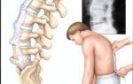
MRI of the knee joints is a unique procedure that consists of obtaining a detailed image of various structures inside and outside the element being studied (tendons, ligaments, fatty tissue, cartilage, blood vessels, muscles, etc.).
This is a non-invasive diagnostic medical method that allows doctors to diagnose and treat various diseases. MRI provides more complete information, unlike X-rays, ultrasound, and computed tomography.
This procedure uses strong magnetic radiation and radio waves.
Indications for MRI of the knee joint
Diseases of the musculoskeletal system can be caused by sprains, injuries, exposure to high humidity, low temperature, and excessively intense physical activity.
In case of an infectious lesion, inflammatory processes of connective tissues and joints may occur. MRI of the knee joints can reveal the disease at different stages.
Indications for the procedure are the following:
- Damage to the meniscus, cartilage, tendons, ligaments.
- Weakness, pain in the knee, bleeding, swelling in the tissues of the joint and around it.
- Sports injuries of the knee (torn, sprained ligaments, tendons).
- Degenerative joint diseases (osteoarthritis).
- Bone fractures that are not visible with x-rays and other imaging methods.
- Infections (osteomyelitis).
- Fluid accumulation.
- Feeling of instability.
- Tumors (metastases, primary tumors) involving soft and bone tissues.
- Pain, kneecap injuries.
- Decreased range of motion of the knee.
- Complications that may be associated with implanted surgical devices.
- Knee joint lock.
- Osteochondrosis, osteomyelitis.
- Bursitis, arthritis.
- Ligament surgery.
- Baker's cyst.
- Pathological changes inside the bone (gout).
- Pinched tendons and nerve endings.
- To determine the need for arthroscopy or other surgical procedures.
Which doctor prescribes this procedure?
MRI of the knee joints is in most cases used in traumatology and orthopedics. In addition, magnetic resonance imaging is prescribed to diagnose rheumatic joint lesions.
The decision on the need for an MRI is made by the attending traumatologist. An ultrasound or radiography may be prescribed in advance.
A contrast study is performed to identify inflammatory diseases and tumors.
What does magnetic resonance therapy show?
MRI images accurately reflect changes of any nature in the condition and structure of individual parts. Signs of injuries and independent diseases are determined. The photo shows different parts of the knee:
- Cartilage - the image reflects defects and cracks.
- Bone tissue – the knee cup, its bruises, tumors, infections, fractures, cysts are visible.
- Meniscus – the images clearly show the medial and lateral menisci and the damage caused to them.
- Ligaments and tendons – internal and external collateral, posterior and anterior cruciate ligaments, patellar tendons, quadriceps, their damage, existing injuries.
How to prepare for the procedure
The study is carried out without any special preliminary preparation. The patient must remove all metal objects from the body and clothing with metal elements.
If you have pacemakers, implants, cardioverters, or insulin pumps, MRI of the knee is not performed. If individual parts of implants and prostheses are made of titanium, then this will not interfere with tomography.
Hearing aids and other electronic devices must be removed. The general preparation sequence looks like this:
- The patient talks with the doctor and fills out the required documents.
- A person takes off clothes, except underwear, and puts on disposable clothes in the changing room.
- The laboratory assistant checks whether the patient has left any metal or ferromagnetic objects.
- The patient's weight is determined.
- A coil is inserted into the device, and the patient is placed on the platform.
- The client is given explanations regarding the further course of the procedure. A bulb is issued to sound an alarm, and its operation is checked.
- The laboratory assistant takes the patient into the tunnel and makes sure that he is comfortable.
How much does an MRI of the knee cost?
This research is not cheap. Prices vary in different regions. However, the high cost is justified, given the information content of magnetic resonance imaging and the complex anatomy of the knee. In the table below you will find out where you can undergo the study and get acquainted with the cost of the procedure in different cities of Russia.
| Clinic name, address | price, rub. |
| Medical Center “Capital”, Moscow, Bolshoi Vlasyevsky Lane, 9 | 3000 |
| Medical center “Prima Medica”, Moscow, st. Academician Chelomeya, 10B | 4500 |
| MRI center, Moscow, Kurkinskoe highway, 30 | 4950 |
| Energo, St. Petersburg, Engels Ave., 33, bldg. 1 | 6000 |
| Center for Medical Diagnostics "Petrogradsky", St. Petersburg, st. Roentgena, 5 | 4000 |
| Diagnostic+, St. Petersburg, st. Zakharyevskaya, 14 | 3500 |
| Tomography LLC, Ekaterinburg, st. March 8, 2012 | 3700 |
| Center for MRI diagnostics LDC MIBS, Ekaterinburg, st. Baidukova, 63 | 3500 |
| Medical Center "Paracelsus", Ekaterinburg, st. Vikulova, 33 | 6300 |
| Novosibirsk Institute "International Tomographic Center" SB RAS, Novosibirsk, st. Institutskaya, 3A | 3000 |
| MRI Expert, Novosibirsk, st. Yakusheva, 41 | 3500 |
| Tomography Center "EuroMed Clinic", Novosibirsk, Krasny Prospekt, 200 | 3500 |
Note: The data provided was obtained by random analysis of prices in clinics in Moscow, St. Petersburg, Novosibirsk, and Yekaterinburg. The information is not promotional in nature and may be out of date at the time of viewing.
Where can I get an MRI?
In Russia, especially in large cities, there are a large number of medical centers that offer clients such a service. You can get an MRI of the knee in Moscow and St. Petersburg at the addresses indicated in the table.
| Clinic name | Address |
| Medical center "Petrovskie Vorota" | Moscow, 1st Kolobovsky Lane, 4 |
| Oncological center “Sofia” | Moscow, 2nd Tverskoy-Yamskoy lane, 10 |
| MRI center "CityScan" | Moscow, 1st Perova Polya passage, 9, building 1 |
| MRI 24 | Moscow, st. Ostrovityanova, 1, building 9 |
| Clinic "Promed Plus" | St. Petersburg, Workers Boulevard, 18, bldg. 5 |
| Diagnostics "Ramsey" | St. Petersburg, st. Chapaeva, 5 |
| Center for Clinical Neurology CMRT | St. Petersburg, st. Lenskaya, 19, building 1 |
Note: The data provided was obtained by random analysis of the addresses of clinics in Moscow and St. Petersburg. The information is not promotional in nature and may be out of date at the time of viewing.
Contraindications
Magnetic resonance imaging shows almost all diseases, but its use is not justified in all cases.
There are reasons why medical examination is contraindicated - the presence of medical devices and metal parts in the patient’s body.
MRI of the knee joints is undesirable for people with renal failure, claustrophobia, and patients who cannot remain motionless for a long time.
Alternative research methods
The knee joint is considered the most complex of all those found in the human body, in terms of biodynamic, biological properties, and anatomical structure.
It is often susceptible to injury due to the number of functions it performs and its open position. Clinical examination is the basis for making a correct diagnosis.
In some cases, the final diagnosis of existing knee injuries can only be carried out in an inpatient setting using additional research methods.
Radiography
An X-ray is obtained by shining a special radiation through the human body. It identifies traumatic, deformational pathologies of various types in the knee area.
X-rays of the knee joint are prescribed in case of damage to the ligamentous apparatus, trauma to the patellas, menisci, condyle fractures, bone cracks, dislocations, subluxations of the joints.
Using X-rays, the following diseases and injuries are detected: indentation, fracture, bone crack, rupture, sprain, dislocations, arthrosis, tumors, rheumatoid arthritis, cysts, osteomyelitis.
Ultrasonography
Ultrasound examination or ultrasound is widely used in rheumatology. For effective and timely treatment of joint pathology, the ability to visualize parts of the joints is very important. Ultrasound of the knee joint is absolutely safe, painless, quick, and its cost is inexpensive, unlike other diagnostic methods. Ultrasound examination does not require preliminary preparation, has no contraindications, and allows assessing the condition of soft tissues, the structure and thickness of articular cartilage.
Under ultrasound control, puncture and arthroscopic manipulations are performed in the cavity of the knee joint, which allows the surgeon to accurately perform all techniques. However, this research method does not visualize the subchondral bone and its changes.
Ultrasound of the knee joint is prescribed for chronic, acute pain, any bruise, injury, swelling, redness of the joint, feeling of stiffness, clicking, inflammation, suspicion of a degenerative process, bursitis, synovitis, hemarthrosis, fractures.
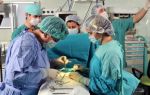
Arthroscopy
Arthroscopy is a surgical operation during which incisions are made on the knee, through which a camera and instruments are inserted to carry out manipulations in order to restore joint functions.
This procedure is recommended for damage or rupture of the posterior and anterior cruciate ligament, meniscus tear, displacement of the kneecap, inflammation of the joint bursa, removal of Baker's cyst, small fragments of cartilage in the knee joint, bone fractures, and injuries.
The operation is performed under spinal anesthesia. The lower limb is fixed in a special device. The procedure begins with two punctures in the knee area through which surgical instruments and an arthroscope are inserted.
The latter is a metal tube with lenses for examining the internal structure of the joint. The location of the incisions protects against damage to blood vessels and nerves.
A saline solution is injected through the pump, which flushes out damaged tissue to improve vision. After the operation, stitches are made.
CT scan
Controlling the condition of the knee joint is considered an important means of preventing diseases that can be easily stopped in time with proper treatment. Computed tomography is one of the leading methods for diagnosing ailments in this area of the body. The tomograph makes it possible to diagnose: arthritis, injuries, osteochondrosis, the condition of the joint space, and oncological processes. During this procedure, the soft tissues surrounding the knee, ligaments, and tendons are poorly visible.
Video
MRI is safe for the patient. During the procedure, the patient lies on his back on a special movable platform. The joint is held motionless for scanning.
The action of waves during magnetic resonance imaging is directed strictly at the knee, and therefore there are practically no contraindications. Before the procedure, the presence of things with magnetic properties is checked.
After the examination, the patient receives 3D graphics on disk, an image, and a transcript. For visual information on what an MRI is, watch the video.
Found an error in the text?
Select it, press Ctrl + Enter and we will fix everything!
Attention! The information presented in the article is for informational purposes only. The materials in the article do not encourage self-treatment. Only a qualified doctor can make a diagnosis and give treatment recommendations based on the individual characteristics of a particular patient.
Source: https://vrachmedik.ru/32-mrt-kolennogo-sustava.html
What does an MRI of the knee joint show???? indications and contraindications
MRI is a highly accurate non-invasive study based on the phenomenon of magnetic resonance.
Its essence is that the nuclei of chemical elements in any tissue of the body can be imagined as magnets rapidly rotating around their axis.
If these nuclei are placed in an external magnetic field, then their rotation axes shift, absorbing part of the radio wave energy (resonance effect). After the cessation of the external field, the atomic nuclei return to their original position (relax).
The accumulated energy is released in the form of electromagnetic oscillations, which can be recorded with special equipment.
Medical tomographs record the resonance of hydrogen protons that make up the water molecule. Due to the fact that this method is very sensitive to even minor changes in hydrogen concentration, it makes it possible not only to visualize tissues, but also to distinguish their normal structure from pathology.
How to do an MRI of the knee joint
When examining the knee joint, it is important to take into account that it has a very complex structure, including:
- epiphyses of the femur and tibia;
- muscles attached to the articulation area;
- tendons and ligaments that provide movement in the joint;
- menisci and cartilaginous surfaces;
- synovial fluid.
To establish the cause of joint dysfunction, it is necessary to carefully examine the condition of all these tissues. Even the most minor damage to any of them can significantly affect the function of the entire lower limb.
MRI accurately visualizes all degrees of cartilage change from swelling to thinning, fibering and cracking. The condition of the subchondral bone tissue, changes in the menisci, inflammation of the synovial membrane, and so on are assessed.
It is possible to calculate the total volume of affected cartilage and altered areas of bone, and the condition of the cruciate ligaments is assessed.
MRI of the knee joint - indications
One of the main reasons for limited working capacity of the active part of people is joint diseases. The popularization of complex sports leads to an increase in the number of injuries and orthopedic problems that arise at a young age. Therefore, it is so important that their diagnosis is safe, accurate and as early as possible.
These requirements are fully met by the magnetic resonance imaging method.
Typically, an MRI is done after consultation with an orthopedic doctor, who performs special tests and makes a preliminary diagnosis. Most often, a tomogram is recommended in the following cases:
- traumatic injuries;
- preparation for surgery;
- signs of joint inflammation;
- suspicion of dystrophic changes;
- congenital developmental anomalies;
- the need to exclude tumors and metastases;
- questionable data obtained during ultrasound, CT or radiography.
The most common form of joint pathology is osteoartosis. This is a disease in which the entire joint is involved in the pathological process, including the subchondral area of the bone, capsule, ligaments, synovial membrane, and periarticular muscles.
Gonarthrosis
This is osteoarthritis of the knee joint. The disease usually develops so imperceptibly that the patient finds it difficult to indicate the duration of the disease.
There is a periodic crunch when bending, episodic dull pain after severe and prolonged load on the joint, which quickly disappears after its cessation.
Gradually, their intensity increases, any load is poorly tolerated, and pain can appear at night.
Clinical manifestations of gonarthrosis usually begin at an older age, after 40-50 years, in women more often during menopause. He can be:
- Primary – degeneration of initially intact articular surfaces. Usually associated with overuse of the knee.
- Secondary - pathology develops after injury, inflammation, vascular or metabolic disorders.
In some cases, there is a combination of several etiological factors. Therefore, the main reason is the discrepancy between the load and the ability of the joint to withstand it.
The changes in cartilage that occur with osteoarthritis are similar to those that occur as a result of aging. They are based on changes in metabolic processes in articular cartilage and a decrease in the content of its main component – proteoglycans. A decrease in their number leads to drying out and degeneration of cartilage tissue.
But with age-related processes, degeneration develops very gradually and does not lead to pronounced disturbances in the structure of nearby tissues.
In the case of osteoarthritis, the pathology progresses much faster, begins at an earlier age, and is combined with changes in the articular surfaces of the bones and synoviocapsular apparatus.
This makes it possible to distinguish gonarthrosis from senile changes and distinguish it as an independent disease.
All stages of this process are clearly visible on MRI. The tomogram reveals synovial fluid effusion and a subchondral bone cyst in osteoarthritis of the knee joint.
First, an uneven surface of the cartilage appears, as it loses its elasticity and elasticity. Further, it becomes less compact and is divided into individual fibers, exposing the area of the underlying bone.
Fragments can separate from the cartilage and end up inside the joint, a phenomenon called “articular mice.” If they get caught and compressed between the articular surfaces, a person experiences sudden acute pain.
With a certain displacement in the knee, the “joint mouse” changes its position and the pain ends as suddenly as it began.
All these changes are most pronounced in the middle part of the articular surface, since it is more heavily loaded.
A decrease in the elasticity of cartilage leads to the fact that it no longer protects the underlying bone, and osteosclerosis is formed. It is also possible for cysts to appear, which break open to form bone erosions.
On the lateral areas of the articular surfaces, cartilage grows compensatoryly and is replaced by bone tissue - osteophytes are formed.
In the absence of timely treatment, fibrous changes in the synovium and joint capsule subsequently occur, which significantly limits its function.
Juvenile idiopathic arthritis
This is a chronic inflammatory disease that develops in children under the age of 16 years.
Accompanied by damage to the joints, often the knees. It can lead to irreversible destruction of cartilage and subchondral bone, including subsequent disability.
The effectiveness of treatment directly depends on the possibility of early diagnosis (before bone erosions form) and correct assessment of the degree of activity of the process.
An important feature of this disease is that about 70% of patients with early juvenile arthritis have no visible changes on radiographs. That is, if a child has knee pain without visible changes on a regular X-ray, it is better not to delay and get an MRI, rather than hope that it will go away on its own over time. Otherwise, you may miss the time needed to stop the process.
In this case, the advantage of MRI is that it makes it possible to assess the condition of the synovium. Most often, these are signs of synovitis in the form of the presence of fluid in the joint cavity, thickening of the synovial membrane.
It is important that based on the analysis of the thickness of the synovial membrane and the volume of effusion, it is possible to distinguish the active process from the remission stage. This radically changes treatment tactics.
Baker's cyst
It is formed when the synovial bursa is stretched as a result of injury and inflammation. The cyst is filled with fluid and communicates with the joint cavity.
At the initial stage, it is visible only on MRI. As it progresses, it can be visually identified as a round, dense formation in the popliteal region. At this stage, it can already cause pain during physical activity and discomfort when bending the leg at the knee.
Its reverse development is possible until it disappears completely, but subject to timely diagnosis and treatment. With its long-term existence, the contents of the cyst harden, and an adhesive process is formed.
It may rupture, leading to acute pain, swelling in the popliteal fossa and lower leg area.
In this case, MRI is of particular importance, as it makes it possible to carry out differential diagnosis with other pathologies.
Contraindications for MRI of the knee joint
The main limitations are associated with the presence of permanent metal structures and foreign bodies in the body.
Based on their properties, they distinguish:
- Ferromagnets. They have their own magnetization and, when placed in a powerful external magnetic field, can behave unpredictably. These include iron, nickel, cobalt, and some alloys. Carrying out MRI if they are present in the body is prohibited.
- Paramagnetic materials. In the absence of an external magnetic field, they do not have magnetic properties. For example, titanium, platinum. They do not pose a danger, but can create interference if they are in the study area.
The final decision is made by the doctor after studying the documents describing the composition of the product.
Absolute contraindications for MRI are:
- presence of electronic devices – pacemaker, cochlear implant, insulin pump and others;
- first trimester of pregnancy;
- severe claustrophobia.
Our medical center does not examine children under the age of 5 years; parents need to go to a specialized children's institution to have an MRI of the knee joint performed under anesthesia. If you weigh more than 150 kg, performing an MRI of the knee joint is also difficult due to the design features of the tomograph.
Preparing for an MRI of the knee joint
No special preparation is required; you can get a tomography procedure in our medical center on the day of your appointment.
Before starting the tomography, you will be asked to remove all accessories and items of clothing containing metal, and remove bank and other magnetic cards.
Be sure to provide the doctor with your medical history and the results of previous studies. This will allow you to pay special attention to controversial issues and choose the optimal protocol for the procedure.
How long does it take to do an MRI of the knee?
The study takes about 20 minutes. The patient is placed on a movable table that moves inside a magnet. At the time of the procedure, the doctor is in the next room, but can maintain contact through an intercom and monitor the patient on the monitor.
A comfortable temperature and lighting are created inside the tomograph. For better relaxation, headphones with pleasant music are provided. Immediately after the examination, you can return to your usual rhythm of life.
Interpretation of MRI of the knee joint
The description of images at the DiMagnit medical center is carried out by doctors with more than 7 years of experience. Our doctors specialize in MRI diagnostics, so their conclusions can be trusted.
MRI technology can detect the earliest changes in various joint structures. For example, it is indispensable in assessing the condition of the menisci. The degree of their changes on MRI:
- 0 degree – normal condition of the meniscus;
- I degree - in the body of the meniscus there is a focal signal of increased intensity, which does not yet reach the edge of the meniscus;
- II degree – the appearance in the meniscus of a linear signal of increased intensity, not reaching its edges;
- III degree – the signal of increased intensity reaches the extreme part of the meniscus.
Only grade III changes indicate a meniscus tear.
The shape of the meniscus is also important for diagnosis. Normally, it has the shape of a butterfly in a certain plane. Any deviations may indicate a possible rupture.
Damage to the meniscus is often detected on MRI when a person has no significant complaints; such cases are not uncommon in older patients.
MRI or CT scan of the knee joint - which is better?
These two research methods are fundamentally different in that CT uses x-rays, while MRI uses a safe magnetic field. Due to the presence of radiation exposure, doctors recommend not performing CT scans more than 2 times a year. The number of MRI sessions is not limited.
Initial soft tissue changes may not be visible on CT scans, making diagnosis of early stages of pathology unreliable. In this case, MRI is more informative, which makes it possible to detect the disease at the very initial stage.
Only magnetic resonance imaging can visualize vessels that are not visible on CT.
In particularly difficult cases, patients are offered arthroscopy to clarify the diagnosis. This is an endoscopic operation that is performed for both diagnostic and therapeutic purposes. Requires special training and rehabilitation. Only MRI can accurately determine the extent to which arthroscopic intervention on the knee joint is necessary.
Accurate and timely diagnosis allows you to reduce treatment time, choose the optimal approach and significantly improve the patient’s quality of life.
Source: https://mrtdon.ru/mrt/info/chto-pokazyvaet-mrt-kolennogo-sustava/
MRI of the knee joint: how to do it for a child and an adult, interpretation of images, preparation for an MRI for knee pain, contraindications
Indications and contraindications
MRI of the knee is a non-invasive examination technique. It is informative not only for damage to the knee joint, but also for changes in bone and soft tissues. The examination shows bone and cartilage tissue, tendons, muscles, menisci, and synovial folds.
Magnetic resonance examination using a tomograph is much more sensitive and informative than radiography. That is why it is often done for injuries and diseases of soft tissues. MRI images even show trabecular edema in the bones.
Tomography can be used to detect evascular necrosis, bone metastases, and oncological processes. MRI shows the following diseases of the knee joint:
- degenerative-dystrophic changes;
- inflammatory and purulent-necrotic processes;
- traumatic damage to bone, muscle tissue, tendons and meniscus (violation of bone integrity, rupture or sprain of ligaments);
- bleeding, hematoma and swelling;
- infectious pathologies;
- hernia in the knee area.
Using tomography, you can identify complications after knee surgery, as well as monitor the effectiveness of treatment. MRI of the knee joint ligaments shows a violation of the integrity of the tendon-ligamentous apparatus.
The examination is prescribed for pain in the knees, swelling or swelling in the knee joints, or limited mobility. MRI of the knee meniscus is performed when it is torn, as well as when oncological processes are suspected.
Despite the fact that tomography is considered one of the safest, the procedure is not suitable for everyone. Contraindications to MRI of the knee joint:
- body tattoos made with metallic inks;
- first trimester of pregnancy;
- the presence of pacemakers, an artificial heart valve, wires that fix bones and other metal objects in the body;
- the patient's weight is more than 120 kg.
Since MRI of the knee does not expose the body to radiation, it can be done on a child, in the second and third trimesters of pregnancy, and while breastfeeding.
When using a contrast agent, contraindications include pregnancy, hepatitis B, as well as kidney pathologies and hypersensitivity to the injected substance.
Preparation
Before you do an MRI of the knee joint, you need to prepare. It is necessary to remove all jewelry and metal objects from the body, including glasses.
If the patient has any diseases (allergic reaction, chronic diseases), then the doctor should know about this before conducting the examination.
When using a contrast agent, you should not eat or drink 5 hours before the procedure. Immediately before the scan, the person should change into a hospital gown.
An MRI of the knee joint in a child under 7 years of age is performed only after using sedatives or sedatives. Children should not move during the examination, so in rare cases the child is placed under general anesthesia.
How is it carried out?
MRI can be performed on closed or open type tomographs. During the examination, the patient is exposed to a magnetic field. The person must lie down on the retractable table of the device, after which the limbs are secured with belts. You can do an MRI of 2 or one joint.
To get good quality pictures, you need to lie still. Even a slight movement will affect the diagnostic result.
After the patient has assumed a horizontal position, the table slides into the tomograph tunnel. During an MRI, a person does not feel any pain or discomfort. Noise from the machine is normal. The tomograph is equipped with a camera and microphone, so the patient hears all the doctor’s instructions.
This examination has many advantages over other methods:
- High information content and reliability. This is one of the most informative diagnostic methods. MRI is effective for chronic knee pain when other methods have failed to find the cause of the pain.
- Clarity of pictures. Thanks to the high quality of images, it is possible to accurately detect the type of pathology and its location.
- Lack of special training. There is no need to diet, take medications or change your usual lifestyle.
- Safety, possibility of use in children and pregnant women.
- Painless, no discomfort.
Duration of the procedure
Scan time depends on the readings. On average, the procedure takes 30-60 minutes. After the examination, the patient can return to everyday life. There are no restrictions.
Price
Compared to other instrumental techniques, the procedure is expensive. The price depends on the number of joints. An MRI of 2 joints costs more than one.
The average cost is from 4.5 to 6.5 thousand rubles. Additionally, you will need to pay for pictures or for recording the scan.
Decoding the result
An MRI of the knee joint is interpreted based on the images obtained during the scan. This should only be done by a specialist, as the images contain a variety of tissue structures.
The description of the images will include the following information:
- condition of cartilage tissue;
- degenerative-dystrophic processes;
- changes associated with metabolic disorders;
- condition of blood vessels;
- volume of synovial fluid;
- the presence of benign or malignant formations.
Most often, the patient receives the results in hand an hour after the examination. The waiting time depends on the doctor’s workload.
After receiving the results of the examination, you should apply for treatment. Identified pathologies must be treated immediately before complications arise.
Oksana Belokur, doctor,
specially for Ortopediya.pro
Source: https://ortopediya.pro/diagnostika/mrt/kolena.html
MRI knee transcript
Have you been trying to heal your JOINTS for many years?
Head of the Institute for the Treatment of Joints: “You will be amazed at how easy it is to cure your joints by taking the product every day for 147 rubles ...
Read more "
Magnetic resonance imaging is based on exposing the area under study to a strong magnetic field and receiving response electromagnetic radiation from hydrogen atoms in water (the human body is 90% water).
OUR READERS RECOMMEND!
Our readers successfully use Sustalaif to treat joints.
Seeing how popular this product is, we decided to bring it to your attention. Read more here...
MRI is carried out using a special device - a tomograph, which creates magnetic radiation and radio waves.
The device scans the area under study, wave vibrations are transmitted to a computer and converted into an image.
After the scanning is completed, the doctor can study the resulting images, which depict all the details of the area under study in various projections, as well as in a layer-by-layer section.
In some cases, for greater information content, the patient is injected with a contrast agent (drugs based on gadolinium) shortly before the examination. MRI with contrast allows you to more accurately visualize the localization of the pathological focus and destructive processes, malignant tissue degeneration.
Indications and contraindications
MRI of the knee is a non-invasive examination technique. It is informative not only for damage to the knee joint, but also for changes in bone and soft tissues. The examination shows bone and cartilage tissue, tendons, muscles, menisci, and synovial folds.
Magnetic resonance examination using a tomograph is much more sensitive and informative than radiography. That is why it is often done for injuries and diseases of soft tissues. MRI images even show trabecular edema in the bones.
Tomography can be used to detect evascular necrosis, bone metastases, and oncological processes. MRI shows the following diseases of the knee joint:
- degenerative-dystrophic changes;
- inflammatory and purulent-necrotic processes;
- traumatic damage to bone, muscle tissue, tendons and meniscus (violation of bone integrity, rupture or sprain of ligaments);
- bleeding, hematoma and swelling;
- infectious pathologies;
- hernia in the knee area.
Using tomography, you can identify complications after knee surgery, as well as monitor the effectiveness of treatment. MRI of the knee joint ligaments shows a violation of the integrity of the tendon-ligamentous apparatus.
The examination is prescribed for pain in the knees, swelling or swelling in the knee joints, or limited mobility. MRI of the knee meniscus is performed when it is torn, as well as when oncological processes are suspected.
Despite the fact that tomography is considered one of the safest, the procedure is not suitable for everyone. Contraindications to MRI of the knee joint:
- body tattoos made with metallic inks;
- first trimester of pregnancy;
- the presence of pacemakers, an artificial heart valve, wires that fix bones and other metal objects in the body;
- the patient's weight is more than 120 kg.
Since MRI of the knee does not expose the body to radiation, it can be done on a child, in the second and third trimesters of pregnancy, and while breastfeeding.
When using a contrast agent, contraindications include pregnancy, hepatitis B, as well as kidney pathologies and hypersensitivity to the injected substance.
Common mistakes in MRI of the knee joint
The Stoller grade of meniscus tear may be graded incorrectly, resulting in unnecessary surgery or improper treatment. Remember that not all meniscal injuries require surgery!
Incorrect diagnosis of meniscus tear. An MRI may diagnose a torn meniscus when in fact the meniscus is normal.
There are normal variations in the structure of the meniscus, which an inexperienced doctor may mistake for damage. As a result, unnecessary arthroscopic knee surgery is prescribed.
Unscrupulous traumatologists also tend to interpret controversial changes in favor of rupture, because then they will be able to make money on an expensive operation.
Meniscal pseudotear on MRI. The initial MRI analysis diagnosed a rupture. In fact, there is a normal variant of the structure caused by the meniscofemoral ligament of Risberg. An impression fracture is often seen by doctors on MRI. The reason is poor familiarity with the x-ray picture of small impression fractures of the condyles.
Prolonged knee pain is often mistakenly associated with injury, when the real cause is inflammatory diseases, such as rheumatoid arthritis, gout and other arthropathies. Rheumatoid arthritis has characteristic MRI features that are often overlooked.
A rupture of the anterior or posterior cruciate ligament may be diagnosed incorrectly: instead of a complete rupture, a partial rupture is indicated, instead of chronic mucoid degeneration, acute edema is indicated, etc.
Sometimes a ligament rupture is missed. Hoffa's disease on MRI.
The radiologist may forget to assess the condition of the patellar fat pad, given that Hoffa's disease is not a very pleasant disease that brings a lot of suffering.
Rare knee diseases may be difficult to diagnose on MRI. The doctor may not see or misinterpret villonodular synovitis, mediopatellar plica syndrome, iliotibial tract syndrome, posterolateral complex rupture, and other rare diseases.
The first diagnosis was a fracture of the patella (kneecap). In fact, the normal development here is patella bipartita (the patella consists of two parts).
Magnetic resonance imaging of the knee joint
An MRI of the knee joint is prescribed if the development of inflammatory complications, destructive diseases and congenital anomalies of the structure of the articular joint is suspected. This research method is non-invasive and safe, therefore, if necessary, it is prescribed even to a child. In order for the examination to be of high quality and the results to be as accurate as possible, it is necessary to prepare properly.
MRI of the knee joint is a non-invasive, informative hardware technique for studying the condition of tissues.
What does the procedure show?
Magnetic resonance imaging is a non-invasive, highly accurate diagnostic method, thanks to which the doctor will be able to see not only the knee joint and bone tissue.
The study can show even minor disturbances in the structure of muscle tissue, ligaments, menisci, and synovial folds.
If an injury occurs and there is a suspicion of trabecular edema in the bones, an MRI of the knee will show these pathological changes, while the result will not be reliable on X-ray images.
This sensitive and specific research method helps to detect avascular necrosis of the femoral head, oncological processes and skeletal metastases in the early stages. Thanks to an MRI of the knee joint, the doctor will be able to diagnose:
- degenerative-dystrophic disorders;
- diseases of inflammatory nature;
- oncological diseases;
- injuries to the right or left knee of traumatic origin.
Indications for the study
MRI of the knee joint is prescribed for injuries, complications after surgery, and prolonged pain.
Tomography of the knee joint is performed if the following disorders are suspected:
- internal bleeding, hematoma formation, swelling;
- complications after installation of implants;
- infectious pathologies;
- oncological diseases with metastases to bone tissue;
- damage to bone and muscle tissue, tendons, meniscus;
- other injuries resulting in impaired functionality of the lower limb.
Contraindications
However, not all patients undergo this test. A tomogram is contraindicated if the patient’s body already has implants, pacemakers, or metal foreign bodies.
Patients suffering from heart pathologies, during pregnancy and during breastfeeding should also avoid the procedure.
The closed space of the tomograph can negatively affect the patient’s psychological well-being, so people suffering from claustrophobia are better off not using this type of diagnosis.
Another contraindication for the study is in patients of young childhood, because during diagnosis you need to lie still, and this is unlikely to happen in an infant. Therefore, in order to avoid dangerous complications, it is worth agreeing with the doctor on all important points before the study.
How is the preparation going?
This diagnostic procedure is non-invasive and completely painless. In specialized medical centers, the equipment is configured so that the patient feels as comfortable as possible during the study. In order for the tomography of the knee to be informative and not cause complications, it is necessary to prepare for the procedure in advance:
- if the patient is in a state of emotional agitation or for some reason is afraid to undergo an MRI, it is worth taking a sedative before the study begins;
- if an inflammatory complication develops in the body, it is necessary to take an analgesic;
- before going into the tomograph, you should remove all metal objects, hearing aids and other parts prohibited during diagnostics;
- During the entire time the diagnosis is being carried out, it is important for the patient to lie still, because unnecessary body movements can distort the results and the doctor’s conclusion will be inaccurate.
How is an MRI of the knee joints performed?
- While an MRI of the knee joint is being performed, the patient will lie motionless on a special table, which will slowly move inside the tomograph.
- Some medical institutions have open-type machines, in which case the scanner will be placed only in the lower part of the body.
- At certain intervals, pictures are taken, which are then compared with each other, thanks to which the doctor receives an image in 3D format.
Sometimes a knee examination needs to be done with contrast.
Therefore, before the procedure, the doctor will introduce a special dye, which will be distributed throughout the body within two minutes, after which the procedure can begin.
But if a person is allergic to the components in the contrast, the doctor should be warned about this.
If the preparation was carried out correctly and no unforeseen complications arose during the examination, the diagnosis lasts on average 1-1.5 hours.
MRI of the meniscus
This cartilaginous formation often suffers during injuries and damage to the knee joint. But X-ray images will not be able to diagnose the disorder and distinguish the affected cartilage from healthy one. Therefore, an MRI of the knee meniscus is prescribed. If the cartilage is damaged, it will appear in the image as a dark stripe on a white background.
Contrast is not used during the examination, so preparatory procedures before the examination are not necessary.
Decoding the results
The results of an MRI of the knee joint make it possible to see pathologies in the tissues.
After the examination of the joint is completed, a conclusion can be made about the disease. The doctor, based on the results obtained, will draw up a description of the pathology and make an accurate diagnosis.
In the photographs, the image appears in three-dimensional space, so the doctor can examine not only bone tissue, but also muscle and cartilage tissue. Since the anatomy of the knee joint is complex, the data obtained will help distinguish pathology in the initial stages of development.
After the interpretation of the MRI of the knee joint is ready, the doctor will select the optimal treatment regimen.
Source: http://EtoSustav.ru/st/procedury/mrt-kolennogo-sustava.html
OUR READERS RECOMMEND!
Our readers successfully use Sustalife to treat joints.
Seeing how popular this product is, we decided to bring it to your attention. Read more here...
The essence of the method
For diagnostics, a tomograph is used - a device that allows you to perform layer-by-layer non-destructive research. Thanks to MRI, doctors receive detailed information about the condition of the soft tissues, cartilage and bones of the organ. The method is as follows:
- magnetic radiation passes through a closed area in the form of a tunnel;
- reaching the organ, the waves return;
- based on vibrations, the device forms an image;
- After the procedure is completed, the patient is given a photo.
The unit of measurement for magnetic field is 1 T (Tesla). Magnetic resonance imaging allows you to see the diseased organ in section and in various projections. To increase the information content, a special substance is injected into the tissue before the procedure, which provides contrast in the photograph.
This identifies pathologies that cannot be tracked by other methods, including visual examination.
What is this procedure?
An MRI examination is considered a non-invasive diagnostic measure, with the help of which it will be possible to examine the condition of bones, cartilage, tendons, the musculo-ligamentous apparatus and the vessels supplying the joint.
Tomography is safe and painless, the results can be viewed on the monitor or obtained ready-made images. During an MRI, a person is not affected by x-rays.
The diagnostic measure involves the use of the properties of a magnetic field, which does not have a negative effect on health.
Interpretation of MRI of the knee joint - important features
There are clinical studies that show that MRI of the joint in overweight people often shows damage to the meniscus, a cartilaginous structure located between the articular parts of the knee bones. It bears the maximum load when walking and an overweight person.
MRI of the joint visualizes blood vessels, which is not possible with computed tomography. The advantages of the study are obvious, but not every specialist is able to competently interpret the tomogram.
The ability to examine structures in detail is created by a large number of tissues saturated with water (menisci, cartilage, ligaments). By adding MR contrast to the circulatory system, it is possible to examine areas of accumulation, identify pathology of the blood supply, vascular abnormalities, atherosclerotic narrowing, and malignant tumors.
Bone tissue receives nutrients exclusively through the small capillaries of the periosteum. When there is insufficient blood supply through large vessels, capillary blood flow insufficiency occurs.
Detailed visualization of the condition of the joint on an MRI using three-dimensional modeling allows you to carefully examine the structures on a computer monitor. Images can be printed on film for consultation with a specialist.
Pathology visualized using MRI of the knee joint:
- Damage to menisci, cartilage, tendons;
- Sports knee injuries;
- Bone fractures;
- Sprain;
- Degenerative-dystrophic diseases;
- Fluid accumulation inside the knee;
- Bacterial infections (osteomyelitis);
- Tumors (including bone tissue).
The treating doctor may prescribe an MRI of the joint after detecting a pathology during arthroscopy (videoscopic examination of the condition of the anatomical structures of the knee) that requires diagnostic confirmation.
Features of visualization of the knee meniscus during MRI:
- Meniscus tear – violation of the anatomical shape or lack of image;
- The triangular structure changes when cracked;
- “Tear structure” is a diagnostic sign of a meniscus tear;
- The MR signal may be abnormal outside the lesion, indicating degenerative changes with destruction of the osteochondral structure. When studying the blood supply in such a situation, dystrophic changes are assumed.
The above-described features of MRI of the knee joint are not diagnostic signs of pathology, since there are many other manifestations of knee pathology.
- Treatment
- Folk remedies
- Symptoms
- Joints
Source: https://kist.asustav.ru/sustavy/mrt-kolena-rasshifrovka/