Modern medicine today offers various methods for diagnosing diseases. However, not all of them are clear to the patient. In this article I would like to talk about what abdominal computed tomography is, under what circumstances it is used, and how you can prepare for this procedure.
Computed tomography of the abdominal cavity: what is it?
Initially, you need to understand what exactly will be discussed. Thus, it is worth noting that patients often come across the abbreviation CT. This is exactly what computed tomography is. In this case, various parts of the body and organs of the patient can be examined.
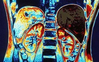
So, what is a CT scan of the abdomen? This procedure carries out layer-by-layer diagnostics of the patient’s body. The basis of the method is the same X-rays. Modern devices are multispiral. This makes it possible to obtain high-quality images with extremely high spatial resolution in the shortest possible time.
The procedure takes a short time. It only takes a few minutes for the patient. However, it should be noted that if the study is carried out using a so-called contrast agent, it can be repeated several times.
A little history
This research method is extremely young. Computed tomography was invented in 1972, and in 1979 it received a Nobel Prize. The merit of this device is very great.
After all, it was with him that the development of other multilayer diagnostic methods (MRI, etc.) began. It should be noted that the first devices were intended exclusively for studying the patient’s brain.
However, soon (already in 1976) they were used to diagnose diseases of the entire human body.
When to use this procedure
In what cases can a computed tomography scan of the abdominal organs be done? Thus, it is worth noting that this is one of the most accurate methods of visualization and examination of various internal organs. In the pictures you can quite clearly see the placement of the spleen, liver and pancreas, as well as hollow structures: intestines, gallbladder and all bile ducts.
What can this diagnostic method show?
- Inflammatory processes.
- Cysts.
- Traumatic changes.
- CT is able to “examine” neoplasms, both benign and malignant. In this case, the boundaries of tumors, the presence of metastases, their germination, as well as other indicators that are important for diagnosis and treatment are visible.
Indications for use of the method
When is a computed tomography scan of the abdomen most often prescribed? So, the list of indications is quite extensive:
- If it is necessary to determine damage to internal organs in a closed abdominal injury.
- When determining diffuse liver diseases, i.e. to detect cirrhosis, hepatitis and other diseases.
- For abscesses in the abdominal organs.
- For vascular disorders that lead to secondary disorders of various internal organs.
- If you suspect cysts, cystomas in the abdominal organs.
- To determine the depth of lymph node damage by metastases.
- To identify various tumors in the abdominal cavity.
However, computed tomography of the abdominal cavity can also be performed in the following cases:
- To clarify the effectiveness of anticancer treatment.
- In preparation for surgery.
- If it is necessary to clarify the results of other studies or confirm a preliminary diagnosis.
- A CT scan is performed if another procedure, magnetic resonance imaging (MRI), is contraindicated for the patient.
About security
Many patients are interested in how safe a CT scan of the abdomen is. After all, it is based on the use of X-rays, which are harmful to the human body. However, there is no need to worry.
In modern devices, the radiation dose is negligible (even compared to conventional x-rays). That is why you should not be afraid of prescribing this procedure.
It will not cause harm to the patient even if it is used repeatedly (which happens during studies with the introduction of a contrast agent).
Preparing for CT
If a patient is scheduled for a computed tomography scan of the abdominal cavity, preparation for the study is a very important step. After all, only by following all the doctors’ recommendations can you get the right results.
- The first and most important rule: a few days before this study, you need to go on a certain diet. You will have to exclude from your diet all those foods that cause increased gas formation, cause constipation, or provoke other problems or diseases in the body. For example, allergy sufferers should exclude all allergenic foods, and those with lactose intolerance should exclude milk and all its derivatives.
- The day before the procedure, be sure to thoroughly cleanse the intestines. To do this you will have to do an enema. In this case, experts advise carrying out the procedure twice. As an alternative, you can drink a certain dose of a laxative (for example, three sachets of a medicine such as Fortrans).
- Keep in mind that this abdominal examination is performed strictly on an empty stomach. Therefore, on the day of the procedure and after the bowel cleansing, you can no longer eat.
MSCT
Recently, in addition to the abbreviation CT, you can often see the designation MSCT. This is a multislice computed tomography scan of the abdominal organs.
Its difference from CT is only that it is a newer study, an improved version of conventional computed tomography, so to speak. This became possible only with the development of technological capabilities and the desire to improve the performance of this diagnostic device.
Let's look at how multislice computed tomography of the abdominal organs differs from conventional CT:
- The image quality is much better.
- Scanning speed has been increased. This significantly reduces patient scanning time.
- The contrast resolution in this case is significantly better.
- The radiation exposure to the patient's body is significantly lower.
Preparation for MSCT
A spiral CT scan of the abdomen requires the same preparation techniques as a conventional CT scan. Those. It is best to follow a diet for a while, and you should not eat anything 8 hours before the test. After all, this diagnostic method will give correct results only if it is carried out on an empty stomach.
Documents and necessary things
To have a computed tomography scan of the abdominal cavity without any problems, you need to take with you:
- Doctor's referral. To carry out the procedure, a referral for examination from the treating doctor is required.
- You will also need an extract from the patient’s outpatient card or medical history.
- You need to take pictures of other studies (or other CT scans) with you.
- Well, you need to have with you documents that are related to the problem being studied.
Stages of CT scanning
Many patients are interested in the question of how the procedure itself goes. So, the stages of its implementation will be as follows:
- Preparation stage. Here you need to remove all metal objects from the body: staples, piercings, etc. If the patient has an implant, the laboratory assistant or the doctor himself must be warned about this.
- During the procedure, the patient is in a horizontal position. He lies on a special pull-out table on his back.
- Even before the study begins, the doctor leaves the room where the tomograph is located. He can communicate with the patient through a microphone and speakers.
- Next, the table with the patient enters the so-called tunnel of the device. The tomograph itself takes a series of specific images using rotational movements.
- If the doctor is satisfied with the quality of the images obtained, the patient can leave the room with the tomograph. Otherwise, another procedure will be required.
CT with contrast
Sometimes the patient is prescribed a CT scan with contrast. Why is this necessary and what can this substance be used for? So, it is simply necessary for improved visualization of certain structures. When it comes to studies of the abdominal organs, the main substance of the contrast fluid is ordinary iodine or barium.
So, this drug is most often injected into a vein to the patient (for studies of the liver, as well as the blood supply system) or simply drunk (if it is necessary to study the stomach). Iodine or barium stains all organs a certain color, which helps improve their contrast and visualization.
The volume of the drug depends on the patient's weight. It is completely eliminated from the body within one and a half days. At the same time, the drug does not have any harmful effects on the body.
If a computed tomography scan of the abdominal cavity is performed with a contrast agent, its duration can take up to 30 minutes (while a regular CT scan takes about 5 minutes).
Contraindications to CT
It is also worth noting that there are a number of contraindications to the use of computed tomography.
- Pregnancy. Even though the body is exposed to extremely low radiation exposure, this procedure is still unsafe for the fetus.
- Hyperkinesis. Those. the procedure cannot be performed if the patient experiences involuntary movements. After all, to get high-quality images you need to lie quietly for a certain amount of time.
- The procedure cannot be performed if there is severe pain.
In some cases (depending on the equipment) there may also be weight restrictions. Most often, the maximum weight for the subjects being studied is 120 kg.
Contraindications to CT with contrast
There are also a number of situations when CT with contrast should not be used:
- Lactation. After all, contrast fluid substances can penetrate into milk. If this procedure is simply necessary, you will have to stop breastfeeding for two days (until the substance is completely removed from the body).
- Kidney failure. After all, when the drug is excreted in the urine, complications may arise (poisoning of the body).
- This procedure is also contraindicated for those people who are allergic to iodine.
Special categories of population
We further consider the topic “computed tomography of the abdominal cavity.” Photos of these devices can be seen in our article. From the information above, it becomes clear that the procedure itself is not at all scary or dangerous.
However, even despite this, there are certain limitations for applying this research method to children. Thus, due to the low radiation exposure, CT can be used only from the age of 14. This procedure is also contraindicated for pregnant women.
There are no other restrictions other than those described above.
results
Multiple reviews indicate that this procedure is completely safe. Computed tomography of the abdominal cavity is a study that is carried out quite quickly and practically does not harm the body.
Images and results can be obtained in an extremely short time. You will have to wait no more than 1 hour. If the CT scan was three-dimensional, the research results can be recorded on a disk or other information medium.
With all the documents, as well as the received images and extracts, you should then go to your doctor.
Source: https://www.syl.ru/article/181482/new_kompyuternaya-tomografiya-bryushnoy-polosti-podgotovka-multispiralnaya-kompyuternaya-tomografiya-organov-bryushnoy-polosti
Preparation for CT scan of the abdomen with and without contrast
Modern medicine increasingly prescribes computed tomography as one of the most modern research methods. During the tomography process, a special three-dimensional visualization of the abdominal organs is performed. The result is a view of its section, which helps to carefully study not only the shape of the abdominal organs, but also their structure.
To obtain a positive result, it is important not only to use modern technology and to properly prepare for the examination. Preparation for a CT scan of the abdominal cavity or for a computed tomography scan with the introduction of a special substance is a fairly important stage of medical research.
With proper preparatory measures, you can obtain guaranteed accurate examination results.
Main purpose of CT
Computed tomography is prescribed in the process of diagnosing various diseases of the urinary and digestive systems, as well as the pelvic area and retroperitoneal lymph nodes.
They resort to this research method in the process of determining pathologies of organs that are located in the retroperitoneal region or in the pelvis.
Other organs that may change shape under the influence of a number of developing formations are also examined.
A modern diagnostic procedure, with proper preparation for a CT scan of the abdominal cavity with or without contrast, allows for the most accurate diagnosis to identify diseases and health problems such as:
- Various inflammatory pathologies;
- Congenital anomalies;
- Cysts and abscesses;
- Malignant and benign formations;
- Damage to abdominal vessels in accidents and injuries;
- Vascular damage due to tumors, liver cirrhosis, and also with a high probability of blood clots.
Modern tomographs make it possible to conduct a virtual examination of the large intestine without causing discomfort to the patient.
The study can be carried out either with or without a contrast agent. This issue is resolved strictly on an individual basis.
It all depends on the patient’s condition or diseases that need to be clarified and investigated.
Before the procedure, it is imperative to prepare for a CT scan of the abdominal cavity without contrast or with the introduction of a substance, which consists of eating before the event.
Nutrition rules before the procedure
Because this is a gastrointestinal examination, much of the preparation process is to maximize overall abdominal imaging. If gases form in the intestines, this will seriously interfere with examinations such as an abdominal CT scan with contrast.
It is for this reason that you need to prepare as properly as possible before performing a CT scan. In about three days you will have to adhere to a certain diet, which will reduce the amount of feces and reduce the amount of gas. Preparing for the procedure requires eliminating foods such as:
- Gas-increasing foods - beans, sauerkraut;
- Fruits and berries;
- Nuts and prunes;
- Flour products made from flour of the first raw material;
- Rice and semolina porridge;
- Alcohol;
- Excessively strong tea or coffee;
- It is worth excluding dairy products if lactose intolerance is present.
Thanks to the high quality of CT images, doctors can determine the presence of pathology with 100% accuracy.
During preparation, a special diet is needed before an abdominal CT scan. The diet should include foods such as lean meats, fish, meatballs and soufflé. Boiled vegetable soups, low-fat cottage cheese, as well as crackers.
Additional preparation rules
You need to prepare for a tomographic examination not only by consuming special foods. It is also worth making sure that the following rules are followed. Any form of computer research is carried out strictly on an empty stomach.
After eating, especially if it was the wrong food, many organs greatly change their shape. All this leads to the fact that the survey results in a highly distorted picture.
To prevent the picture from being greatly distorted, the last meal should be taken approximately 7-8 hours before the examination.
Important! During this time period, it is not recommended to drink water, as it can stimulate intestinal motility.
To obtain a more accurate result, the doctor may prescribe special medications that will improve the condition of the intestinal tract and allow you to obtain the most accurate results. These may be medications such as:
- Activated carbon, Lactofiltrum or Smecta, which are carried out to reduce gas formation;
- Preparation includes the use of laxatives, which will help to effectively empty the intestines, which will automatically improve the visual inspection of the gastrointestinal tract;
- If the motor function of the gastrointestinal tract is increased, before the procedure it is worth taking antispasmodics, for example, No-shpa.
These medications should not be taken on your own, but only as prescribed by the doctor who performs the CT scan.
If an X-ray examination was performed before the CT scan, at least 5 days should pass between these procedures.
The reason is that the remains of special radiopaque substances may affect the overall results of the tomography, and the abdominal image may be incorrect. Visualization of blood vessels and organs is greatly distorted.
CT is considered an absolutely safe research method.
Before the procedure itself, the patient must remove all metal objects, such as jewelry, glasses, watches and piercings. Also, avoid wearing underwear, such as bras, that have metal underwires. The patient's hearing aids and cell phones are also prohibited.
Preparing for contrast injection
The procedure of computed tomography with the introduction of contrast is performed if it is necessary to study hollow organs. An iodine solution or medications containing barium can be used as a contrast. Almost immediately after administration, they paint the abdominal organs a different color from other organs.
This is what makes it possible to clearly visually examine certain formations, as well as features of structural changes. These substances are completely harmless to the body and can be used to identify areas with increased blood supply.
Before performing a CT scan, you should definitely consult with a specialist. This could be a radiologist. The reason is that the generally accepted scheme for administering contrast is slightly modified in direct dependence on the goal and the nuances of the study.
Written consent from the patient for the administration of a special radiocontrast agent is also required.
The doctor will need to find out whether or not the patient is allergic to drugs that contain iodine or to various seafood that contain significant amounts of iodine.
If a contrast agent is administered orally, the patient must first prepare, and the drug itself must be prepared, that is, diluted in full accordance with the instructions.
Usually the total amount of the resulting suspension is prepared, the amount of which is 1.5 liters. Take it 300 ml per day for three or four days. If the contrast agent is administered intravenously, this should also be done on an empty stomach.
Administration can also be done rectally. Then the day before an enema is done and laxatives are taken.
Main contraindications
Modern computed tomography is a relatively safe examination for the patient. The radiation dose level for the longest procedure does not exceed the normal X-ray rate. Despite this, it is worth familiarizing yourself with the main contraindications to this procedure. Among these factors are:
- Excess weight – more than 120 kg;
- Pregnancy;
- Patient anxiety;
- Age up to 14 years;
- Inflammatory kidney diseases;
- Lactation period;
- Severe diabetes mellitus;
- Allergy to contrast agents;
- Severe diseases of the liver, heart and blood vessels;
- Blood diseases.
Also, CT is not performed if the patient has undergone gastrointestinal tract examination using contrast in less than a week. In some cases, other preliminary examinations may be required. This could be an ultrasound of the abdominal cavity, FGS of the stomach, colonoscopy, regular x-ray or examination by an oncologist.
Important Tips
Preparation for the procedure involves fulfilling certain conditions. In almost all cases, an enema is performed. To cleanse the intestines very thoroughly, the procedure can be performed twice.
In most cases, this event is combined with preliminary use of laxatives.
This procedure must be carried out strictly on an empty stomach, that is, the last meal is taken strictly 8 hours before the medical event.
If you plan to conduct a kidney examination, no special preparation is required. If this part of the body requires the administration of a contrast agent, you do not need to eat for 4 hours first.
When examining the bladder, you will need to drink a contrast mixture approximately 5 hours before.
Just before the start of the study, urine is removed through a special catheter and air is introduced into the bladder through it, which will significantly improve visualization and contrast, and accordingly, higher-quality images will be obtained.
In some cases, diabetes mellitus may be a contraindication.
During a female examination, when it is necessary to diagnose the appendages and uterus, contrast may not be used. The doctor makes the exact decision. If the decision was made to introduce contrast, the woman will need to prepare approximately 5 hours in advance.
The X-ray substance is taken orally. Before the procedure itself, another type of contrast agent is injected into the bladder cavity, and a gauze swab is injected into the vagina.
When studying a man's reproductive system, he will need to take a contrast agent approximately 5 hours before the procedure.
How is the CT scan procedure performed?
A modern computed tomograph is a device that consists of a movable table and a fairly large cube. There is a tunnel in its central part.
During the examination, the patient is placed on his back and remains completely motionless throughout the entire procedure. During the operation of the tomograph, the scanner, which is located inside the device, rotates around the person in a special way. In this case, no discomfort is felt, there is no pain.
You must have a referral from your doctor and the results of other diagnostic tests with you.
During the operation of the tomograph, a person hears the faint noise of a normal operating computer device, and also follows the doctor’s instructions regarding the characteristics of the respiratory process.
X-ray radiation, which is used for tomography, is emitted in very small doses.
As for time, this procedure takes an hour on average, then a certain amount of time will be required to process the data.
Conclusion
Properly performed computed tomography with the introduction of contrast involves intravenous administration of a contrast agent. Due to it, the general visualization of the abdominal organs is significantly enhanced.
The examination process will allow timely detection of quite complex diseases that can greatly harm the body and even lead to death.
A CT scan will help identify diseases such as pathological narrowing of blood vessels, the development of tumors, the general functional state of the renal pelvis and the quality of blood flow in emerging tumors. The use of a contrast agent is prescribed in most cases when a tumor is suspected.
The result obtained allows you to get a complete picture of your health status.
Source: https://tomografa.net/kt/bryushnaya-polost/podgtovoka-i-vypolnenie-s-kontrastom.html
Computed tomography of the abdomen
Computed tomography of the abdominal cavity is a diagnostic method that describes the condition of internal organs along with lymph nodes and vessels, and the retroperitoneal space. Scanning is carried out layer by layer, with small steps. The goal is to analyze the condition and find pathology.
The method allows you to explore the structure:
- Liver, gallbladder, spleen and pancreas and surrounding tissues, ducts and vessels;
- Stomach and intestines;
- Bone tissue;
- Vascular and nervous system of the abdominal region;
- Lymph nodes in this area.
What the diagnostics show:
- The location of the abdominal organs and their sizes;
- Condition of tissues, vessels and walls of hollow organs;
- The presence of inflammatory processes, their localization and phase of development;
- Presence of parasites;
- Tumor formations;
- Traumatic changes and prospects for their development;
- Anomalies in the structure of organs;
- Changes in the nervous tissue that ensures the functioning of the digestive, urinary and skeletal systems;
- The structure of the spine, sternum and ribs.
Indications for diagnostics
- Inflammatory liver diseases, cirrhosis, parasites and neoplasms.
- pathologies of the pancreas and gallbladder.
- Weakness, spleen diseases, tumors.
- Purulent and cystic pathologies.
- Tumors and inflammation of the lymph nodes.
- Vascular thrombosis.
- Injuries with hemorrhages in internal organs.
- Developmental anomalies of the peritoneal organs.
How to prepare for the procedure?
A doctor prescribes an examination if there is a good reason for this, since X-ray radiation is harmful.
When preparing for a CT scan, you must:
- Warn your doctor about the medications you are taking;
- Since the procedure can be performed with contrast, it is necessary to exclude an allergy to the contrast agent; a special test is done for this;
- The intestines should not be overcrowded or bloated, so a few days before the test it is necessary to adjust your diet.
Your doctor may order a blood test.
How does the process work?
- The patient should remove metal jewelry and electronic devices so as not to interfere with the operation of the device.
- Clothes should be loose and not restrict movement.
- It is important to tell your doctor if you have metal dental crowns or a pacemaker.
- The patient lies down on a retractable table and is secured with straps and bolsters.
- The table slides into the tomograph and scanning begins.
During operation of the tomograph, noise is heard and the scanning device moves.
Communication with a specialist is carried out via a special connection.
Scanning with contrast
A contrast agent is administered if the clinical picture is ambiguous. CT scan with contrast agent:
- Detects tumors;
- Allows you to distinguish between malignant and benign processes; Shows the peculiarities of the functioning of the peritoneal organs.
In addition, the boundaries of organs are indistinguishable without the use of enhancement in the form of a contrast agent, which provides a visible difference in the density of the object of study.
The amount of contrast agent is selected individually in each case. After a day, the harmless liquid leaves the body.
Result
Typically, decoding information takes about an hour. As a result, the patient is given the resulting images and a doctor’s report signed and stamped.
Contraindications
- Pregnancy.
- Lactation.
- High degree of obesity.
- Kidney failure.
- Intolerance to contrast agent.
- Deviations in mental state.
- The presence of metal objects in the body.
What they say in reviews
Patients who have undergone this procedure note:
- The study is carried out to definitively confirm the diagnosis;
- Information content – high;
- The method helps to make the correct diagnosis and decide on treatment;
- For the abdominal cavity, this method is more suitable than MRI and ultrasound.
Source: https://www.ckbran.ru/diagnostic/x-ray-1/komputernaya-tomografiya-kt/kt-bryushnoi-polosti
CT scan of the abdominal cavity: preparation for a study with contrast, taking urografin, which shows
Computed tomography of the abdominal cavity is a painless procedure that creates cross-sectional images and volumetric images of the abdominal cavity to detect pathology.
Abdominal CT is performed with or without contrast. The diagnostician examines the patient on a table in a supine position.
To ensure the accuracy of the images before the procedure, tomography requires preparation in the form of a diet and sometimes medication.
Preparing for CT
The abdominal space and parts of the peritoneal space are interconnected with each other, located close to each other, and therefore deformation and changes in one affect the other. The attending physician who prescribes a CT scan talks about the preparatory stage in advance. The characteristics of the patient's body are taken into account.
For computed tomography:
- They do a preliminary examination - ultrasound, FGS, colonoscopy, x-ray diagnostics.
- The patient is consulted by an anesthesiologist about the side effects of the drugs. Signs documents on the administration of a contrast agent.
- It is necessary to test blood biochemistry for the predominance of creatinine and liver enzymes. Deciphering the tests allows us to identify organ pathology.
You need to prepare for a CT scan of the abdominal cavity in advance. A clogged intestine reduces the information content when the procedure is performed. The administration of contrast may cause vomiting and volvulus. For three days before the procedure, you must follow a strict diet and not eat certain foods.
Assess the condition:
- pancreas;
- liver;
- stomach;
- gallbladder;
- lymph nodes;
- pelvis;
- intestines;
- spleen;
- lungs;
- vessels;
- kidneys;
- bladder.
A diet is followed to prepare for a CT scan with contrast. It helps reduce gas formation and normalizes intestinal function in terms of stool movement. The pictures will be better quality and the results will be more accurate.
The diet lasts from two to three days. The patient can eat:
- cottage cheese;
- steamed vegetables;
- lean poultry, fish, meat;
- steamed dishes;
- hard cookies and dry white bread;
- oatmeal cooked in water;
- steamed chicken;
- empty broths;
- lean steamed fish;
- omelette without oil.
Prohibited: baked goods, peas, dairy products, vegetables and fruits, flour, kefir, prunes, chocolate. If you follow and adhere to the recommendations, complications from the stomach and intestines can be avoided. The procedure can be done on an empty stomach.
You need to prepare your intestines for the examination:
- Enemas are given in the evening and in the morning.
- Within 18 hours, the intestines are cleansed with drugs, for example, laxatives, sorbents.
The procedure is carried out on an empty stomach. Don't eat for 6 hours. A patient with a contrast examination undergoes blood tests for biochemistry before a CT scan simultaneously with a study of creatinine and urea.
The contrast is administered by solution, enema, and intravenously (done on an empty stomach). Substances containing iodine are used. For example, it is part of Urografin and Omnipaque.
These solutions can detect tumors in organs and metastases.
If the contrast agent is administered orally, take Urografin long before the procedure. One ampoule dissolves in 1 liter of water. Patients are often recommended a certain regimen with the reagent: Take 250 ml at 18-00, 23-00, 3 hours before the procedure, before the start of the CT scan.
The medications are prescribed by the doctor. Before the procedure, the patient takes a laxative, enterosorbent and antispasmodic orally before CT. This will cleanse the intestines. Independent use and use of medications will lead to ineffective tomography and a distorted diagnosis.
Before a CT scan with contrast, your doctor may stop taking your medication. The procedure is comfortable if you follow the doctor's recommendations. Having started the procedure, the patient needs to relax and comply with the doctor’s requirements.
The patient does not feel any discomfort or discomfort.
The contrast agent, penetrating into the diseased area, significantly enhances the contrast and clear contours of the images. This allows you to see violations. The diagnostician shows the shape, structure of organs and blood vessels.
Main purpose of the method
Using computed tomography, problems of the retroperitoneal space are diagnosed to identify:
- focus of the disease;
- level of pathology;
- prevalence of the disease;
- lymphatic and circulatory permeability;
- study of the organ.
If a pathology occurs - tissue proliferation, inflammation, acute abdomen syndrome, any lesion - the doctor prescribes a computed tomography scan.
The procedure reveals:
- oncology;
- polyps, fibromas;
- infection;
- atherosclerosis, vascular pathologies;
- inflammation of the lymph nodes.
For people who are contraindicated for magnetic resonance imaging, but require clarification of the diagnosis, computed tomography of the abdominal cavity can be used.
With its help, you can determine infection of organs with parasites, cirrhosis, hepatitis.
This diagnostic method cannot be avoided if there are: closed abdominal injuries, inflammation of the appendix, preoperative preparation, evaluation of surgical intervention.
Indications for the procedure
A computed tomography scan of the abdominal cavity is an X-ray examination. The insides are scanned and the condition, the image of the internal organs is transferred to a computer. The native procedure is performed without contrast. Accurate data is needed, then a tomography of a certain organ is prescribed using contrast (a special drug is infused).
Symptoms for examination:
- yellowness;
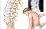
- abdominal pain;
- weight loss;
- dysfunction of the digestive and genitourinary systems;
- bleeding.
To diagnose the respiratory tract, a CT scan of the sternum is performed, which complements tomography of the aorta. The method should be carried out according to indications, through a doctor’s referral. When performing computed tomography with contrast, developmental pathologies are determined from the images.
Indications for examination using a contrast solution
For abdominal CT scans with contrast, iodine-containing drug formulations are used. Penetrating into the circulatory system, they accumulate in cells and are painted a certain color. In pathological tissues, diseased organs are outlined. They are eliminated after two days and are safe for health. Drinking more fluid may speed up elimination.
Conducting research allows you to identify problems:
- Narrowing of the lumen of blood vessels.
- Tumors in the first stages.
Doctors check organs, condition, and make accurate diagnoses.
CT with contrast is recommended for:
- determination of metastases;
- clarifying the results of therapy;
- surgical intervention;
- diagnosis when MRI is not possible.
CT scan of the abdomen with contrast – administration of iodine-containing drugs in the following ways:
- With intravenous. Thanks to the injection, you can view changes in blood vessels and organs.
- The patient drinks the solution. After a while, areas of the image are highlighted - the liver, pancreas, spleen, kidneys.
- Contrast is administered through the rectum. To determine the condition of the intestines.
- With bolus. For a full examination of organs, a contrast agent is administered by drip, automatically. Scanning can be completed in 5-10 minutes. It will take half an hour.
The information is then processed by a computer.
Spiral examination is a separate direction of tomography. The X-ray emitter constantly rotates, synchronously with the movements of the table on which the patient is fixed in the center.
When turned, the mechanism draws a spiral around the patient. The method explores the area. It is characterized by high speed and a low radiation dose.
Based on the results, the doctor prescribes a specific treatment or additional examination.
Contraindications to the use of tomography
As with any method of examination and diagnosis, there are contraindications. There are prohibitions for CT with contrast:
- pregnancy;
- allergy to a solution with a contrast agent;
- thyroid dysfunction;
- acute kidney and liver diseases;
- oncology;
- diseases of the heart and blood vessels;
- children under 14 years of age;
- heavy weight.
If the benefit of the procedure is greater than the harm, the council of doctors allows it to be carried out. The current method without contrast is not effective in CT of the abdominal aorta. Contrast is a safe drug, but CT scans can detect side effects.
Radiation exposure is dangerous for humans; the patient should not be scanned frequently. The radiation dose for abdominal CT is 3-7 MSV. The doctor compares the risk of a health threat and the severity of the disease.
You can attend a CT scan once every 2 years. Upon request, possible in six months. After the procedure, it is recommended to drink at least two liters of water.
A summary is described in the conclusion, which the doctor must decipher and indicate which areas are affected.
At the end of the procedure, after treatment, doctors look at the order of the pathological state of the organs. A conclusion about pathologies is drawn up.
It includes data on the patient’s condition, which became known and informative thanks to X-ray pulses and contrast agent. The doctor can see not only the condition of the sick, but also potentially healthy organs.
The patient is given the photographs. A diagnosis cannot be made based on the conclusion. This is an informative confirmation.
It is CT that can find the affected areas, take pictures of certain zones and spaces of the abdominal cavity for further diagnosis and treatment. Patients may feel slightly dizzy and weak. The procedure is painless and does not cause any inconvenience. When iodine is administered, there may be an aftertaste that will soon go away.
Source: https://GastroTract.ru/obsledovanie/kt-bryushnoj-polosti.html
Abdominal CT
At the Yauza Clinical Hospital, a CT scan of the abdominal cavity and retroperitoneal space is performed with or without a contrast agent.
For scanning, a Philips Ingenuity Elite digital computed tomograph with 128 slices is used, equipped with iMR technology, which reduces radiation exposure and at the same time increases the quality of images by 60–80% .
Multispiral computed tomography of the abdominal organs can be performed either with or without the use of a contrast agent. It provides complete and reliable information about the location of internal organs, the condition of tissues and the presence of pathologies.
It is important to note that only a study with intravenous contrast allows one to simultaneously evaluate many organ parameters, blood/lymphatic vessels and accurately make a diagnosis.
CT is most effective in diagnosing tumor diseases and can detect even small pathological formations.
CT scan of the abdominal organs evaluates:
- abdominal organs - gall bladder, liver, spleen, pancreas, biliary tract, gall bladder, large and small intestines;
- organs of the retroperitoneal space - ureters (except for the lower third), kidneys;
- part of the lower lobes of the lungs and heart;
- surrounding bone structures (bones from the 10th thoracic to 4th lumbar vertebrae);
- vessels of the systemic circulation (in the area of study);
- The lymph nodes.
About the procedure
- The duration of the study is from 5 to 40 minutes.
- Allows you to find formations with a diameter of 5 mm or more.
- The reliability of the information obtained during diagnosis tends to 90%.
- Rare research methods are available - 3D reconstruction of the urinary system using dynamic contrast CT angiography, virtual colonoscopy and others.
Indications for use of the method and advantages of the study
Our hospital has a modern multislice computed tomography scanner for scanning the whole body.
The device significantly reduces radiation exposure and allows the examination to be carried out as quickly as possible without discomfort. The result of the research is finished images and files recorded on electronic media.
Tomography done in our clinic is recognized by all medical institutions in Russia and abroad.
The advantages of multislice tomography performed in our clinic over standard CT:
- a more contrasting picture due to a reduction in the amount of noise using a new generation of iterative reconstruction;
- reduction in radiation exposure by 30-80%;
- possibility of using less contrast agent;
- increasing the area of the study area;
- reducing the thickness of the section, giving a chance to track down even the smallest pathological focus;
- the ability to create 3D models of organs and foci of disease, as well as their multiplanar reconstructions.
Contrast perfusion allows you to cover more parameters, as well as detect bleeding, neoplasms and hemorrhages, so it is recommended for most studies. In 80-87% of cases, the examination is carried out using contrast.
In what cases is a CT scan of the abdominal organs prescribed:
- the presence of pain in the abdomen, bleeding, both due to injury and for unexplained reasons;
- suspicion of tumors;
- the presence of metastases in internal organs, lymph nodes, bones;
- the presence of stones in the kidneys, biliary and urinary tracts;
- upcoming surgery requiring precise examination of the intervention area;
- the need to assess the dynamics of the process and treatment results;
- peritonitis, abscesses;
- liver diseases;
- kidney inflammation;
- the presence of a hernia (area of the anterior abdominal wall, diaphragm);
- developmental anomalies.
First of all, CT scan of the abdominal cavity is aimed at detecting volumetric processes in internal organs, which include polyps, cysts, abscesses, tumors, and stones. CT scan reveals benign and malignant tumors, metastases, stones, cysts, kidneys and liver, echinococosis, atherosclerosis, various changes in the structure of organs (cirrhosis, hepatosis).
Preparing for a CT scan
Preparation for the study begins several days before and includes a special diet, tests and additional procedures. Before the session, you must provide the doctor with tests indicating the level of urea and creatinine (no later than 2 weeks). You can take a rapid test directly in our clinic. Below all the preparatory stages are presented in chronological order:
Desirable:
- A few days before the session, start following a diet that excludes foods that cause constipation and increased gas formation;
- 2 days before the session - start drinking adsorbents (usually activated carbon);
- On the eve of the procedure, perform a cleansing enema, repeat if necessary before starting the study.
Necessarily:
- 4 hours before the procedure - refuse to eat or take medications, only light drinks are allowed.
- If ultrasound, gastroscopy, colonoscopy, CT, or X-ray examinations have been previously performed, provide the results to the doctor for the most correct interpretation of the study.
- Before starting the study, a conversation is held during which the course, safety, painlessness of the method, and rules of behavior during the session are discussed.
Before starting the procedure, you must remove all metal objects - jewelry, piercings, belts, etc. You must be prepared to lie still and calm, breathing evenly, and strictly follow the radiologist's commands.
Before the start of the study, a written receipt for the examination is taken. During the session, an unpleasant taste in the mouth and a feeling of heat in the body may appear during the administration of the contrast agent.
Contrast enhancement
Contrasting provides a more informative picture of the condition of the patient’s internal organs and is performed using an iodine-containing substance. The contrast is administered intravenously using an automatic syringe. The device sets the time and speed of administration depending on the diagnostic tasks assigned.
Contraindications to CT
CT scan of the abdominal organs at the Yauza Clinical Hospital is a safe examination with virtually no contraindications. The only category of people for whom alternative types of studies may be recommended are pregnant women (for any CT studies) and lactating women (for studies with intravenous contrast).
The following are considered relative limitations for contrast-enhanced CT:
- renal failure;
- severe diabetes mellitus;
- contrast intolerance.
Research results
Evaluation of the examination results takes from 1 to 24 hours, depending on the complexity of the examination. At the end of the specialist’s work, you will receive a final conclusion, recommendations for contacting certain doctors and a full set of images printed or recorded on a DVD, film or flash drive.
Alternative methods for diagnosing abdominal organs
For patients who, for some reason, are not indicated for abdominal CT, several types of alternative studies are available:
- Survey radiography of the abdominal organs - allows you to detect x-ray positive gallstones and stones in the urinary tract, free gas in the abdominal cavity (with perforation of a hollow organ), signs of intestinal obstruction;
- Ultrasound is a good screening method for most patients, for most of the most common pathologies;
- MRI is a highly sensitive method for diagnosing diseases of the abdominal cavity and retroperitoneal space, which is a full-fledged alternative to CT when performed at a high level. However, a significant limitation of MRI is the inability to detect small stones in the kidneys and gall bladder.
How to sign up for a study
Leading expert doctors will perform a CT scan of the abdominal organs according to international protocols, provide a professional opinion and give recommendations on choosing a treating physician.
Our specialized specialist will prescribe the optimal treatment for you and develop effective therapeutic programs. To sign up for a study, call us or fill out an application using the form on the website.
Before the study, you can sign up for a consultation with a radiologist or the head of the radiology department (regular and extended).
Advantages of our clinic
- ultra-modern diagnostic equipment - unique Philips digital tomographs, which allow identifying many pathologies, including neoplasms and metastases, with high accuracy without high radiation exposure;
- highly qualified diagnostic doctors - specialists in the field of radiology are trained and trained in leading clinics in Russia, Europe, the USA, Israel on a regular basis;
- automated research quality control system - implemented on the basis of a modern IT platform, the system allows for triple control of results, the research results are sent to a single service center and become available to doctors caring for the patient;
- high-quality equipment of the department - creating a comfortable environment for the patient, the ability to conduct a wide range of studies.
Price
When studying with contrasting, the cost of contrast is added if contrasting is not immediately included in the cost of services (then this is reflected in its name).
You can see prices for services
The article was checked by radiologist S.A. Vilkov. , is for general informational purposes only and does not replace consultation with a specialist. For recommendations on diagnosis and treatment, consultation with a doctor is required.
Source: https://www.yamed.ru/services/kt/kt-organov-bryushnoy-polosti/