The development of helminthiases is caused by parasitism of various worms. When infected with round parasites - dirofilaria, dirofilariasis occurs in humans. In most cases, the disease affects domestic animals - cats and dogs.
Recently, parasitic pathology has begun to be diagnosed in people more often. This is based on human habitation with animals, as well as on the high percentage of helminth larvae in the environment.
Let's look at the disease dirofilariasis, what it is, what forms there are and methods of infection.
What is dirofilariasis?
Dirofilariasis is a helminthiasis caused by roundworms belonging to the genus Dirofilaria. The worms are large in size; males can reach a length of up to 30 centimeters. The risk group includes people living near rivers and swamps, summer residents, tourists, people who work in forestry and fishing.
For your information, heartworms have another name - “evil thread”. These helminths pose a serious danger to humans. Lack of timely and adequate therapy leads to damage to internal organs, and death cannot be ruled out.
Common types of parasites:
- Repens. This type of helminth leads to the development of skin pathology. Most often found in Europe. This is the only species of heartworm that is found in the Russian Federation;
- Immitis. This type of parasite provokes visceral disease. Worms live in countries with hot climates.
The final hosts are dogs (more often) and cats (somewhat less often). The blood of a sick animal contains filariae, which do not pose a danger to humans or other animals. When an insect bites a sick animal, the filaria transforms into a dangerous larva in its body.
The main carriers of larvae include mosquitoes, bedbugs, horseflies, fleas, etc. After their bite, the larva enters the human circulatory system and begins to grow rapidly. But the individual does not change into a sexually mature individual, and the process of reproduction is not observed.
The incubation period after infection varies from a month to a couple of years, which significantly complicates the diagnostic process. Often, drug treatment is prescribed already at a severe stage of the pathological process.
Causes of dirofilariasis
A person can become infected through blood. The larva is transmitted through the bites of mosquitoes and other insects. The source of infection is primarily pets. According to statistics, more than 30% of pets are infected. In rural areas this percentage is even higher.
When dirofilariasis is diagnosed in a person, this implies the activity of parasites in the patient’s body. They can be localized anywhere - heart, lungs, bronchi, large blood vessels.
The female secretes millions of filaria larvae, which have microscopic parameters. They spread throughout the body and begin “subversive” activities. The main causes of infection are the following:
- Bite of a blood-sucking insect;
- During pregnancy, the larvae enter the fetal tissue.
Facts: humans cannot become infected directly from infected cats and dogs; if people have a strong immune system, then infection may not occur - the larva is attacked by the immune system and destroyed.
Many patients are looking for photos of filariasis and heartworm photos. It is necessary to emphasize that filariasis is a group of diseases, and dirofilariasis is a specific disease that belongs to this category. Therefore, “symptoms of filaria” in the body is a somewhat incorrect statement, since this is the name of parasites, not a disease.
Symptoms and signs
The bites of blood-sucking mosquitoes pose a certain danger. After a bite, the larva remains under the skin or in the subcutaneous tissue for about three months. Afterwards, it sheds, moves into the circulatory system, and spreads throughout the body. Thus, the development of the worm in the human body occurs.
Symptoms of dirofilariasis are varied; they are caused by the primary localization of the larva and its subsequent migration. Diagnosis of dirofilariasis in humans at an early stage is associated with a number of difficulties, since during this period only the inflammatory process manifests itself, and there are no other subjective sensations.
Also, signs of dirofilariasis infection depend on the “end point” of the parasite - if the development of dirofilaria larvae is observed in the lungs, then the clinic is associated with the respiratory system; when parasites have settled in the eyes, ocular manifestations are detected, etc.
Ocular dirofilariasis
Manifestations of ocular dirofilariasis are associated with damage to the organs of vision. Most often, the worm settles in the conjunctiva or subcutaneous tissue of the eyelid. Somewhat less commonly, it affects the tunica albuginea or the vitreous body.
If the encapsulated parasite is located in the subcutaneous tissue of the eyelid, then in appearance it is similar to a boil or chalazion. This dirofilariasis in humans has symptoms (photos) of mild inflammation.
It is worth knowing: if the parasite capsule is localized in the conjunctiva, then lacrimation, hyperemia, and conjunctivitis appear; when the capsule is located in the orbit, symptoms are erythema, swelling, pain, tenonitis.
Patients often complain of the feeling of helminth movement under the skin. There is drooping of the lower eyelid. Symptoms of general intoxication are also revealed. In most pictures it manifests itself as headache, weakness, nausea, and increased body temperature. Lymph nodes almost never enlarge, which makes it possible to distinguish the ocular form of the disease from the pathology of lymphatic filariasis.
Cutaneous dirofilariasis
The cutaneous form of dirofilariasis is characterized by the appearance of a tumor growth under the skin or mucous membrane. There is a characteristic itching on the affected area of the body. The seal may be painful or not cause discomfort.
A characteristic sign is the movement of the parasite, which can be seen after some time. In 48 hours, the larva can move 20-30 centimeters.
Often people turn to their doctor, in particular a surgeon, with a lump. Various diagnoses are suggested, but they can only be confirmed by surgery or ultrasound.
This is usually what happens; often the doctor discovers an unexpected finding during surgery.
Favorite places for dirofilaria:
- Eyes;
- Face;
- Neck, torso;
- Upper limbs;
- Mammary gland;
- Scrotum.
Subcutaneous dirofilariasis is accompanied by nonspecific symptoms. The patient complains of constant weakness, headaches, increased body temperature, and pain in the affected area.
Pulmonary dirofilariasis
Manifestations of pulmonary dirofilariasis are accompanied by signs from the respiratory system. There are painful sensations in the sternum, a nonproductive cough (more often), sometimes sputum with blood appears, shortness of breath, an increase in body temperature to subfebrile levels, chills, and general malaise. There is always a seal on the skin through which the parasite has entered the body.
Treatment of dirofilariasis
The primary diagnosis of a parasitic disease has certain difficulties. When contacting your doctor, lipomas, fibromas, atheromas, benign tumors, lymphadenitis, etc. are suspected.
If the doctor does not prescribe an ultrasound examination, then the parasitic disease is detected only during surgical intervention. But subcutaneous localizations can be detected by ultrasound. Blood tests for dirofilariasis, including polymerase chain reaction - PCR, cannot detect parasites in biological fluid.
Important: treatment of dirofilariasis is carried out surgically, during which the helminth is mechanically removed from the tissues. The parasite's body is then examined in the laboratory.
It happens that surgical intervention is difficult because the parasite is actively moving, in this case the drug Ditrazine is prescribed. The product is produced in tablet form and is prescribed in a course. Mode of application:
- Per 1 kg of weight, the dosage is 2 mg.
- Duration of treatment is 10 days.
If the effect is insufficient, then take a 10-day break and repeat taking the drug again. The product is quite effective, helping to destroy parasites, including microfilariae, found in the blood. A significant drawback is an allergic reaction resulting from the death of the helminth.
Then surgical removal is performed. The doctor dissects the area of skin above the inflammatory seal and removes the worm. Subsequent medications are taken on an outpatient basis.
Treatment of dirofilariasis in a person with a pulmonary form of the disease involves the procedure of thoracotomy - penetration through an incision in the chest. Afterwards, the inflamed node containing the worm is removed.
After the operation, the patient is in the hospital under constant medical supervision. Conservative treatment includes anti-inflammatory, antihistamine and sedative medications.
As a preventative measure, it is recommended to use personal protective equipment (sprays and creams against mosquitoes and other insects), protect pets from blood-sucking bites, and deworm pets annually.
Source: https://parazitron.ru/zabolevaniya/simptomy-dirofilyarioza-u-cheloveka-i-metody-lecheniya.html
Dirofilariasis in humans - symptoms, treatment, diagnosis, photos
Dirofilariasis in humans (Dirofilariasis) is a helminthiasis caused by parasitism of immature stages of nematodes of the genus Dirofilaria, most often D. repens and D. immitis. Cases of infection with the species D. Ursi, D. tenuis and some others have also been recorded.
The definitive hosts of helminths are animals; in the human body they usually do not develop to sexually mature individuals, but can grow for up to five months and then remain alive for up to several years, leading to the appearance of nodules in the subcutaneous tissues, eyes, lungs and other places.
Pathogen
Depending on the type of parasite, the length of worms removed from a person is usually from several to 20 cm. Males are shorter than females. In appearance, heartworms are thin whitish worms.
An immature female heartworm removed from a subconjunctival (ocular) nodule. Length 85.1 mm, thickness – 0.545 mm.
The larva enters the human body most often through the bite of an infected mosquito of the genus Aedes, Culex or Anopheles. It is assumed that fleas, ticks and lice may also act as carriers.
In most cases, with dirofilariasis, the parasite is localized in the subcutaneous tissue of a person, but can affect other organs (in particular the eyes, mammary glands, lungs, and brain).
Kinds
Depending on the location of the pathogen in the body, the main types of dirofilariasis are distinguished:
- Pulmonary - causes D. immitis, which are also called heartworms, since they live in this organ in dogs and their relatives, and less often in cats.
- Extrapulmonary:
- subcutaneous - the most common, it is most often caused by D. repens, but also by D. tenuis and other species.
- ophthalmic – most cases are caused by D. tenuis, some by D. repens.
- cardiovascular - a very rare form caused by D. immitis, in which immature worms are found not in the lungs of a person, but in the heart and blood vessels near it;
- Visceral – D. immitis is sometimes found in internal organs such as the liver, uterus and abdomen.
Story
The first case of dirofilariasis in humans was described in 1566, when a doctor from Portugal removed a worm from a girl’s eye. Further infection was recorded in 1767. In an adult man, the parasite was found in the subcutaneous tissue.
In Russia, the disease was first identified in 1915, when a helminth was removed from a patient’s eye. The second case (if we consider post-Soviet countries) was recorded 15 years later in Ukraine. In 2005, dirofilariasis was diagnosed in a patient living in the Republic of Belarus.
Statistics
To date, there are only 4 known cases where heartworms (D. immitis) were found in the human heart (they usually affect the lungs in humans).
In the first case, females and males were found in the left ventricle of a Brazilian boy in 1887.
Another case involved two heartworms in the heart and inferior vena cava of a 36-year-old Japanese man, both worms were identified as adult D. immitis. The remaining 2 cases were reported in New Orleans in the USA.
One case has been described in which microfilariae were found in human blood, i.e. the parasites were able to reproduce.
According to scientists, there is a direct connection between the level of morbidity in animals (especially dogs and cats) and humans. But this applies to the region as a whole, and does not mean that a person who does not keep pets is less likely to get sick.
It is believed that the number of people affected by pulmonary dirofilariasis is underestimated. Since most often it is asymptomatic, and according to statistics, the common mosquito feeds on the blood of dogs 70% of the time, and humans - 26%, which indicates a high chance of infection.
Epidemiology
The highest incidence of dirofilariasis in humans is observed in regions with a warm, humid climate - at temperatures below 14 ° C, dirofilaria stop developing.
Cutaneous dirofilariasis
Cutaneous dirofilariasis (caused by D. repens) in humans is most common in southern and eastern Europe. Italy is in first place (66%), followed by France (22%), Greece (8%) and Spain (4%). Currently, 45% of the European population and their pets are at risk of heartworm infection.
Endemic foci of subcutaneous dirofilariasis in humans are found in Asia Minor, Central Asia and Sri Lanka.
Between 1995 and 2012 in Russia, more than 200 cases of D. repens infection were recorded in the south of Russia (Rostov region, Krasnodar region, Astrakhan region, Republic of Adygea), 9 cases in the Novgorod region from 2010 to 2011. Source.
Cases of subcutaneous dirofilariasis predominantly in Eurasia and sub-Saharan Africa are explained by the fact that the main causative agent of dirofilariasis in humans (D. repens) is considered a parasite of the Old World, and is not found in America and Australia. But on other continents, this form of the disease also occurs, only it has other pathogens - D. tenuis, D. ursi, D. striata, etc.
Pulmonary dirofilariasis
Most human cases of pulmonary heartworm disease caused by D. immitis have been reported in the United States and Japan, but the disease has also been described in many other countries on all inhabited continents, including South America (Brazil), Europe (France, Italy, Spain, Ukraine), Asia (Japan, India), Africa and Australia.
Ocular dirofilariasis
The incidence of ocular heartworm disease is higher in some parts of India, especially in Kerala. Cases of this form of the disease have also been reported in isolated communities of Colombian Indian tribes in the Amazon rainforest.
Recently, cases of dirofilariasis infection are more often recorded in countries for which the disease is not typical. This is caused by climate change, as well as the migration of definitive hosts (mostly infected canines). This phenomenon affected Central and Northern Europe, incl. post-Soviet countries.
Data as of 2012
Age demographics
Pulmonary heartworm infection is more common in people aged 50–60 years, but this may also be explained by sampling bias as adults are more likely to have a chest x-ray to screen for cancer or other lung diseases.
The peak incidence of the subcutaneous form occurs at 40-49 years of age. The exception is Sri Lanka, where children under 9 years of age are more likely to get sick.
Symptoms and signs
In humans, dirofilariasis most often manifests itself in the form of subcutaneous nodules, sometimes as parenchymal disease of the lungs. In many cases it is asymptomatic. Much less often, parasites can enter the organs of vision, abdominal cavity, and brain.
Pulmonary dirofilariasis is asymptomatic in 60% of cases, but can manifest itself as:
- chest pain;
- cough;
- hemoptysis;
- wheezing;
- low-grade fever (prolonged moderate increase in temperature);
- chills;
- malaise.
An example of subcutaneous dirofilariasis is a soft, painless, subcutaneous swelling measuring 3 × 2 cm on the left forearm of a 25-year-old man
With subcutaneous localization, patients notice painful bumps in the affected area that can move. Most often affected:
- face and eyelids;
- rib cage;
- hands;
- upper legs;
- stomach;
- genitals (in men).
This localization of subcutaneous nodules is explained by the preference of mosquito bite sites.
Ocular dirofilariasis - a nodule in the left eye of a man from Norway
When the eyes are affected, lacrimation and conjunctivitis are observed. Patients complain that something is bothering them, as if there is a foreign object in the eye. What is noteworthy is that visual acuity remains unchanged in most cases, but sometimes patients notice interference described as shadows. Intraocular pressure may increase, causing double vision.
There is also a description of acute abdominal pain when the pathogen is localized in the abdominal cavity.
Incubation period
In the case of subcutaneous heartworm disease, if the infective larva is not destroyed by the immune system, it gradually grows over a period of about five months.
The worm remains confined within the inflammatory nodule, where it survives for several months or years, and eventually dies and is destroyed.
Since subcutaneous dirofilariasis often leads to the appearance of painless nodules that are asymptomatic, the infection may not be noticed very soon.
In human pulmonary heartworm disease, the infective larvae migrate through the venous system and die in the right ventricle, obstructing the pulmonary artery, and causing fibrous nodules in the lungs. The shortest documented time between a negative chest x-ray and the appearance of a pulmonary nodule is 5 months.
Ocular dirofilariasis has the shortest incubation period - the disease soon causes severe irritation, which prompts urgent surgical removal of the worm.
Diagnostics
The diagnosis is established by examining the patient, determining characteristic symptoms, and also by test results. In most cases, a person cannot say anything about the mosquito bite itself, since it usually takes several months before symptoms appear.
Compared to the pulmonary form, diagnosing subcutaneous dirofilariasis usually does not cause much difficulty. But it is still easy to miss due to the irregularity of cases around the world and, in particular, in the Western Hemisphere. For example, breast nodules associated with D. repens are usually diagnosed as potential tumor masses.
Spot on an x-ray with pulmonary dirofilariasis
Pulmonary dirofilariasis almost always causes concern among doctors about a possible malignancy. To exclude such a diagnosis, a thoracotomy (autopsy) with an open biopsy or wedge resection of the lung is performed.
The heartworm test is used primarily in animals because it reacts to a protein secreted typically by adult females.
Diagnosis of dirofilariasis in humans may include the following studies:
- blood test - eosinophilia is detected in 20% of cases;
- cytological examination of sputum - the presence of eosinophils confirms the diagnosis;
- serological studies - the ELISA method gives positive results in 75% of patients;
- PCR;
- chest x-ray;
- ultrasonography;
- biopsy;
- MRI.
Diagnostic scheme for dirofilariasis
Treatment
For subcutaneous and ocular dirofilariasis, surgical removal of the worm is performed if possible.
When treating a patient for dirofilariasis, the doctor sets the following goals:
- destruction of parasites;
- reducing the level of intoxication in the body;
- treatment of the consequences of helminth parasitism.
Treatment for subcutaneous heartworm can usually be done on an outpatient basis with one-day surgical procedures. The pulmonary form often requires hospital care if a thoracotomy was performed for diagnosis.
Therapy is mainly carried out surgically. After successful removal of nodules, drug treatment is usually not recommended. More often it is prescribed before surgery, but also not in all cases.
Medications used include ivermectin and diethylcarbamazine.
A relatively new approach to treating heartworm disease involves the use of tetracycline, an antibiotic that can kill Wolbachia bacteria secreted by filariae. This leads to the death of the worms themselves, for which these bacteria are intracellular symbionts (mutually beneficial organisms).
Possibility of incorrect treatment
An incorrect diagnosis can lead to irrational treatment. There is a described case where a woman underwent a mastectomy (removal of the mammary glands) due to suspected cancer. Therefore, examination by an infectious disease specialist is necessary in all cases where dirofilariasis is suspected.
Forecast
The overall prognosis for patients with dirofilariasis after diagnosis and resection of the affected tissue is favorable.
Complications are rare. The most common are nonspecific respiratory symptoms or small pulmonary infarction. Intraocular, subconjunctival, and retro-orbital infections have also been reported. One case of meningoencephalitis with aphasia (speech impairment) caused by D. repens has been described.
There are no reported cases of fatal human dirofilariasis in the medical literature that are directly related to this disease.
The worst thing that pulmonary dirofilariasis leads to is the need for an open biopsy or wedge resection of the affected area of the lung using a thoractomy (autopsy) to rule out serious diseases. The visceral form can cause internal hemorrhages.
Prevention
General preventive measures for dirofilariasis in humans are as follows:
- protection against mosquitoes (special means for repelling insects) and their mass destruction using insecticides in the region;
- the use of special collars for cats and dogs, as they can contribute to the spread of the disease;
- timely examination upon arrival from countries with high incidence rates.
Source: https://gelmintoz.net/gelmintozy/dirofilyarioz/dirofilyarioz-u-cheloveka.html
Dirofilariasis: causes, symptoms, 5 diagnostic procedures, treatment and prevention
The disease in question is caused by a species of parasites that belong to the family of round white worms of the Nematode class of the genus Dirofilaria . In humans, heartworm is an occasional intermediate guest, but it causes a lot of problems.
The main host of this parasite is a dog, less often a cat. The larva of the worm enters the human body, where it calmly reaches a mature individual. With the bloodstream, the parasite is carried into the eye tissue or subcutaneous tissue, less often into the heart or lungs.
The carrier of heartworm larvae is the Anopheles mosquito. It happens that the larvae migrate from the body of animals, using other blood-sucking animals (ticks, fleas). It is quite clear that the danger of infection is not associated with pets, but with an invasion of mosquitoes. Outbreaks of this disease are possible where it is warm and humid.
Dirofilaria die at temperatures below +14 degrees.
Over the past 10 years, this disease has been recorded in the Rostov, Astrakhan regions and Krasnodar Territory. Moreover, it was not at all necessary that the sick person kept pets.
The path of dirofilaria in the human body
When a heartworm larva enters the human body, conditions for an immune response are immediately created. Symptoms of a specific immune reaction increase in the blood within a couple of weeks.
While the parasite larva is looking for a way to stay, the number of leukocytes and eosinophils in the blood increases.
Accordingly, the first signs of infection appear in a person, although they are not as bright as they usually are during intoxication.
Usually, having reached the size of an adult individual, dirofilaria already finds a place of localization, creating a kind of capsule around itself. That’s when the inflammatory process begins, with which a person will consult a doctor.
Most often, dirofilaria prefers a subcutaneous location, then a tumor-like formation becomes clinically noticeable, which can reach the size of a chicken egg. Of course, this is not the size of the worm, but the volume of the inflammatory process.
With such symptoms, the patient will go to the surgeon.
Ocular localization will manifest itself as a clinical manifestation of conjunctivitis, but 1.5 - 2 months after the invasion. This is where the first difficulties in diagnosis appear, because the hot days are behind us, and no one will remember about mosquito bites.
Symptoms of dirofilariasis
Dirofilariasis in humans can have varied symptoms depending on the location of the larvae. The clinical signs of this helminthiasis have nothing in common with the manifestations of other helminthic infestations.
The ocular form of dirofilariasis has symptoms that will depend directly on the location of the pathogen.
The most common location of the helminth is in the sclera or subcutaneous tissue of the eyelid .
In the first case, after 4 - 5 weeks the patient will begin to feel a foreign body sensation with local redness. As the larva grows, the clinic will also grow.
Any inflammatory process is characterized by redness, purulent discharge, and painful sensations. The sclera of the affected eye becomes hyperemic, multiple small superficial hemorrhages are found in it, there may be purulent discharge, but in small quantities, as an indicator of the addition of a secondary infection.
But the most important manifestation that will bother a person is the appearance of a convex yellow or whitish formation on the surface of the sclera. This tumor creates a foreign body sensation, causing restlessness and discomfort when blinking.
Most likely, the patient will complain of a feeling of movement of this nodule. As a rule, such symptoms always lead the suffering person to an ophthalmologist. Further management of the patient falls on the shoulders of a specialist.
If the helminth is localized under the skin of the eyelid, the symptoms will not be so bright. In the skin of the eyelid, where the “nest” of adult dirofilaria is located, a soft but painful tumor-like formation is formed. It can bother you for a long time only as a cosmetic defect, which can still move!
But given that the life cycle of this parasite in the human body most often ends in self-destruction due to an active immune reaction, its destruction can provoke a boil or even an abscess. Therefore, you should not start treating any tumor on your own, no matter how harmless it may seem.
In addition to local symptoms at any location, the patient will complain of general malaise for some time, drowsiness, and increased fatigue even with minor exertion . Sometimes the patient notes a slight periodic increase in temperature to 37.2 - 37.5 degrees, but does not cough or suffer from rhinitis.
Important points in diagnosing dirofilariasis
For an ophthalmologist, to whom a patient comes with complaints of any inflammatory process in the eye, it is always important to collect an anamnesis. A person’s complaints about the sensation of someone moving in the eye and redness of this eye, the absence of complaints about decreased vision, the presence of a lesion on only one side will always alert the specialist to a specific infection.
Of course, given the rather rare proportion of such helminthiasis, not every doctor will begin to suspect dirofilariasis. But when collecting an anamnesis, you will definitely notice the fact that two months ago the person was in the south, or there was an invasion of mosquitoes where the supposed patient was.
The further examination method in the ophthalmology office will never allow you to make a mistake. A slit lamp examination of the eye will reveal a nodule in the sclera that is different in shape from a normal abscess or a “cyst” on the eyelid containing an adult worm. A set of magnifying lenses on an ophthalmoscope will allow the doctor to see even the movements of the helminth.
If dirofilaria is detected, the examination should be supplemented with additional methods.
Blood tests for a specific immune response and PCR will confirm that this is dirofilariasis and not another helminthization of the body.
The presence of leukocytes and eosinophils in human blood will help determine treatment tactics. Considering that parasites can be located in the lungs and heart, it is necessary to do a survey x-ray .
Despite the fact that a person comes to an ophthalmologist, the doctor must examine the patient’s skin for the presence of subcutaneous localization.
Treatment of dirofilariasis
Any neoplasm in the human body must first of all be examined by a doctor! Self-medication of tumor processes is unacceptable!
As a rule, among the population there is a certain wariness when any formation appears in their body. Almost always, the presence of any tumor-like process leads to the clinic without delay. However, eye damage from such formations for some reason does not alarm a person.
Mistaking the swelling associated with dirofilariasis for a stye or chalazion, a person begins self-medication and, most often, on the advice of friends, warms up the swelling. This treatment has a beneficial effect on parasitic inflammation. The process is accelerated due to the destruction of the worm, and inflammation reaches the abscess of the eyelid. Therefore, it is better to entrust the treatment to an ophthalmologist immediately.
The surgical method of treatment is common in medical practice . Any unknown tumor located in any part of the eyeball must be removed. When localized in the sclera or in the skin of the eyelid, the operation is performed under local anesthesia on an outpatient basis. The parasite extracted in this way should be sent for research.
In the 16th century, a similar operation was first performed by a Portuguese doctor. At the same time, the first case of such a disease was first recorded, when dirofilariasis was discovered and registered in humans.
Once the worm is removed, no further treatment is usually required. But the presence of inflammatory phenomena in the sclera and conjunctiva requires instillation of antibiotics .
Considering that tetracycline is a common drug in the treatment of dirofilariasis, tetracycline ointment immediately after removing the parasite.
Eye drops containing broad-spectrum antibiotics are used for concomitant secondary infections.
An important point in both diagnosis and treatment is consultation of such a patient with an infectious disease specialist. If there are signs of intoxication, this specialist will determine the need to take antihelminthic drugs.
Prevention of this parasitic disease consists of methods of combating helminth carriers and their larvae. First of all, you need to protect yourself and your home from mosquitoes, regardless of climatic conditions. Nowadays, a wide range of different repellents that are safe for people are offered.
It is advisable for pet owners to provide their pets with special collars in order to combat heartworm carriers from this side. An important preventive point is to conduct a medical examination after returning from countries where outbreaks of this disease have been recorded.
Conclusion
We live in a modern world where the environmental situation changes every minute. Our capabilities allow us to visit different corners of the globe, enjoying the beauty and unusual views of distant countries. But we should not forget about the dangers that may await our body in a new environment.
Source: https://UstamiVrachey.ru/ophtalmologiya/dirofilyarioz
Dirofilariasis and other vector-borne infections
In this article, I would like to talk in detail about one of the many vector-borne diseases (that is, diseases transmitted by a vector when it bites a person) - heartworm disease . This disease is relatively new to the Black Earth Region and central regions of Russia. But first, I will outline the problem of vector-borne infections in general.
The most well-known vectors of disease are mosquitoes. Other carriers may include ticks, flies, mosquitoes, fleas, lice, triatomine bugs, horseflies, midges, midges and some other arthropods. And all of them are carriers of not one, but often many human infectious diseases.
Just imagine: more than 17% of all infectious diseases are vector-borne (let me remind you that humanity currently knows (according to minimal estimates) a total of about 300 infectious diseases, i.e. the number of diseases transmitted by the bite of arthropods exceeds 50!) .
More than 700 thousand people die annually from vector-borne diseases (more than half (60%) of these deaths are due to malaria). It’s hard to even imagine the number of people who get sick every year! Almost half of the world's population suffers from malaria and dengue fever alone every year.
Currently, more than 80% of the world's population is at risk of vector-borne diseases, with half of these people at risk from two or more diseases.
Rapid urbanization, significant increases in international travel and trade, changing agricultural practices and other environmental changes are helping to spread disease vectors around the world, putting more people at risk.
- Malaria
- Dengue fever
- West Nile fever
- Dirofilariasis
- Yellow fever
- Lymphatic filariasis or elephantiasis (vucheriosis, brugiosis)
- Chikungunya
- Zika disease
- Japanese encephalitis
- Rift Valley Fever
- Setariasis
- Leishmaniasis
- Mosquito fever (phlebotomy fever)
They carry more than 15 infectious diseases - see their full list here
- Chagas disease (American trypanosomiasis)
- Sleeping sickness (African trypanosomiasis)
- Plague
- Endemic or flea (rat) typhus
- Trench (Volyn trench) or five-day fever
- Onchocerciasis (river blindness)
- Mansonellosis
- Streptocerciasis
- Acanthocheilonematosis (syn. dipetalonematosis)
- Typhus
- Epidemic relapsing fever
- Loiasis (another human filariasis, but not lymphatic, but bloody)
Perhaps, compared to malaria or dengue fever, dirofilariasis does not seem to be such a terrible disease, and there is practically no mortality associated with it, but you must agree that the suffering of a person who has had a worm crawling under the skin or in the eye for 3 years seems no less severe !
Dirofilariasis is a disease caused by parasitism of the roundworm (nematode) Dirofilaria repens in the subcutaneous tissue of various parts of the body, mucous membranes and conjunctiva of the organ of vision, in the genitals (scrotum, testicle, etc.), mammary glands, internal membranes of tissues and organs of the human abdominal cavity.
This is tissue helminthiasis, characterized by slow development and long-term chronic course. By the way, this is the only helminthiasis in temperate climates with a transmissible transmission mechanism.
Human infection occurs through the bites of infected blood-sucking mosquitoes of the genera Aedes, Culex and Anopheles (remember these names - I will write about the unusual and sometimes frighteningly intelligent behavior of mosquitoes below).
In turn, the source of mosquito infection is infested domestic dogs, as well as cats, and less commonly wild carnivores (wolves, foxes, etc.).
Typically, a person is infested during agricultural work, during outdoor recreation - summer cottages, fishing, hunting, tourism and in other places where there are significant populations of mosquitoes and infected animals - accordingly, with dirofilariasis there is a distinct spring-summer-autumn seasonality of infection. However, in the conditions of a city apartment, transmission of dirofilariasis in the presence of a sick dog or cat can be carried out year-round by “basement” mosquitoes of the genus Culex (C. p. molestus) and there is no seasonality of infection. The mosquito is the main, but not the only, distributor of the disease: isolated cases of invasion after bites of ticks, horseflies, lice and fleas have been described.
The causative agent of dirofilariasis widely circulates in the wild environment among cats, dogs, ferrets, bears and Amur tigers.
And due to the lack of appropriate measures for identifying and deworming infected animals - the final hosts of the parasite (domestic dogs and cats) - it also circulates widely in anthropourgic foci in close proximity to humans - in cities and villages.
According to the veterinary service, up to 30% of dogs living in cities suffer from dirofilariasis. In rural areas this figure is even higher.
Most often, parasites are found in purebred animals with short and smooth hair (most likely, this is due to the fact that such animals are more often bitten by mosquitoes).
For those who like to travel, let us mention that the highest percentage of dogs infected with dirofilariasis is observed in Greece and Iran - 25-60%!!!
In dogs, clinical signs of dirofilariasis infection caused by D. repens most often appear in the form of various skin lesions: thickening of the skin, the appearance of subcutaneous nodules and swellings, multiple pustules with serous or purulent contents, dermatitis.
Symptoms of dirofilariasis begin to appear clinically in dogs when they have at least 25 adult dirofilariae, so sometimes clinical signs of the disease may be absent for several years, while microfilariae are detected in the blood, which contributes to the spread of infestation as a result of infection of mosquitoes that drink blood from dogs. dogs like this for years.
The true incidence of dirofilariasis in people is unknown, since it is not officially registered.
Over the course of a year, in non-southern regions of Russia, up to 5-15 such patients can pass through an average regional hospital. In the southern regions of Russia, an order of magnitude more patients are treated through hospitals of similar size.
But these figures do not show the real number of sick people, of whom there are much more than those who went to hospitals.
As for the infection of mosquitoes with dirofilariasis, it depends on the degree of infection of the animals and in Russia, according to various authors, ranges from 5.9% in the Astrakhan region to 10% in the Rostov region.
In areas of individual development, mosquito infestation is, as a rule, 2-3 times higher than in areas of multi-storey residential buildings.
The highest infestation with heartworm larvae was detected in mosquitoes of the genus Aedes (31%) and Culex (17%), less so in species of the genus Anopheles (2.5%).
In northern latitudes, the prevalence of dirofilariasis is less, however, due to climate warming and the migration of new species of mosquitoes to the north, dirofilariasis has ceased to be exotic even in the northern regions.
If only 20 years ago the northern limit of the disease was considered to be 53-54°N latitude.
, then today foci of invasion with local transmission in the temperate climate zone are detected down to 55-57°C. w.
! Other factors contributing to the increase in the transmission of dirofilariasis from wild carnivores to domestic animals and humans are the increase in the number of stray animals and their mass migration in nature and populated areas.
In Russia, dirofilariasis occurs in residents of Astrakhan, Volgograd, Nizhny Novgorod, Saratov, Rostov, Ryazan, Voronezh, Vladimir, Moscow, Lipetsk, Kursk, Tambov, Tula, Kurgan, Penza, Gorky, Ulyanovsk, Chelyabinsk regions, as well as in Altai, Krasnodar, Primorsky, Stavropol, Khabarovsk territories, North Ossetia, Dagestan and the Jewish Autonomous Region. Dirofilariasis is characterized by focal spread. Foci of invasion with local stable transmission (i.e. non-imported cases) in the temperate climate zone up to 55-57° N. detected in residents of the Moscow, Ryazan, Tambov, Tula, Voronezh, Lipetsk, Chelyabinsk, Novosibirsk, Tyumen regions, in the Altai Territory, the Republics of Bashkortostan, Mari-El and Tatarstan.
The distribution of vector-borne diseases is determined by a complex of demographic, environmental and social factors.
Globalization of trade, increased international travel, spontaneous urbanization, changes in agricultural practices due to fluctuations in temperature and precipitation, and environmental issues such as climate change can all influence vector populations and pathogen transmission. As a result, the transmission season for a disease may become longer, seasonal disease transmission may become more intense, and some diseases may appear in countries where they have never previously been detected. We see all this in the example of dirofilariasis, which over the past 20 years has spread far to the north in Russia and the incidence of this disease has at least tripled during this time (according to some estimates, a tenfold increase has been noted).
Dirofilariae develop with a double change of hosts (the intermediate host is a mosquito; the final host is an animal). Heartworms do not lay eggs - they are viviparous.
Dirofilariae have a thread-like body covered with a thin striated cuticle of a milky or grayish-yellow color. Mature females of D. repens have a length of 13-15 cm, a diameter of up to 0.5 mm, males are slightly smaller - their length is 10-11 cm, a diameter of up to 0.4 mm.
This is a typical representative of roundworms - nematodes. The name of the parasite translates as “evil thread.”
In the animal's body, adult sexually mature heartworms (male and female) parasitize the heart, lungs, bronchi and large blood vessels, or in the subcutaneous tissue (depending on the type of heartworm: D. immitis and D. repens, respectively).
Females hatch thousands of microfilaria larvae into the blood of their final host, which are microscopic in size - up to 0.3 mm in length.
Microfilariae, without changing morphologically, circulate in the animal’s circulatory system for up to 2.5 years or until they reach a blood-sucking insect, while first the microfilariae enter the mosquito’s intestines with the blood, then they migrate into the cavity of its body and develop to the invasive stage (L3) in the Malpighian vessels.
L3 larvae are concentrated in the head and lower lip of the insect; during subsequent blood sucking, they actively penetrate the skin of the next animal, where they continue to develop until the mature stage. However, before sexual maturity, dirofilaria develops only in the body of animals.
Humans are a dead-end host: in most cases, microfilariae that enter the human blood die.
As a rule, only one microfilaria develops into an adult in the human body, and in 99.7% of sick people only one copy of the adult parasite is detected, and this, as a rule (more than 97%), is an unfertilized developing female. Why female? Why alone? Mystery! Due to the absence of a male, fertilization does not occur and microfilariae do not hatch.
However, this single female can live in the human body for up to 3 years, causing great suffering with her movements and inflammation that develops in response to helminth invasion.
Humans are not a source of infection for mosquitoes, since due to the small number of adult parasites in the human body (1 piece) and the absence of simultaneous parasitism of males and females in the same person, the females remain unfertilized and do not hatch microfilariae into the blood, but completely eliminate the possibility of microfilariaemia in humans is not possible.
Dirofilariasis is detected among people of various age groups - from 3 to 75 years. The maximum number of patients is aged 30-39 years, but no significant differences in age-related incidence are observed.
Surprisingly, among infected people, females predominate (64.6%). At the moment there is no explanation for this fact either. And among those who have sought medical help, there are already 3 times more women with dirofilariasis than men.
But there is no mystery here - women monitor their health more closely.
Two species of dirofilariae are pathogenic for humans: D. repens and D. immitis, and the latter variety is found only in isolated observations. D. repens most often causes subcutaneous dirofilariasis (“subcutaneous worms”), and D.
Immitis – visceral dirofilariasis (“heartworms”).
In recent years, according to various authors, in various regions of the world there has been an increase in infections and invasions transmitted to humans through blood-sucking mosquitoes through vector-borne transmission, including dirofilariasis.
The development of an adult heartworm from the microfilaria that the mosquito left under the human skin at the time of the bite takes from 1-3 to 8-9 months (option with fast and option with slow development of the parasite).
In the case of a short incubation of 1-3 months, the patient seeks medical help in June-July or September-October of the current year.
In the case of long-term incubation, amounting to 8-9 months, the patient seeks medical help as early as next year (in January-May). According to our data, patients with short and long incubation of heartworms were distributed approximately equally – i.e.
52% of our patients applied in winter or spring, which suggested that they were infected in the previous year (however, in this case it was impossible to completely exclude infection from year-round active basement mosquitoes of the genus Culex (C. p. Molestus)).
Since the helminth often does not leave the site of introduction and is located under the skin at the site of the mosquito bite, the clinic of dirofilariasis will depend on the site of the mosquito bite and, accordingly, on the location of the adult helminth: the parasite can be on the scalp and neck, in the eye, in the genitals (penis, scrotum), in the perineum and in the subcutaneous fatty tissue of any other areas of the body.
About 50% of all registered cases are dirofilariasis with localization of the pathogen on the head - under the skin of the eyelids, in the mucous membrane of the eyes and under the conjunctiva, less often - in the eyeball.
According to our own observations, the detection of a parasite on the head is not always associated with a primary mosquito bite in this area.
For unknown reasons, very often the worm tries to crawl to the head area from other places of primary localization (from under the skin tissue in the chest, side surface of the body, shoulder blade, abdominal wall, etc. it migrates to the eye socket).
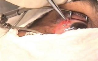
In the ocular form of dirofilariasis, the eyelids, conjunctiva, anterior chamber, sclera, and orbit are affected (Figure 1, 2). When the skin of the eyebrows and eyelids is affected, angioedema develops, which is associated with the parasitism of female (rarely male) dirofilaria in the subcutaneous tissue.
The eyelids are sharply swollen, pasty, inactive, close the eye, sometimes there is itching of varying intensity and lacrimation from moderate to very strong, pain at rest and on palpation. Some patients experience a sensation of a foreign body in the eye, movement in the area of the seal, and protrusion of the eye. Hyperemia of the skin of the eyelids, ptosis and blepharospasm are characteristic.
Dense nodules, granulomas, or tumors form under the skin. Some patients notice the presence of heartworm in the conjunctiva of the eye when looking in the mirror (Figure 1, 2).
Figure 1. Heartworm under the skin of the upper eyelid (the worm is actively moving).
Source: https://www.vsevrachizdes.ru/blog/dirofilyarioz-i-drugie-transmissivnye-infekcii