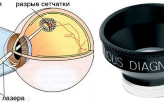
The retina provides primary information processing. It is on it that the image is formed after the refraction of rays in the optical media of the eye. Moreover, the retina is also the thinnest structure of the eyeball. Even a minor injury can lead to its detachment. Let us consider the features of this pathological process.
The retina, or inner layer, lines the inside of the eyeball. Light rays, refracted after passing through the vitreous body, cornea and lens, fall on the retina and form an image.
These light pulses are converted into nerve impulses and transmitted to the brain. This is how the process of vision can be described in general terms. The retina is also the thinnest structure of the eye. Various factors can lead to a violation of its integrity.
Any damage to the retina of the eye is accompanied by impaired visual function.
Retinal detachment (detachment) is the separation of the retina from the choroid. Light-sensitive receptors, which are located in the inner shell, do not receive nutrients due to disruption of connections with blood vessels.
As a result, these receptors cannot perform their functions, that is, ensure the normal functioning of vision. Gradually, the places where the retina has detached fill with fluid. Ultimately, this pathological process can lead to complete and irreversible blindness.
Why does it start? Let us list the causes and symptoms of the disease.
Retinal detachment: causes
The main, or primary, cause of retinal detachment is its rupture. This subsequently leads to the accumulation of fluid, which causes detachment. Rupture can be caused by various diseases - systemic and ophthalmological. They can be considered secondary causes, or predisposing factors. These include:
- Myopia. In patients with myopia, the eyeball is larger than in people without visual impairment. Because of this, light rays, after refraction, end up not at the center of the retina, but in front of it. The reasons for abnormal growth of the visual organs can be different. The main one is a genetic predisposition to this refractive error. As the eyeball increases in size, the retina stretches. Unlike the cornea, vitreous body and other structures, the retina does not grow. With a high degree of myopia, it is in constant tension. Gradually it becomes thinner. As already noted, this is the thinnest membrane of the eye. Any minor injury, physical activity, or stress can lead to its rupture and subsequent detachment.
- Diabetes. This disease is one of the main culprits of retinal detachment. Diabetics are often diagnosed with a disease called diabetic retinopathy. It develops due to a violation of the integrity of blood vessels. An increase in blood glucose levels affects the condition of the entire vascular system. It becomes more vulnerable, and the vessels become fragile. When they rupture, blood enters the retina. In other words, hemorrhage occurs in the inner lining of the eye. The accumulation of blood is accompanied by the formation of scars. They become the cause of detachment.
- Retinal dystrophy is the process of death of the tissues of the inner membrane. There are different causes and types of this degenerative disease. Almost all of them ultimately lead to retinal detachment.
Detachment can also occur due to other factors. This pathological process can be triggered by:
- mechanical injury to the eyeball;
- eye surgery;
- diseases affecting the circulatory system;
- inflammatory, infectious, viral ophthalmological diseases;
- excessive physical activity;
- pregnancy and childbirth;
- stress.
Now we will find out how this disease manifests itself.
Retinal detachment: symptoms
If you consult a doctor when the first symptoms of detachment appear, the risk of a quick recovery and a favorable treatment outcome increases significantly. The disease is accompanied by the following symptoms:
- the appearance of a veil before the eyes;
- shadows in the field of view, especially when moving the head;
- floating black dots/spots that indicate vitreous hemorrhage;
- flashes, lightning, sparks in the eyes;
- curvature of shapes and outlines of objects.
Also, with this pathology, people complain of constant fatigue. For some, vision improves in the morning, but becomes worse in the evening. This is due to the fact that at night the liquid that accumulates in the areas of detachment partially dissolves.
Symptoms of retinal detachment can be severe or moderate. This depends on the type of pathological process and its location.
So, with a detachment in the upper part of the eye, vision deteriorates quickly, due to the fact that the fluid begins to descend down, provoking the separation of the lower sections of the inner membrane from the choroid.
As the pathological process develops from below, signs of detachment appear gradually. The disease is asymptomatic for a long time. This form of pathology is more dangerous.
Types of retinal detachment: symptoms
Depending on the causes, provoking factors, degree of development, localization of detachment or ruptures, and duration of the disease, several types of retinal detachment are distinguished. Depending on the type of occurrence, there are the following types of this pathology:
- primary;
- secondary;
- traumatic.
Primary, or dystrophic, is a consequence of a violation of the integrity of the inner membrane, as a result of which fluid from the vitreous body penetrates into it. In this case, degenerative processes in the retina become a provoking factor in the development of the disease.
Another name for this type of detachment is rhegmatogenous detachment. Translated from Greek, the word "regma" means gap. In most cases, this is what causes the layers of the inner shell to separate. More often this happens in older people.
This is due to the fact that with age, the vitreous body loses its elasticity.
Traumatic detachment is a consequence of mechanical damage to the eye or surgery on the eyeball. The pathological process can occur immediately at the moment of tissue damage and even several months or a year after the injury. Therefore, symptoms of detachment may not appear immediately, which is why the disease is not diagnosed in a timely manner.
Secondary detachment develops against the background of various systemic ailments or ophthalmic pathologies. Along with signs of retinal detachment, symptoms of the underlying disease are observed. The secondary form is further divided into two types:
Tractional detachment of the retina is caused by abnormal adhesive processes between it and the vitreous body. In other words, detachment occurs in places where the retina is stretched in the area of the vitreous substance, which is provoked by the proliferation of blood vessels.
Serous detachment is the result of fluid penetrating into the inner part of the eye from the vessels of the retina. This usually occurs with arterial hypertension, vasculitis, swelling of the optic nerve head and other severe pathological processes.
Detachment is accompanied by symptoms characteristic of the diseases that cause it.
According to the severity of the pathology, there are 4 types:
- Local. Detachment is characterized by damage to ¼ of the entire retina.
- Common is a pathological process that covers 1/2 of the inner membrane.
- Subtotal leads to the spread of the disease by ¾.
- Complete detachment, or total, involves the entire plane of the ocular structure in question.
There are other classifications. So, if the fluid under the retina has collected in a bubble, a vesicular detachment is diagnosed. The formation of folds indicates flat peeling. According to the mobility of the pathological process, they are distinguished:
- mobile detachment, localized in the lower layers;
- rigid retinal detachment, in which there is no attachment to the lower or upper layers.
Determining the type and form of the disease is the main task of diagnosing retinal detachment. The treatment method depends on its results. Let's find out what procedures are prescribed if this pathology is suspected.
Diagnosis of retinal detachment
Diagnostics takes place in several stages, including three types of research methods:
- standard;
- special;
- laboratory
Standard procedures include:
- visual acuity testing - visometry;
- measurement of intraocular pressure - tonometry;
- examination of the anterior part of the eye - biomicroscopy;
- fundus examination - ophthalmoscopy;
- examination of visual fields and detection of scotomas - perimetry.
Special methods may also be prescribed, for example, ultrasound of the eyes, which allows you to examine them in detail, assessing the functional features of the retina. In some cases, electrophysiological procedures (EPI) are required - electrooculography (EOG), electroretinography (ERG) and electroencephalography with the study of the potentials of the visual cortex.
Laboratory methods include blood tests - general, syphilis, hepatitis, biochemistry, as well as urine tests (general and sugar).
The main one of these studies is fundus ophthalmoscopy. Using this diagnostic method, the doctor can examine the retina in great detail, assess the extent of the disease, and the extent of the detachment. Under an ophthalmoscope, retinal detachments appear grayish-white against a red-pink background.
If the pathological process has been developing for months, scars and folds are observed. Areas of ruptures appear in red. However, they have non-standard shapes. After the ophthalmologist determines the cause and type of pathology, a treatment method is selected.
Let's consider the main ones and move on to the prevention of retinal diseases.
Retinal detachment: treatment
In case of retinal detachment, surgery is prescribed. However, surgical intervention is not always required. There are several ways to treat this pathology, which can be grouped into three groups:
- extrascleral;
- endovitreal;
- laser
Extrascleral methods include extrascleral ballooning and extrascleral filling. The essence of the first operation is as follows. A balloon with a catheter is inserted behind the eyeball. With its help, pressure is created on the scleral membrane. After this, laser fixation of the retina is carried out, that is, coagulation. After 5-7 days the balloon is removed. Contraindications to the procedure:
- extensive tears with their location in the back of the eye;
- folds occupying almost the entire fundus of the eye;
- hemorrhages into the vitreous body.
Extrascleral filling is also performed on the surface of the sclera. The purpose of the operation is to bring the detached part of the retina closer to the pigment epithelium.
The surgeon makes incisions on the conjunctiva and on certain areas of the scleral membrane, in places of detachment, and applies fillings from a silicone sponge. They are biocompatible. If necessary, the doctor removes accumulated fluid from the eye.
Vision returns after such an operation within a few months. Sometimes complications arise - cataracts, changes in refraction towards myopia, increased intraocular pressure.
Endovitreal methods of treating retinal detachment
These techniques involve treating the eye from the inside. One commonly prescribed procedure of this type is a vitrectomy, which involves removing part or all of the vitreous humor. This gives the doctor access to the back wall of the eye.
After this, he performs laser coagulation - soldering the detached mesh shell. The removed substance is replaced by a viscous transparent material - saline solution, gas, polymers.
There are a number of contraindications to vitrectomy:
- optic nerve pathologies;
- corneal clouding;
- severe retinal diseases.
Vitrectomy is one of the most effective ways to treat detachment of the inner lining of the eye. However, even after such an operation there are complications:
- retinal hemorrhages;
- increased intraocular pressure;
- change in corneal curvature;
- repeated detachment;
- development of inflammatory processes;
- damage to the lens.
Treatment of retinal detachment: laser type operations
Laser coagulation can be a stage of one of the procedures and a separate method for treating retinal detachment. This procedure is called peripheral restrictive laser coagulation.
It can also be considered as a prevention of retinal detachment. During the procedure, the doctor acts on the thinned areas of the inner shell with a laser, soldering it to the underlying tissues.
This achieves:
- increased blood flow speed;
- normalization of blood supply;
- nutrition of injured parts of the retina;
- blocking the flow of fluid under the inner shell.
If the patient has myopia, then laser correction can be performed within two weeks after such an operation. Peripheral restrictive laser coagulation is not prescribed for:
- gross changes in the fundus of the eye;
- the presence of films on the retina;
- clouding of optical media.
Prevention of detachment
So, the causes of detachment are primary and secondary. If there are predisposing factors, a person must take all measures to prevent the occurrence of a pathological process. It is necessary to limit physical activity, visit an ophthalmologist more often, and wear corrective means for existing myopia.
No specific prevention of retinal detachment has been developed. At the first signs, you should consult a doctor. Usually, the first symptom that appears is floating spots in the field of vision. They are noticeable when you look at light objects. This sign cannot be ignored.
It may also indicate other severe pathologies, including systemic ones.
Now you know what retinal detachment is, the causes, symptoms, treatment methods and measures to prevent this disease. A few words about its consequences. The most dangerous of them is blindness. There may be other complications:
- hypotony of the eye;
- cataract;
- subatrophy of the eyeball;
- chronic iridocyclitis;
- loss of some visual functions.
MagazinLinz.ru team
Source: https://magazinlinz.ru/otsloenie-setchatki.htm
Early signs and symptoms of retinal detachment
A complication of retinal detachment is complete loss of vision. This part of the visual apparatus perceives surrounding rays and sends the received information to the brain.
In the early stages of the pathological condition, flashes before the eyes, progressive loss of vision and a shadow in one of the visual fields are observed.
The retina receives nutrients from the eyeball. When any of the sections are detached, hypoxia of the nerve fibers and cones in the organ is observed, which leads to their complete death.
Early symptoms
The main symptomatic manifestations of the onset of the process of rhegmatogenous detachment are:
- Formation of floaters before the eyes. Such symptoms do not always signal retinal problems. If the symptom does not go away within a few hours, it is recommended to visit an ophthalmologist to prevent the development of the disease in the early stages.
- Flashes appear before the eyes. They develop as a result of disturbances in the perception of rays emanating from objects.
- Cloudy or blurry picture. With pathology, a veil appears before the eyes that cannot be eliminated on your own. Traditional methods are also powerless in the presence of fogging. For a quick diagnosis, the doctor needs to determine the period of onset and development of the disease. In the initial stages of the disease, clouding of the picture is observed after a night's sleep, which is due to the horizontal position of the body. When rising after waking up, there is a lag in the area of the organ of vision from the eyeball.
- Shadow in the central zone of vision. In this pathological condition, a ring is formed in the center of the gaze, which is called a Weiss ring.
- Complete loss of vision in one eye. Occurs due to disturbances in the metabolism of the organs of visual perception.
- Rapid loss of lateral vision. Due to the detachment of a section of the retina from the eyeball, the death of cones and nerve fibers is observed, which cannot be completely restored. Reduction or loss of lateral vision is also possible after the appearance of hemorrhages in the vitreous body.
- Deformation of objects. When the central zone of the eye surface is detached, visible objects may be distorted. There is also a change in letters when reading or the loss of several fields from the visible zone.
Complete detachment very rarely occurs rapidly. The manifestations of pathology can only be eliminated by surgical intervention, which must be carried out in a timely manner. In the initial stages of exudative detachment, an increase in visual defects is observed. When the macula is involved in the process, there is a noticeable decrease in the quality of the perceived image.
Diagnostics
Timely detection of the pathological process makes it possible to prevent complications that develop as a result of detachment. The following methods are used for diagnosis:
Having studied the anamnesis and the data obtained as a result of the diagnosis, the doctor makes a presumptive diagnosis and prescribes the necessary therapy.
Treatment
Therapy to eliminate detachment is based on surgical intervention. Detachment is treated using methods:
- Extrascleral method. Includes laser correction and cryotherapy. The method is based on gluing the areas of delamination.
- Endovitreal technique. During surgery, the eyeball is affected.
Preparing for surgery
Emergency surgery is performed only in the presence of injuries to the apple of the eye and retinal detachment. Preparation for surgery consists of the following stages:
- 7 days before the procedure, complete abstinence from anticoagulants is necessary;
- before the intervention, it is forbidden to eat for 6 hours;
- It is not recommended to drink alcohol and smoke tobacco;
- If there are prescriptions for the constant use of medications, you must notify the treating ophthalmologist about this.
Surgical intervention
Retinal detachment can only be eliminated through surgery. When detachment of the epithelium with nerve fibers requires the following manipulations:
- Pneumoretinopexy. Air is used to eliminate the detachment. Using pressure, complete contact of the retina with the eyeball is created. Air bubbles resolve on their own within a month.
- Laser coagulation. The peeled tissue is sealed using a laser.
- Scleroplasty. The ophthalmologist glues the detached areas with a silicone strip and presses them down. The procedure allows you to return the retina to its place.
- Vitrectomy. During the surgical procedure, the vitreous body is removed and the space is filled with gas or a special silicone substance. The manipulation is performed under local anesthesia, which is especially important in complicated forms of detachment of part of the visual organ. A small amount of silicone substance is introduced into the vitreous cavity. The duration of the procedure is from 2 to 4 hours.
Rehabilitation and recovery
As a result of the intervention, the patient remains under medical supervision for several hours. Patients are prohibited from leaving the hospital on the day of the procedure. After surgery, side symptoms may appear:
- attacks of nausea;
- blurring of the picture;
- pain syndrome.
After the manipulation, a sterile bandage is applied to the organ of vision, which is necessary to prevent injury and reduce the load on the visual apparatus.
Only a doctor can remove the bandage for examination or when indicated. It is not recommended to visit a sauna, steam bath or other thermal procedures during the rehabilitation period.
The recovery period due to the manipulation ranges from 1 to 2 weeks.
Complications
Possible complications after surgery include:
- continuation of detachment;
- scar formation;
- secondary infection of the organ of vision;
- development of the inflammatory process;
- swelling of the eye;
- hyperthermia.
Complications develop due to neglect of the attending physician’s instructions, additional diseases of the visual system, or the weakened condition of the patient.
Prevention
There are no specific methods for preventing retinal detachment. However, by adhering to certain rules, you can diagnose the pathological process in time:
- if the first symptomatic manifestations occur, seek medical help;
- in case of injuries to the visual apparatus, visit an ophthalmologist;
- in the presence of any infectious processes in the area of the visual organs, carry out timely treatment;
- if you are nearsighted or farsighted, you must visit an ophthalmologist at least once a year;
- avoid sudden lifting of heavy objects;
- beware of blows to the head;
- It is not recommended to overstrain the visual apparatus;
- control physical activity.
It is impossible to completely prevent detachment using prophylaxis, but it will be possible to notice the pathological process in time.
Source: https://proglazki.ru/simptomy/rannie-priznaki-otsloeniya-setchatki-glaza/
Retinal detachment
The retina is the thinnest multilayer structure of the eye, covering 70% of the area of the inner surface of the eyeball. The retina of the eye is connected to the visual analyzers of the brain, into which it transmits information and is responsible for converting light impulses into nerve impulses.
The inner side of the retina is adjacent to the vitreous body, and the outer side is adjacent to the choroid: the choroid from which it receives nutrients and oxygen.
Sometimes, under the influence of one reason or another, part of the retina moves away from the choroid. In this case, cells deprived of nutrition and oxygen quickly die.
Without surgical intervention, the process leads to disturbances up to complete loss of vision. This condition is called retinal detachment.
Retinal detachment is a severe pathological process requiring emergency surgical care, in which the retinal layer of the eyeball is separated from the vascular layer.
The term itself was first proposed at the beginning of the 8th century, but its clinical confirmation was made only in 1851 after the invention of the ophthalmoscope.
According to research, the incidence of the disease has increased significantly over the past decade: previously, 1 in 15,000 people faced detachment, now it is 1 in 10,000.
Patients with myopia and aphakia often suffer from retinal detachment, especially if part of the vitreous body was removed during surgery simultaneously with the lens. In the latter case, the risk of developing pathology increases to 10%.
Causes of retinal detachment
The most common cause directly leading to detachment is a retinal tear. As long as the shell is intact and sealed, it remains motionless. If a gap appears on it, its edges begin to lag further and further behind the choroid. The more extensive the process, the more severe the consequences for the patient, and the more difficult it is for the doctor to restore the patient’s vision.
Among the main causes of the disease are:
- Congenital or acquired myopia (myopia): 40-50% of all cases. With myopia, the size of the eye changes: this leads to tension in the tissues of the eye and, as a result, thinning of the retina, its fragility and vulnerability. The affected retina easily bursts, which leads to detachment.
- Injuries to the organ of vision: 10-20% of all cases.
- Surgical interventions (for example, cataract removal - 30-40% of all cases). It happens that retinal detachment recurs in an eye that has already been operated on for this reason.
- Oncological diseases of the eyes.
- Inflammatory diseases of the retina, degenerative processes in it.
- Hemorrhages, retinopathy, and retinal dystrophy significantly increase the risk of detachment.
- Systemic diseases of the body (endocrinological, hematological, cardiological, vascular).
The risk of the disease increases with intense sports (especially if it is associated with impacts, jumping), as well as pregnancy and childbirth - due to strong tension in pushing. If one eye has already had an episode of retinal detachment, the likelihood of a recurrence in the other increases by 15%. Heredity also influences the possibility of developing the disease.
Types of retinal detachment
Depending on the reasons that caused the detachment and the nature of the lesions, rhegmatogenous, traction and exudative (serous) types of the disease are distinguished.
Rhegmatogenous retinal detachment (RRD)
Primary detachment associated with a rupture (from the Greek rhegma - rupture) in the retinal membrane.
Medical statistics indicate that men are more likely to experience RRD, with the peak of the disease occurring at 65-69 years of age.
With RRD, the vitreous body begins to penetrate through the breaks formed in the retina, promoting increasingly severe detachment. The condition of the vitreous body loses its homogeneity with age, dividing into two fractions: thick and liquid, which explains the fact that the peak incidence occurs in old age.
Tractional retinal detachment (TRD)
Due to various pathological processes, adhesions can form between the retina and the vitreous body. Strands or formed vessels, when the position of the vitreous body changes, literally pull back and tear the retina away from the choroid. There is no retinal tear.
This type is associated with diseases such as diabetes, sickle cell anemia, retinopathy of prematurity, etc.
Exudative retinal detachment (ERD)
Exudative, or, as it is also called, serous retinal detachment also develops without breaks. With EOS, fluid accumulates in the lower parts of the subretinal space. With an increase in the volume of this interstitial fluid, the retina begins to lag behind the choroid.
The cause of the accumulation is a violation of vascular permeability. This indicator is affected by inflammatory and infectious diseases, genetic characteristics, oncology, complications of surgical interventions, systemic diseases, and kidney diseases.
The prognosis for any type of retinal detachment is associated with:
- how acutely the disease begins and how quickly the affected area increases;
- area and location of the detachment (if the central areas are affected, it will be more difficult to restore vision);
- speed of medical care.
Symptoms of retinal detachment
The main symptoms indicating retinal detachment are:
- the appearance of a veil, flies, floating spots before the eyes. The veil is not eliminated when using eye drops; “foreign objects” cannot be removed;
- blurred vision, decreased sharpness;
- “curtain” effect: peripheral vision suddenly disappears, as if part of the field is covered with a gray curtain. The situation improves in the morning: it seems to the patient that the area hidden behind the “curtain” has become smaller. By evening, the viewing area decreases again;
- flashes in the form of lightning or sparks;
- distortion of the objects in question.
Diagnosis and treatment of retinal detachment
If one or more symptoms appear, immediately consult a specialist: this will allow you to diagnose retinal detachment and begin treatment at an early stage.
To diagnose the disease, the doctor will check:
- visual acuity;
- fields of view;
- intraocular pressure.
In addition, the fundus of the eye will be examined and examined using an ophthalmoscope - a slit lamp.
- Slit lamp
- The ophthalmologist may also prescribe additional studies: ultrasound, electrophysiological (cardiogram of the eye), X-ray, tomography, MRI.
- The disease cannot be corrected with drops, ointments or tablets.
Treatment of retinal detachments is surgical. During the operation, the detached part is returned to its place, the breaks are blocked, and, if necessary, the cords connecting the retina with the vitreous body are removed. The retina, returned to its original position, is strengthened with fillings or a laser.
If the outcome is successful, significant improvements will occur immediately, and vision will be restored within another 3 months.
However, you may not be able to get rid of some of the consequences of retinal detachment - especially its advanced forms: blurry spots, blurriness and blurriness may remain forever.
Prevention of retinal detachment
To prevent retinal detachment, you can use all the recommendations that accompany a healthy lifestyle. Give up bad habits, eat a variety of foods rich in vitamins, follow the rules of eye hygiene, protect them from external influences - bright sunlight, shocks, and injuries at work.
Monitor the general condition of the body: control blood pressure and blood sugar levels. And, of course, do not forget about regular – at least once a year – visits to the ophthalmologist.
Source: https://ultralinzi.ru/articles/otsloenie-setchatki/
Retinal disinsertion
Retinal detachment is a pathology of the retina of the eye, in which it separates from the underlying choroid (choroid). Retinal detachment is accompanied by a sharp deterioration in vision, the appearance of a veil in front of the eye, a progressive narrowing of the field of vision, flickering of “floaters”, “sparks”, “flashes”, “lightning”, etc. Diagnosis is carried out using visometry, perimetry, tonometry, biomicroscopy, ophthalmoscopy, ultrasound of the eye, electrophysiological studies. Treatment is carried out surgically (scleral filling, scleral ballooning, transciliary vitrectomy, vitreoretinal surgery, cryocoagulation, etc.) or laser methods (laser coagulation of the retina).
Retinal detachment is a dangerous and most complex pathological condition in surgical ophthalmology, which is diagnosed annually in 5-20 people per 100 thousand population. Today, retinal detachment is the leading cause of blindness and disability; Moreover, 70% of cases of this pathology develop in people of working age.
With retinal detachment, the layer of photoreceptor cells (rods and cones) for certain reasons is separated from the outer layer of the retina - the pigment epithelium, which leads to disruption of the trophism and functioning of the retina. If specialized assistance is not provided in time, retinal detachment can quickly lead to vision loss.
Retinal disinsertion
According to the mechanism of pathology formation, rhegmatogenous (primary), traumatic and secondary (exudative and traction) retinal detachment are distinguished.
- The development of rhegmatogenous retinal detachment is associated with a rupture of the retina and the penetration of fluid from the vitreous body under it. This condition develops when the retina thins in areas of peripheral dystrophy. With various types of retinal dystrophies (latticed, racemose, retinoschisis, etc.), a rupture in a degeneratively changed area can be triggered by sudden movements, excessive physical stress, traumatic brain injury, falls, or occur spontaneously. According to the type of defect, primary retinal detachment can be bubble-shaped or flat; according to the degree of detachment - limited or total.
- Retinal detachment of traumatic origin is caused by eye injuries (including surgical injuries). In this case, retinal detachment can occur at any time: immediately at the time of injury, immediately after it, or several years later.
- The occurrence of secondary retinal detachment is observed against the background of various pathological processes of the eye: tumor, inflammatory (with uveitis, retinitis, chorioretinitis), occlusion (occlusion of the central retinal artery), diabetic retinopathy, sickle cell anemia, toxicosis of pregnancy, hypertension, etc.
- Secondary exudative (serous) retinal detachment is caused by the accumulation of fluid in the subretinal space (under the retina). The traction mechanism of detachment is caused by tension (traction) of the retina by fibrinous strands or newly formed vessels growing into the vitreous body.
Factors that increase the risk of retinal detachment include myopia, astigmatism, degenerative changes in the fundus, eye surgery, diabetes mellitus, vascular pathology, pregnancy, cases of similar pathology in close relatives, etc.
In most cases, retinal detachment develops in one eye; in 15% of patients there is a risk of bilateral pathology. In the presence of bilateral cataracts, the risk of bilateral retinal detachment increases to 25-30%.
At the onset of the disease, warning symptoms appear - so-called light phenomena. These include flashes of light (photopsia) in front of the eyes and zigzag lines (metamorphopsia).
When a retinal vessel ruptures, “floaters” and black dots appear before the eyes and pain in the eye.
These phenomena indicate irritation of the light-sensitive cells of the retina caused by traction from the vitreous body.
With further progression of retinal detachment, a “veil” appears before the eyes (according to patients, “a wide curtain, a curtain”), which increases over time and can occupy most or the entire field of vision.
Visual acuity decreases quickly. Sometimes in the morning, visual acuity improves for a while, and the visual field expands, which is associated with partial resorption of fluid during sleep and the independent reattachment of the retina.
However, within a day, the symptoms of retinal detachment return again.
Temporary improvement in visual function occurs only with recent retinal detachment; If the defect persists for a long time, the retina loses its elasticity and mobility, which is why it cannot fit into place on its own.
When the retina ruptures in the lower parts of the fundus, the detachment progresses relatively slowly, over several weeks or months, without causing visual field defects for a long time.
This type of retinal detachment is very insidious, since it is detected only when the macula is involved in the process, which aggravates the prognosis in relation to visual functions.
When the retinal tear is localized in the upper parts of the fundus, on the contrary, retinal detachment progresses quite quickly, over several days. The fluid accumulating in the subretinal space, with its weight, detaches the retina over a large area.
If help is not provided in time, detachment of all quadrants of the retina may occur, including the macular area - complete, total detachment. When the macula is detached, curvature and vibrations of objects occur, followed by a sharp drop in central vision.
Sometimes, with retinal detachment, diplopia occurs due to decreased visual acuity and the development of hidden strabismus. In some cases, retinal detachment is accompanied by the development of sluggish iridocyclitis and hemophthalmos.
If retinal detachment is suspected, a complete ophthalmological examination is necessary, since early diagnosis can avoid irreversible vision loss. If there is a history of TBI, the patient must be consulted not only by a neurologist, but also by an ophthalmologist to exclude ruptures and signs of retinal detachment.
The study of visual functions is carried out by checking visual acuity and determining visual fields (static, kinetic or computer perimetry). Loss of visual fields occurs on the side opposite the retinal detachment.
Using biomicroscopy (including using a Goldman lens), the presence of pathological changes in the vitreous body (strands, destruction, hemorrhages) is determined, and peripheral areas of the fundus are examined. Tonometry data are characterized by a moderate decrease in IOP compared to a healthy eye.
The key role in recognizing retinal detachment belongs to direct and indirect ophthalmoscopy.
The ophthalmoscopic picture allows us to judge the location of the breaks and their number, the relationship of the detached retina with the vitreous body; allows you to identify areas of dystrophy that require attention during surgical treatment.
If it is impossible to perform ophthalmoscopy (in case of opacities in the lens or vitreous body), an ultrasound of the eye in B-mode is indicated.
The diagnostic complex for retinal detachment includes methods for studying entopic phenomena (the phenomenon of autoophthalmoscopy, mechanophosphene, etc.).
To assess the viability of the retina and optic, electrophysiological studies are carried out - determining the threshold of electrical sensitivity and lability of the optic nerve, CFSM (critical flicker fusion frequency).
Detection of pathology requires immediate surgical treatment.
Delay in treating this pathology is fraught with the development of persistent hypotension and subatrophy of the eyeball, chronic iridocyclitis, secondary cataracts, and incurable blindness.
The main goal of treating retinal detachment is to bring the layer of photosensitive receptors closer to the pigment epithelium and create an adhesion of the retina with the underlying tissues in the area of the rupture.
In surgery for retinal detachment, extrascleral and endovitreal techniques are used: in the first case, the intervention is performed on the scleral surface, in the second - inside the eyeball. Extrascleral methods include filling and ballooning of the sclera.
Extrascleral filling involves suturing a special silicone sponge (filling) to the sclera, which creates an area of scleral depression, blocks retinal breaks and creates conditions for the gradual absorption of fluid accumulated under the retina by capillaries and pigment epithelium. Options for extrascleral filling for retinal detachment can be radial, sectoral, circular (circlage) filling of the sclera.
Ballooning of the sclera in case of retinal detachment is achieved by temporarily suturing a special balloon catheter into the area of projection of the rupture; when inflated, an effect similar to filling occurs (a shaft of scleral depression and resorption of subretinal fluid).
Endovitreal treatments for retinal detachment may include vitreoretinal surgery or vitrectomy. During the process of vitrectomy, the altered vitreous body is removed and special preparations (liquid silicone, saline solution, special gas) are introduced instead, which bring the retina and the choroid closer together.
Gentle methods of treating retinal detachment include cryocoagulation of breaks and subclinical retinal detachments and laser coagulation of the retina, which allows for the formation of chorioretinal adhesions. Cryopexy and laser coagulation of the retina can be used both for the prevention of retinal detachment and for therapeutic purposes alone or in combination with surgical techniques.
The prognosis depends on the duration of the pathology and the timeliness of treatment. Surgery performed early after the development of retinal detachment usually promotes a favorable outcome.
In most cases, retinal detachment can be prevented. For this purpose, patients with myopia, retinal dystrophy, diabetes mellitus, head and eye injuries need regular preventive examinations by an ophthalmologist.
An examination by an ophthalmologist is included in the standard of pregnancy management and helps prevent retinal detachment during childbirth.
Patients at risk for retinal detachment are contraindicated in heavy physical activity, heavy lifting, and playing certain sports.
When areas of retinal dystrophy are identified, cryopexy or laser coagulation of the retina is performed for preventive purposes.
Source: https://www.KrasotaiMedicina.ru/diseases/ophthalmology/retinal-detachment
Why is retinal detachment dangerous?
Retinal detachment is the separation of the retina from the underlying vascular layer. There is a disruption in the nutrition of the exfoliated area of light-sensitive neurons, the death of which can become irreversible if the pathological process lasts more than 72 hours.
Some anatomical details
To understand what it is, what a detachment is, let’s understand the anatomical structure of the eyeball.
The cavity of the organ of vision is filled with the vitreous body (vitreum), which serves to maintain its tone and shape and transmit light rays to the retina. It resembles a clear jelly and is 90% water. On the outside, it is formed by a shell - the hyaloid membrane. Normally, the vitreum has several fixing adhesions, including those with the retina.
The retina (retina) covers the inner surface of the eyeball. It perceives and processes visual information, converts it into nerve impulses and transmits it through the optic nerve to the central nervous system. Consists of ten cell layers. One of them is the neuronal layer of photoreceptors (rods, cones).
The reticular layer is adjacent to the choroid, which, together with the central artery, performs the function of blood supply.
Causes of retinal detachment
The most common cause is the presence of a tear in the retina, through which the liquid contents of the vitreous body enters under the retina and separates it from the underlying tissues. It is rhegmanthogenic (Greek.
regma - gap) detachment mechanism: with age-related or degenerative changes, rough adhesions are formed between the hyaloid membrane of the vitreous body and the retina.
When the eyeball moves (and in a static state), a traction (pulling) effect occurs on the reticular layer and leads to its rupture.
More than 40% of cases of rhegmantogenic detachment are caused by cataract surgery. Most often occurs in men. The average age of patients is 40-70 years. At a young age, a retinal tear is caused by eye injury.
The development of traction is caused by:
- blunt (concussion) or penetrating eye injuries, head injuries,
- long-term dystrophic retinopathy and pathologies of the vitreous body (diabetic retinopathy, sickle cell anemia, thrombosis of the ophthalmic veins),
- inflammatory diseases in the posterior parts of the eye,
- progressive malignant myopia,
- liquefaction of the vitreous.
Retinal detachment can be caused by sudden physical stress: coughing, straining, lifting heavy objects.
The mechanism of exudative detachment is due to the pathology of the vascular layer. The permeability of blood vessels is disrupted, fluid comes out of them and accumulates under the mesh layer, separating it and depriving it of blood supply.
The cause of exudative retinal detachment can be:
- malignant neoplasms,
- thrombosis of the central ophthalmic vein,
- arterial hypertension,
- local vasculitis,
- genetic pathologies.
Symptoms of retinal detachment
The prognosis for the disease depends on timely medical care received. Therefore, patients at risk should know the initial signs of retinal detachment:
- the appearance of a dark “curtain” before the eyes above, below, right or left (corresponds to the place of detachment),
- objects look like they are through water,
- lightning flashes, sparks, other photo effects (occur from the opposite side),
- the appearance of floating black flies,
- reduction of field of view,
- a sharp decrease in visual acuity.
There are no external signs of retinal detachment. But with a large degree of it, the color of the pupil may change - it becomes gray.
Morning improvements in the condition are explained by partial resorption of the fluid overnight, when the patient is in a horizontal position. But then the condition continues to worsen.
First aid for suspected detachment
Immediately transport the victim to the hospital, preferably to the eye department. Transportation should be gentle. If the road is bumpy, you need to drive slowly: strong shaking will increase the detachment.
Ensure bed rest, only get up to go to the toilet. Eat food while sitting in bed. Ensure immobility of the affected eye: a thick, light-proof bandage is applied to both eyes.
The rest mode helps to reduce the accumulation of intraocular fluid under the retina, improve its resorption, and helps straighten the separated area.
Diagnosis of retinal detachment
A complete ophthalmological examination is indicated:
- visual acuity test,
- checking visual fields (perimetry),
- examination using a Goldmann lens (the condition of the vitreous body and fundus of the eye is examined),
- ocular tonometry (with detachment, eye pressure decreases slightly),
- ophthalmoscopy,
- Ultrasound (for vitreous opacities, when ophthalmoscopy is inconclusive),
- Electrophysiological examination determines the viability of the optic nerve and photosensitive layer.
Treatment
No conservative methods - tablets, injections - will help with detachment. Emergency surgery is the only way to save the eye and restore vision. Therapeutic measures are selected individually, depending on the specific type of detachment.
The main goal of the operation is to bring the sensory layer closer to the underlying tissues.
To close the gap, inflammation is artificially caused around it (exposure to cold or laser). Subsequent scarring fuses the retina to the underlying layer. For a more reliable seal, a silicone seal is sutured to the eye from the outside, which, pressing into the wall of the eye, presses the vascular layer to the retina.
Vitrectomy - removal of the pathologically altered vitreous body, replacing it with liquid silicone or special gas to press the mesh layer from the inside to the vascular wall.
Prevention and prognosis for retinal detachment
Patients at risk (severe myopia, diabetes mellitus, history of head and eye injuries) should undergo regular examinations by an ophthalmologist. Pregnant women undergo routine examinations in the eye office to prevent possible abruption during childbirth.
The prognosis is determined by the duration of the disease and the urgency of the emergency measures taken.
Source: http://ShkolaZreniya.com/bolezny/otsloyka-setchatki
Retinal detachment
The retina is a thin tissue that fits tightly to the choroid of the eye. The retina is responsible for the interaction between the optical system of the eye and the visual parts of the brain; it receives and transmits visual information.
Detachment of the retina from the choroid that feeds it is a serious problem that requires a surgical solution. If this happens, it is necessary to seek qualified help as soon as possible; delay can lead to irreversible loss of vision.
When it comes to retinal detachment, the risk group primarily includes those who suffer from moderate or high myopia, diabetes mellitus, circulatory disorders, various inflammatory diseases, as well as those who have suffered head injuries, eye injuries and surgical interventions on the organs of vision.
The causes of rupture or detachment of the retina can be excessive physical exertion and severe shaking of the body when jumping and falling , so even people with excellent physical data and excellent vision, who engage in, for example, contact sports, ski jumping, etc., as well as Representatives of some extreme professions must be attentive to the health of their eyes.
Types and symptoms of retinal detachment
Retinal detachment can be primary or secondary.
Primary is a pathology in which detachment is preceded by a rupture followed by fluid flowing under the retina and detachment of this important membrane of the eye.
Secondary detachment develops as a complication of some pathological process - for example, due to the appearance of a neoplasm between the retina and choroid of the eye.
The following types of retinal detachment can be distinguished:
- rhegmatogenous (from the Greek rhegma, rupture), resulting from a rupture of the retina;
- traction , which occurs due to tension in the retinal tissue from the vitreous body;
- exudative , characterized by the penetration of serous fluid into the space under the retina and caused by increased vascular permeability and other hemodynamic disorders of the eye;
- mixed - for example, traction-rhegmatogenous type, in which a rupture forms against the background of traction of the vitreous body.
Retinal detachment
Normal vision
Retinal detachment is a painless . You can suspect that the retina needs urgent attention from specialists by noticing the following symptoms:
- flashes, lightning, flickering;
- black spots floating before the eyes, cobwebs;
- cloudiness before the eyes in the shape of a ring;
- deformation and vibration of the objects in question;
- decreased visual acuity;
- a veil before the eyes;
- sudden loss of lateral vision.
Flashes and lightning are usually present during the initial stages of rhegmatogenous retinal detachment. The development of exudative and tractional detachment may have more sparse symptoms, when a noticeable deterioration in vision is noted only after the central region of the retina, the macula, is involved in the pathological process.
Healthy retina
Retinal detachment
Detachments affecting the central region of the retina have a worse prognosis for vision preservation than pathology localized in the periphery.
Retinal detachment can be acute and take just a few hours or even minutes - or it can take years. In the initial stages, changes occurring in the visual system may be completely invisible. Therefore, at least once a year it is necessary to undergo an examination by an ophthalmologist.
Diagnosis of retinal detachment
In order to confirm or refute the diagnosis of retinal detachment, it is necessary to undergo a thorough ophthalmological examination.
At the Excimer clinic, diagnostics are performed using a complex of modern computerized equipment; the level and quality of such studies allow our specialists to create the most complete and objective picture of the condition of the patient’s retina.
Evaluation of patients with suspected retinal detachment may include:
- examination of the fundus (ophthalmoscopy);
- optical coherence tomography of the posterior segment of the eye;
- study of visual fields (perimetry) in order to assess the condition of the retina in its periphery;
- electrophysiological study - determination of the viability of nerve cells of the retina and optic nerve;
- ultrasound examination of the internal structures of the eye and other diagnostic procedures.
Important! Retinal detachment can only be diagnosed by an ophthalmologist. If a pathological process develops, treatment must be started as early as possible, this significantly increases the chances of a successful outcome.
The main goals of treatment for retinal detachment are to restore the integrity of the retinal tissue and ensure their adherence to the choroid. For treatment, methods such as vitrectomy, extrascleral (episcleral) filling, and laser coagulation can be used.
Vitrectomy
The vitrectomy operation involves partial or complete removal of the vitreous body of the eye. In case of retinal detachments, this allows you to gain access to the affected tissues and carry out the necessary actions to restore the viability of the retina.
The vitreous body removed during vitrectomy is replaced with a special gas mixture, silicone oil or water saturated with fluoride. Subsequently, the tamponing substance is removed surgically or resolves on its own, being replaced by natural intraocular fluid. The anatomy of the eye is restored .
For patients at the Excimer Clinic, we offer vitrectomy surgery in the most gentle way possible, through micropunctures. This microinvasive vitrectomy has a number of advantages compared to standard surgery, including less trauma, a reduced risk of bleeding, and, of course, a shorter postoperative recovery period.
Extrascleral (episcleral) filling
A procedure during which maximum adherence of damaged retinal tissue to the choroid of the eye is achieved by applying fixing fillings to the sclera, the outer “white” membrane of the eye, in places of retinal detachments and ruptures.
At the Excimer Clinic, such microsurgical interventions are performed using high-precision equipment of the latest generation. Our doctors have extensive experience in performing all types of extrascleral filling, achieving the best possible results for retinal damage of any type.
Laser coagulation
Using a laser beam, creating point adhesions between the retina and choroid of the eye, thereby strengthening the retina. Timely laser coagulation can prevent further damage to the retina.
The operation is non-contact and bloodless, under local drip anesthesia, which is usually easily tolerated by patients of all ages. In our clinic, such intervention is performed using high-precision equipment of the latest generation.
Benefits of treating retinal detachment at the Excimer Clinic
- All operations and procedures aimed at treating retinal detachment at the Excimer ophthalmology clinic are carried out in a convenient “one-day” mode for patients, without hospitalization .
- An obligatory component of quality treatment is the use of the wide capabilities of the latest generation microsurgical equipment. It is this equipment that allows our specialists to act effectively and achieve results even in situations of complex retinal problems.
- The qualifications and vast practical experience of our doctors allow us to perform microsurgical interventions at the highest professional level. We regularly exchange current experience in the treatment of retinal diseases with Russian and foreign colleagues and offer patients of our clinic all the best and most effective of what modern ophthalmology is proud of.
Prevention of retinal detachment
The main medical method for preventing retinal detachment today is PPLC, a procedure of peripheral preventive laser coagulation. The essence of the method is to strengthen the retina by creating point fusions between the retina and choroid of the eye. PPLC is performed using a laser, non-contact and bloodless .
As a general prevention of retinal diseases, experts recommend:
- stop smoking and excessive drinking of alcohol;
- diversify the menu with products that have a beneficial effect on the functioning of the visual system - nuts, fish, egg yolk, vegetables, fruits, berries, herbs, etc.;
- maintain a sleep-wake schedule;
- pay attention to harmonious physical development, avoid traumatic sports;
- observe the rules of visual hygiene.
It is necessary to regularly monitor blood sugar levels and blood pressure, and for people at risk, make routine visits to the ophthalmologist more frequent.
As a medicinal prevention of problems with the retina, the doctor may recommend various vitamin complexes, as well as courses of special medications for the eyes.
For vascular diseases, these can be Quinax, Emoxy-Optic, Emoxipin drops.
As part of the complex treatment of retinal dystrophy - Taufon, Aisotin and other drugs, which are used only after examination of the visual system and as prescribed by a doctor.
Leading ophthalmic surgeon and medical director of the Excimer clinics, doctor of the highest category, Doctor of Medical Sciences, Professor,
Academician of the Russian Academy of Natural Sciences
Pashinova Nadezhda Fedorovna Chief physician of the Moscow ophthalmological clinic "Excimer", ophthalmic surgeon of the highest category, Doctor of Medical Sciences, Associate Professor,
Academician of the Russian Academy of Natural Sciences
, A. Alexander Isakovich, Moscow
I, Alexander Isakovich Alterman, am simply shocked by the attention and sensitivity of Dr. Kalashi Zurab Yusupovich and Irina Srgeevna Yusupova. I am a disabled person of the 2nd group, a labor veteran born in 1938.
Ë
È
The doctor diagnosed retinal edema. What to do?
Retinal edema accompanies inflammatory and dystrophic changes in retinal tissue. Treatment should not be delayed under any circumstances, as this may result in irreparable loss of vision.
- Ë
- È
- Is it possible to play sports with high degrees of myopia?
Myopia itself is not a reason to stop playing sports. The cause can only be dystrophic foci found on the retina, which are dangerous in terms of the appearance of breaks. We recommend that you undergo a full diagnostic examination and consult a retinologist.
- Ë
- È
- I have diabetes, how does this affect my vision?
Diabetes mellitus is a fairly common disease. In diabetes mellitus, due to insufficient insulin production, metabolic disorders occur, which also has a negative effect on the visual system. One of these manifestations of diabetes is diabetic retinopathy.
In almost half of the cases, diabetic retinopathy is not diagnosed and is often discovered only after complications have developed. In diabetes, changes occur in the blood vessels of the retina, which leads to a disruption in the supply of oxygen to the retinal vessels.
Hemorrhages, deposits of metabolic products, and retinal edema occur on the retina. Later stages are characterized by the formation of new vessels that grow from the retina into the vitreous body, causing hemorrhages in it and deterioration of vision.
Diabetic retinopathy develops gradually, and even its pronounced stages may be imperceptible to the patient. Therefore, patients with diabetes need to regularly visit a retina specialist and undergo a retinal examination.
| Ask a Question | All questions |
Article rating: 4.6/5 (308 ratings)
Source: https://excimerclinic.ru/retina/retinaldetachment/