Optic neuritis (optic neuritis) is a pathology that is characterized by the course of the inflammatory process in the optic nerve and damage to its tissue and membrane.
There are two forms of the disease - intrabulbar and retrobulbar.
More often, the disease develops against the background of destruction of fibers caused by neurological disorders and causes deterioration of visual functions and a number of other symptoms.
Structure and functions
In order to imagine the process of neuritis of the optic nerve, it is necessary to consider its structure and functions. It consists of neuron axons (processes) that extend from the retina. The nerve, consisting of more than 1 million fibers, transmits signals in the form of impulses to the visual center of the brain. It originates behind the disc of the organ of vision.
The area inside the retina where the optic nerves are located is called intrabulbar, or intraorbital. The area where the fibers enter the cranium is known as the retrobulbar.
In neurology, the optic nerve performs several functions:
- ensures the ability of the eye to distinguish objects of different sizes (visual acuity);
- provides the ability to distinguish colors;
- determines the visibility area (the boundaries of the field of view).
If this inflammatory disease develops, the functional abilities of the eye simultaneously decrease.
Neuritis of the eye cannot be completely cured. This is due to the fact that decreased visual function causes degeneration of nerve fibers that cannot be restored.
Kinds
The classification of optic nerve pathology is based on the etiology of the development of the disease and the localization of the inflammatory process. According to the first sign, neurosis is divided into:
- toxic;
- ischemic;
- autoimmune;
- parainfectious;
- infectious;
- demyelinating.
The parainfectious form develops against the background of an infectious lesion of the body or an abnormal reaction to the vaccine. The ischemic type of pathology occurs due to an acute disorder of cerebral circulation caused by diseases of the cardiovascular system (pressure, hypertension), diabetes mellitus, atherosclerosis, blood diseases and others.
Toxic optic neuritis is caused by poisoning with chemical compounds, methyl alcohol, pesticides of various classes and other toxic and volatile toxic substances.
Depending on the location of the inflammatory process, intrabulbar (papillitis) and retrobulbar forms of the disease are distinguished. The first type of neuritis occurs with changes in the optic nerve head.
In rare cases, simultaneously with papillitis, the layer of nerve fibers that makes up the retina becomes inflamed. This condition is known as neuroretinitis. This form of optic neuritis is usually associated with Lyme disease, syphilis, viral pathologies, or cat scratch disease.
Retrobulbar neuritis is localized behind the eyeball. The disease does not cause changes in the optic nerve head and therefore this form of neuritis is usually detected after the inflammatory process has spread to the intraocular part.
Causes of the disease
The causes of the development of optic neuritis in children and adults are mainly due to infectious infection of the body. Pathology can occur when:
- keratitis, iridocyclitis and other eye diseases;
- meningitis, encephalitis;
- periostitis, phlegmon;
- chronic tonsillitis, sore throat, sinusitis and other diseases of the nasopharynx.
Systemic infectious pathologies can cause optic neuritis:
- tuberculosis;
- typhus;
- malaria;
- brucellosis;
- diphtheria;
- gonorrhea and others.
Inflammation of the optic nerve is often observed during complicated pregnancy and traumatic brain injury. Neuritis can be provoked by prolonged alcoholism, diabetes mellitus, blood pathologies, and autoimmune diseases.
The course of the pathology provokes inflammatory edema, which causes compression of the optic fibers, leading to their degeneration.
As a result, visual acuity decreases. At the same time, over time, the intensity of the pathological process decreases and eye functions are restored. In advanced cases, the inflammatory process causes the breakdown of fibers, in place of which connective tissue is formed. Because of this, atrophy of the optic nerve occurs, which cannot be restored.
The risk group for developing ocular neuritis includes people aged 20-40 years. More often the disease is diagnosed in women. A high risk of pathology is observed in multiple sclerosis. This pathology promotes demyelination (destruction of the myelin sheath) of nerve fibers.
Symptoms of optic nerve neuralgia
With inflammation of the optic nerve, symptoms and treatment are determined by the form of the disease. The most common is intrabulbar ocular neuritis, which is characterized by intense manifestation and rapid development.
The following symptoms are observed with this disease:
- Scotomas (blind spots). The main symptom of neuritis. Due to damage, the optic nerve does not conduct all signals generated by the eye. As a result, the patient does not see individual zones, the size of which varies depending on the severity of the case.
- Myopia (decreased visual acuity). Diagnosed in 50% of cases. With neuritis, visual acuity decreases by 0.5-2 diopters. In extreme cases, the patient stops seeing in one eye. Depending on the causative factor and the intensity of development of the inflammatory process, blindness can be reversible or irreversible.
- Decreased quality of vision at night. Patients with damage to the optic nerve begin to distinguish objects in the dark with a delay of 3 minutes, when normally it is 40-60 seconds.
- Partial or complete lack of color perception. With intrabulbar lesions, patients cease to distinguish shades of colors.
In addition, due to the disease, patients see blurry spots instead of objects. At the same time, the boundaries of the visual fields remain normal.
Symptoms of chronic retrobulbar neuritis of the eye manifest themselves in different ways. This is explained by the structural features of the nerve, which runs freely in the cranial cavity.
In case of damage to the inner part of the fibers, signs characteristic of the intrabulbar form of the disease are noted.
The development of inflammation on the outer layer provokes painful sensations in the eyes and a decrease in the field of vision while maintaining the same sharpness.
Diagnosis of inflammation of the optic nerve
Symptoms of pinched optic nerve are characteristic of various pathologies, and therefore this problem is considered in neurology and ophthalmology. To diagnose the disease, a consultation with an ophthalmologist is often sufficient. In this case, it is necessary to differentiate optic neuritis from other pathologies with similar symptoms.
With minor lesions, the disease causes subtle changes in the structure of the disc and mild visual disturbances. In this case, fluorescein angiography of the fundus will be required. Using this procedure, neuritis is differentiated from diseases of the optic nerve (congestive disc and others). Additionally, lumbar puncture and echo-encephalography are prescribed for a similar purpose.
When developing treatment tactics, it is important to take into account the specific causes of the disease. To establish it, MRI of the brain, enzyme immunoassay, blood culture and other examination methods are used.
How to treat the optic nerve?
If optic neuritis is detected, treatment is started if the causative factor is diagnosed. If the disease develops against the background of a viral infection, then antiviral drugs such as Amiksin are included in the treatment regimen.
If the bacterial microflora that causes inflammation of the optic nerve is identified, antibiotics are used in treatment. More often, the pathogen strain cannot be diagnosed, so for optic neuritis, antibiotics are used that act on different forms of pathogens. In this case, penicillin or cephalosporin medications are recommended.
To reduce swelling of the optic disc, glucocorticosteroids are used: Dexameson, Methylprednisolone, Hydrocortisone. In the retrobulbar form of the disease, medications of this type are injected through a syringe into the tissue located behind the eye. Intrabulbar neuritis is treated with general glucocorticosteroids.
In case of toxic damage to the body, Reopoliglyukin, Hemodez and other detoxification drugs are administered through a vein through droppers.
An important condition for successful restoration of a nerve when it is pinched in the eye is taking vitamins B1, B6, PP (nicotinic acid), and Neurobion. These drugs normalize metabolic processes. Taking vitamins improves the conduction of nerve impulses. In hospital settings, medications are administered intramuscularly, and when treated at home, they are taken in tablet form.
Damage to the optic nerve is also relieved with the help of drugs that improve blood microcirculation: Nicergoline, Trental, Actovegin. These medications are prescribed after the end of the acute period.
In addition to drug treatment, physiotherapeutic correction methods are used. Inflammation of the optic nerve is treated with laser eye stimulation, magnetic or electrotherapy.
Treatment of neuritis carried out at home is often combined with the use of traditional medicine, but it must first be agreed with a doctor.
Shilajit shows good results for the treatment of neuritis. This substance in an amount of 5 g must be mixed with 90 ml of pure water and 10 ml of aloe juice. The resulting mixture should be injected one drop into both eyes.
It is recommended to repeat the procedure twice a day.
To speed up the recovery of the eye in case of inflammation of the nerve fibers, use a compress of aloe juice (1 tsp) and water (5 tsp). Soak cotton swabs with the resulting product and apply them to your eyes for 15 minutes. Repeat the procedure up to 8 times a day.
Methods of prevention and prognosis of the disease
Prevention of inflammation of the optic nerve is based on early relief of pathological processes caused by infectious or viral infection.
To do this, it is necessary to start treatment at the initial stages of the development of inflammation of the brain, nasopharynx tissue, and eyes.
It is also recommended to constantly use drugs that suppress systemic diseases such as diabetes or tuberculosis.
Due to the fact that toxic damage leads to pathology of the optic nerve, it is recommended to limit consumption or completely abstain from alcohol. In addition, it is important to adhere to the principles of healthy eating.
The prognosis for neuritis directly depends on the degree of neglect of the case and the speed of development of the inflammatory process.
With timely intervention, eye function is restored within 30 days. However, the patient recovers completely within a few months.
The consequences of optic neuritis are varied. In extreme cases, the nerve fibers atrophy, causing the patient to go blind in one or both eyes.
Source: https://bereginerv.ru/nevrozy/glaznogo-nerva.html
Treatment of optic neuritis
Optic neuritis is an inflammatory process that causes a significant decrease in visual acuity. This is an extremely dangerous condition because the main pathway for transmitting visual information is affected. Optic neuritis requires urgent and extensive treatment, otherwise complete blindness is possible.
Optic nerve structure
The optic nerve is made up of retinal nerve cells, each of which has more than 1 million axons. Axons carry visual information from the retina to the cerebral visual centers. All retinal nerve fibers are collected in the optic disc.
The part of the nerve located inside the orbit is called intrabulbar. Extending beyond the orbit, the optic nerve penetrates the cranial cavity (retrobulbar part). The end of the nerve is located in the visual centers of the midbrain and diencephalon.
Throughout its entire length, the optic nerve is protected by membranes that are connected to the structures of the orbit, brain and cerebral membranes. Therefore, inflammation from these structures easily penetrates the optic nerve. This is how optic neuritis develops.
Classification of optic neurites
The first signs may appear unexpectedly and abruptly. Usually the process begins with a decrease in visual acuity or pain in the orbital area. Depending on which part of the visual pathway is affected, retrobulbar and intrabulbar neuritis are distinguished.
Kinds:
- intrabulbar (true) neuritis, when the inflammatory process affects the optic nerve and optic disc inside the orbit;
- retrobulbar neuritis, when inflammation is concentrated behind the eyeball and affects the axial bundle of nerve fibers.
Causes
The exact reasons for the development of neuritis remain unknown. Research shows that under certain conditions, the immune system begins to mistakenly attack myelin, the substance that covers the optic nerve.
This leads to its damage and the spread of inflammation. Myelin serves to conduct electrical impulses (visual information) from the eyes to the brain.
Neuritis is dangerous by disrupting this process and, accordingly, vision itself.
What diseases are associated with optic neuritis:
- Multiple sclerosis. In multiple sclerosis, the autoimmune system begins to attack the lining of the brain and spinal cord. It is believed that after one case of optic neuritis, the risk of developing sclerosis increases by 50%. An MRI scan can detect brain damage after neuritis.
- Neuromyelitis optica. This is an inflammation that simultaneously affects the optic nerve and spinal cord. A condition similar to multiple sclerosis, but with less damage to the nerves in the brain.
- Inflammatory processes in the brain and its membranes (meningitis, encephalitis, arachnoiditis).
- General diseases (diabetes mellitus, gout, blood diseases, sarcoidosis, lupus).
The development of the disease is often associated with acute or chronic general infections (syphilis, Lyme disease, measles, herpes, mumps, tuberculosis, brucellosis, gonorrhea, malaria, erysipelas, smallpox, typhus, diphtheria, tonsillitis, influenza). Even nasopharyngeal infections such as tonsillitis, otitis, sinusitis, sinusitis, and pharyngitis can play a role.
Ophthalmological reasons:
- inflammation of the orbit (phlegmon, periostitis);
- inflammation of the eyeball (iridocyclitis, keratitis, retinitis, panophthalmitis).
Often the cause of the onset or recurrence of neuritis is intoxication due to alcoholism. Women with pathological pregnancies and people with traumatic brain injuries are also at risk. Sometimes inflammation of the optic nerve is associated with taking medications (especially quinine and some antibiotics).
What happens with optic neuritis
Optic neuritis begins with infiltration and proliferation. The inflammatory process passes from the soft membrane of the brain to the nerve fibers. Inflammation can develop in the optic nerve trunk or sheaths.
Tissue inflammation occurs with the involvement of leukocytes, lymphocytes and plasma cells. Infiltration and swelling will compress the fibers, impairing their functions and causing vision impairment. Subsequently, neovascularization develops and connective tissue is formed. Secondary damage to nerve fibers occurs.
After the acute phase, individual fibers can recover, but severe inflammation leads to total breakdown of the fibers and proliferation of glial tissue. Persistent neuritis leads to optic nerve atrophy with irreversible deterioration of visual function.
If the cause of neuritis is multiple sclerosis, the disorder is based on demyelination of the fibers, that is, destruction of the myelin sheath. Despite the fact that the process of demyelination is not inflammation, such a lesion is identical to neuritis in clinical symptoms. Therefore, destruction of the optic nerve in multiple sclerosis is equated to retrobulbar neuritis.
Clinical picture
Symptoms of neuritis usually appear in only one eye. Inflammation is characterized by impaired visual functions and a narrowing of the visual field.
The degree of visual impairment with neuritis depends on changes in the papillomacular bundle. Typically, the narrowing of the visual field is concentric, and central and paracentral scotomas (blind spots) occur.
There may be a complete lack of color perception. The optic disc protrudes only when neuritis is combined with edema.
Symptoms of intrabulbar neuritis
Intrabulbar neuritis lasts 3-6 weeks. If treatment is started in time, complete restoration of vision is possible. In severe and advanced cases, there is a risk of complete loss of vision in the affected eye.
Damage to the optic nerve in the area located inside the eyeball always begins acutely. The first symptoms appear within 1-2 days, the patient’s condition deteriorates very quickly. The severity of symptoms will depend on the degree of damage to the optic nerve.
Characteristic symptoms of intrabulbar neuritis:
- Scotomas (blind spots in the field of vision). This symptom is observed in most patients with neuritis. With inflammation of the optic nerve, central scotomas predominate.
- Deterioration of vision (mainly myopia). Visual impairment is diagnosed in every second person with inflammation. Initially, visual acuity decreases by 0.5-2 diopters, but as the optic nerve is damaged, complete blindness may occur. Depending on the quality of treatment and the aggressiveness of the inflammation, blindness may be reversible or incurable.
- Deterioration of twilight vision. The eyes of a healthy person quickly adapt to poor lighting conditions (40-60 seconds), but with inflammation, the visual system begins to distinguish objects in the dark only after 3 minutes or more. Typically, deterioration of twilight vision occurs on the side of the affected eye.
- Change of color perception. With optic neuritis, patients lose the ability to distinguish certain colors. Due to nerve irritation, colored spots may appear in the field of vision.
Symptoms of retrobulbar neuritis
Inflammation of the nerve outside the orbit is diagnosed less frequently. Since in this zone the nerve is not surrounded by fiber and lies freely, the infection can affect the outer surface or the inner surface - peripheral and axial neuritis, respectively.
Symptoms:
- Axial: decreased visual acuity by 3-6 diopters, one-sided blindness, central scotomas.
- Peripheral: visual acuity remains normal, a decrease in the visual field from the periphery, dull pain in the orbit (intensified by movement of the eyeball, relieved by non-steroidal anti-inflammatory drugs and glucocorticosteroids).
Transverse neuritis, when the infection affects the entire thickness of the nerve, combines signs of two types of retrobulbar inflammation. Retrobulbar neuritis is characterized by the appearance of a central absolute scotoma. At first the spot is large, but as vision improves it becomes smaller and may disappear. Sometimes a central scotoma can turn into a paracentral annular scotoma.
At first, with retrobulbar neuritis, the fundus of the eye remains in a normal state, but slight hyperemia and blurring of the boundaries of the optic disc are possible. Usually the inflammation affects one eye, the disc enlarges, the boundaries are not defined, the veins expand and twist. Sometimes the clinical picture of retrobulbar neuritis resembles a congestive disc.
There are acute and chronic retrobulbar neuritis. In acute neuritis, a decrease in visual acuity occurs over 2-3 days, and in chronic neuritis, it occurs gradually. Acute inflammation is accompanied by pain behind the eyeball. Vision begins to return after a few days, although in rare cases this may not occur.
Toxic neuritis
Typically, toxic neuritis is caused by methyl alcohol poisoning. In addition to the symptoms of poisoning, after 1-2 days the vision in both eyes drops sharply (up to complete blindness).
The patient's pupils dilate and the reaction to light is weakened or absent.
Mild hyperemia of the optic disc is possible; in rare cases, symptoms of ischemic neuritis occur (pale disc, blurred borders, narrowed arteries).
Serious disorders in the papillomacular bundle develop with chronic alcoholism and smoking strong tobacco with a high nicotine content. Alcohol and tobacco intoxication is diagnosed mainly in men over 30 years of age.
The disease occurs as a chronic retrobulbar inflammation. Changes in the fundus are not observed, only occasionally there is slight hyperemia of the optic disc. A central scotoma appears, but the peripheral boundaries of the visual fields remain normal. By giving up bad habits, vision improves and blind spots decrease.
Neuritis in tuberculosis
In this case, the development of ordinary neuritis or solitary optic disc tubercle is possible. Tubercle is a tumor-like formation on the surface of the disc that spreads to the retina.
Neuritis in diabetes
Neuritis associated with diabetes mellitus is usually diagnosed in men. Chronic inflammation is often bilateral, and vision deteriorates slowly. Central scotomas occur, although the peripheral boundaries of the visual fields remain normal. Initially, the optic disc is normal, but with the development of inflammation, temporal pallor is observed.
Neuritis in neurosyphilis
Against the background of relapse of neurosyphilis, an edematous form of neuritis is usually diagnosed. There are two possible options for the development of inflammation: mild (hyperemia and unclear boundaries) and severe papillitis (sharp deterioration of vision). Rarely, papulous neuritis develops when the disc is hidden behind a massive gray-white exudate that protrudes into the vitreous.
Diagnosis of optic neuritis
It is easy to recognize a typical case of optic neuritis. It is much more difficult to diagnose mild neuritis or inflammation with edema, which are similar to pseudoneuritis and congestive disc. The main difference is the preservation of visual functions.
If there are symptoms of increased intracranial pressure, a spinal tap is performed to confirm a congestive disc. It is most difficult to distinguish neuritis from edema or complications of a stagnant disc, since in both cases there are visual disturbances.
The diagnosis of neuritis can be confirmed by the presence of small hemorrhages or foci of exudate in the disc tissue or retina. The most informative method for diagnosing optic neuritis is fluorescein angiography of the fundus.
Signs of neuritis according to the severity of the process:
- mild: moderate hyperemia of the optic disc, unclear boundaries of the optic disc, dilatation of arteries and veins;
- pronounced: sharp hyperemia of the disc, the boundaries of the disc merge with the retina, white spots and many hemorrhages appear in the peripapillary zone;
- transition to atrophy: disc blanching, narrowing of arteries, resorption of exudate and hemorrhages.
If neuritis is suspected, the patient must be hospitalized immediately. Until the reasons are clarified, drugs are prescribed to suppress infection and inflammation, as well as measures for dehydration, desensitization, immunocorrection and improvement of metabolism.
Treatment regimen:
- For 5-7 days antibiotics (injections or droppers). Do not use drugs with ototoxic effects (Streptomycin, Gentamicin, Kanamycin, Neomycin).
- Retrobulbar administration of corticosteroids is indicated (dexamethasone solution 0.4%, 1 ml for 10-15 days). Orally Prednisolone 0.005 g 4-6 times a day for 5 days with a reduction in dosage.
- Diacarb orally 0.25 g three times a day (after three days of taking two days off), while taking Panangin 2 tablets three times a day.
- The dosage of Glycerin is calculated from the ratio of 1-1.5 g/kg body weight.
- 10 ml of magnesium sulfate solution 25% is injected intramuscularly.
- Intravenous glucose 40%, hexamethylenetetramine solution 40%.
- Intranasal tampons with 0.1% adrenaline solution every day for 20 minutes with constant monitoring of blood pressure levels.
- B vitamins orally.
- Piracetam up to 12 g per day.
- Solcoseryl intramuscularly.
- Dibazol 10 mg twice a day for 2-3 months.
After determining the diagnosis, the underlying disease is treated and the patient’s condition is normalized for chronic disorders. In the case of toxic retrobulbar neuritis, similar treatment is carried out, excluding antibiotics. Additionally, detoxification of the body is required: taking a 30% ethyl alcohol solution (first 90-100 ml, then 50 ml every 2 hours).
Optic neuritis cannot be ignored. Any inflammation in the visual system without treatment can lead to blindness, so at the first signs of neuritis you should urgently consult a therapist or ophthalmologist.
Source: https://BeregiZrenie.ru/rogovitsa-setchatka/nevrit-zritelnogo-nerva/
Neuritis (inflammation) of the optic nerve
Optic neuritis is an inflammatory process during which the nerve fibers of the organ of vision are damaged. Most often it affects one eye and is accompanied by pain inside the organ, loss or deterioration of visual acuity, and the appearance of spots in the center of the image.
If you do not pay attention to the symptoms of the disease in a timely manner, it progresses and can lead to irreparable complications, in particular blindness.
Causes
Optic nerve neurosis is caused by inflammation in the body and subsequent swelling and compression of the nerve fibers. As a result, its nutrition deteriorates, which leads to atrophy, disruption of the passage of nerve impulses and a sharp decrease in vision.
The exact cause of neuritis has not been established. Suspected provoking factors:
- infectious processes occurring in the membranes of the brain (meningitis, encephalitis), parts of the eye (iridocyclitis, keratitis, periostitis), nasopharynx (tonsillitis, pharyngitis and frontal sinusitis);
- advanced dental diseases (gingivitis, periodontitis, caries);
- general weakening of the body and infection due to HIV, AIDS, tuberculosis, malaria, lupus, gonorrhea;
- inadequate treatment of influenza and ARVI, diphtheria, typhus, etc.;
- diseases of the endocrine system (diabetes mellitus);
- damage to the nervous system and in particular the optic nerve in multiple sclerosis;
- blood diseases, collagenosis, gout;
- weakening of the body due to a difficult, complicated pregnancy;
- various injuries of the eye, skull;
- abuse of toxic and narcotic substances, alcoholism.
Risk group
Neuralgia and inflammation of the optic nerve most often occurs between the ages of 20 and 40 in women. Risk groups include:
- people with reduced immunity, diabetes;
- persons who abuse alcohol, toxic and narcotic substances;
- patients with advanced infections.
Classification
The division of the disease into types is based on the location and nature of neuritis.
From the site of the lesion, intrabulbar and retrobulbar forms are distinguished. The first type is more common in children. It is characterized by changes in the optic nerve head. In the second type, the nerve is affected after leaving the orbit, there are no changes in the disc. Depending on the provoking factor, the following types of neuritis are distinguished:
- infectious;
- parainfectious (due to a viral infection or as a complication after vaccination);
- ischemic;
- toxic;
- demyelinating;
- autoimmune.
Symptoms
In any form of the disease, only one eye is affected, unlike neuropathy. Symptoms depend on the type - intrabulbar or retrobulbar neuritis.
Signs of the intrabulbar form of the disease:
- acute severity of symptoms during the first few days and rapid progression;
- blind spots in the visual field, otherwise called scotomas (mainly in the center);
- development of myopia, vision deterioration in most cases by 0.5-2 diopters;
- altered color perception, the appearance of blurry colored spots in the field of vision;
- deterioration of twilight vision; to see objects in the dark, an adaptation of three minutes or more is required.
The duration of this form varies from 3 to 6 weeks and depends on the timeliness of diagnosis and treatment. In this case, vision may be completely restored or the patient will remain unilaterally blind.
Retrobulbar neuritis is much less common. If the infectious process is localized in the center of the nerve, then the patient is worried about scotomas, one-sided blindness or severe visual impairment of up to 6 diopters.
When foci of infection are located on the outer fibers of the nerve, lateral vision deteriorates, visual acuity is preserved almost completely, and pain in the orbit is bothersome.
Pain may intensify when turning the head.
Photo
Diagnostics
If the optic nerve hurts, the quality and acuity of vision has decreased, or other unpleasant symptoms bother you, you need to visit two specialists - a neurologist and an ophthalmologist.
To diagnose the disease, the following measures are taken:
- visual acuity test;
- ophthalmoscopy to check the condition of the fundus, determine swelling and hyperemia (redness) of the eye disc and the presence of foci of hemorrhage;
- perimetry;
- fluorescein angiography to differentiate the diagnosis from other diseases with similar symptoms and assess the patient’s eye condition.
In difficult cases, they consult other specialists and check the blood for sterility, do MRI, PCR studies, and refer the patient to an immunologist, infectious disease specialist, rheumatologist and other doctors to determine the nature and provoking factor of the disease.
Neuropathy has similar symptoms to optic neuritis. It is a consequence of poisoning the body with methyl alcohol, large amounts of strong alcohol, salts of heavy metals, and some drugs. The disease affects the nerves of both eyes, and ophthalmoscopy is ineffective.
Treatment
Therapy is carried out in a hospital and includes a complex of medications in the form of intravenous infusions, intramuscular injections and tablet forms.
The main cause of the disease is an infectious process. To relieve it, broad-spectrum antibacterial drugs are prescribed in injections (Ceftriaxone, Cefotaxime, Cefazolin) and tablets (Amoxiclav, Flemoclav, Tsiprolet). The course is determined individually and averages 7-10 days.
If the cause is established, then antiviral and anti-tuberculosis drugs are used.
The disease is accompanied by severe swelling. To relieve it, a course of corticosteroids is used intramuscularly or parabulbarly (Methylprednisolone, Dexamethasone, Hydrocortisone).
To improve blood supply to the organ of vision, tablets or injections of the drugs Pentoxifylline (Trental), Actovegin, Nicergoline (Sermion) are prescribed. To improve the transmission of impulses to nerve endings, B vitamins are used separately or in special complexes in the form of tablets or intramuscular injections (Neurobion, Milgamma, Combilipen, Compligamma B).
To relieve signs of intoxication (nausea, vomiting, diarrhea, fever), droppers with Hemodez and Reopoliglucin are used.
For the fastest recovery, drug therapy is supplemented with physiotherapy (magnetic and electrotherapy, laser stimulation of the organ of vision).
For pain, NSAIDs in tablet form are recommended (Ibuprofen, Ketorolac, Nimesulide).
Complications
If optic neuritis is not diagnosed in a timely manner and treated incorrectly (or without therapy), there is a high risk of complications:
- death of nerve endings and damage to the optic nerve;
- blurred vision, blurred boundaries, blindness;
- severe swelling of the conjunctiva;
- the occurrence of relapses of the disease.
Forecast
Restoration of the organ of vision and complete cure depend on the stage at which the disease was detected, the characteristics of its course, and the timeliness of anti-inflammatory therapy.
If the provoking factor is not removed, in 25% of cases of visits to the clinic there are complaints of relapses.
One of the symptoms of the disease is deterioration of visual acuity or blindness. In some cases, it goes away spontaneously within a couple of months after starting treatment.
Prevention
Pathology is often a consequence of advanced infection. A preventative measure is the timely detection and comprehensive treatment of diseases, preventing the spread of inflammation in the body. Particular attention should be paid to colds and flu, infections of the tissues and structures of the eye, nasopharynx, and skull.
Other preventive measures:
- avoid injuries to the eyeball; if a traumatic incident occurs, visit a doctor to prevent complications;
- follow all recommendations of a specialist for infectious eye diseases without self-medicating;
- timely sanitize foci of infections in the nasopharynx, treat caries;
- when the first symptoms appear, visit a neurologist and ophthalmologist;
- visit an ophthalmologist for preventive purposes twice a year and when visual acuity decreases, as necessary.
Source: https://proglazki.ru/bolezni/nevrit-zritelnogo-nerva/
Optic neuritis: causes, symptoms, diagnosis, treatment. Delivery of contact lenses and glasses in Moscow and Russia
Optic neuritis is an acute inflammatory process affecting the optic (optic) nerve. This pathology is most often observed in middle-aged people aged 20 to 50 years.
There have been isolated cases of the disease manifesting itself in childhood and old age. The inflammatory process affects the optic disc and the nerve fibers themselves and usually affects one side.
What happens with optic neuritis?
The optic nerve is covered with a myelin sheath, the main function of which is to transmit nerve impulses from the retina to the visual thalamus of the brain. With inflammation, this membrane is gradually destroyed and replaced by scar tissue. This phenomenon is called demyelination and it leads to a gradual deterioration of vision until the development of permanent irreversible blindness.
Causes of optic neuritis
The causes of the development of optic neuritis are very diverse. Neurological diseases are frequent accompaniments of demyelination. Therefore, a full examination of the patient is required to make a correct diagnosis.
Damage to the optic nerve is often observed against the background of some acute and chronic infections, such as brucellosis, influenza, malaria, tuberculosis, etc.
With meningitis and encephalitis - serious inflammatory diseases of the brain and its membranes - the pathological process can spread and affect the fibers of the optic nerve, thereby becoming the cause of neuritis.
In addition, visual neuritis of the brain can be provoked by kidney disease, various pathologies of the hematopoietic system, and diabetes mellitus.
Intoxication, such as methyl alcohol poisoning, tonsillitis, sinusitis, and traumatic brain injury, can also lead to the development of this disease.
But its most common cause is eye diseases - inflammatory processes in the inner membranes of the orbits and eyes.
Symptoms of optic neuritis
The symptoms of optic neuritis are largely determined by the degree of activity of the inflammatory process. But the most important symptom of the disease is a progressive decrease in visual function. The more active the inflammation, the faster vision deteriorates and the worse the prognosis of the disease. Patients complain of decreased acuity and decreased visual fields. They often note the appearance of paracentral or central scotomas. Contact lenses and glasses do not allow normalization of visual acuity.
A characteristic symptom of optic neuritis is a violation of color vision, first in relation to red, and then in relation to other colors. Another sign of the disease is “hot bath” syndrome, in which the symptoms of the disease increase as the ambient or body temperature rises.
Diagnosis of optic neuritis
The disease is diagnosed quite accurately and easily during an ophthalmological examination of the patient. In accordance with the main symptoms of the disease, small foci of hemorrhage in the tissues of the disc or retina are visually determined. A characteristic sign in the presence of appropriate symptoms is the absence of changes in the fundus, determined by fluorescein angiography. Be sure to conduct a study of the reaction of the pupils to light stimulation. In this case, the diseased eye does not react to light or its reaction is expressed in a weakened form, but in the healthy eye it remains. If necessary, additional research is carried out.
Treatment methods
It must be remembered that, like any disease, optic neuritis is easier to cure with early diagnosis and, accordingly, early initiation of therapy. First of all, hospitalization of the patient is necessary.
Initially, measures are taken to eliminate infection and stop the inflammatory process. In addition, drugs that normalize metabolic processes are necessarily used, and the immune system is adjusted.
For bacterial etiology of the disease, antibiotics are prescribed.
Corticosteroids, which have a powerful anti-inflammatory effect, are widely used in the treatment of optic neuritis. Prednisolone or dexamethasone are usually used. These medications can be used either topically (as eye drops) or systemically (as injections or tablets).
Unfortunately, the disease is not always treated successfully. Dead nerve fibers cannot be restored. Therefore, residual effects may reduce the patient's quality of vision.
In severe cases, complete or partial atrophy of the nerve may occur.
But I would like to repeat - the earlier therapy for optic neuritis is started, the higher the chances of maintaining visual function at a sufficiently high level.
Team of doctors Ochkov.Net
Source: https://www.ochkov.net/informaciya/bolezni-glaz/nevrit-zritelnogo-nerva.htm
Optic neuritis
Optic neuritis (optic neuritis) is an inflammatory lesion of the optic nerve. This disease also includes nerve damage in demyelinating diseases. Within the framework of optical neuritis, intra- and retrobulbar neuritis are distinguished, which differ significantly in the ophthalmoscopic picture. Common symptoms are: decreased vision and the appearance of scotomas; In some forms, pain in the eye is possible. Ophthalmoscopy plays a primary role in diagnosis. Treatment is based on a combination of anti-edematous, anti-inflammatory, desensitizing, antibacterial or antiviral, immunocorrective, detoxification and metabolic therapy.
The optic nerve (n. opticus) consists of processes (axons) of retinal neurons. The latter perceive the image and transmit information about it in the form of nerve impulses traveling along axons to the cerebral visual centers.
Each optic nerve consists of more than 1 million axons. It begins with the optic disc, located on the retina and accessible to ophthalmological examination. The part n located inside the orbit. opticus is called intrabulbar (intraorbital).
After leaving the orbit, the optic nerve passes into the cranial cavity, this part is called retrobulbar. In the area of the sella turcica, the optic nerves cross (chiasma), where they partially exchange their fibers.
The optic nerves end in the visual centers of the midbrain and diencephalon.
Throughout its entire length, the optic nerve is enveloped in membranes that are closely connected with nearby structures of the orbit and brain, as well as with the cerebral membranes. This causes the frequent occurrence of optic neuritis in inflammatory diseases of the orbit, brain and its membranes.
Optic neuritis
Among the factors that provoke optic neuritis, the most common are inflammatory processes of the orbit (periostitis, phlegmon), eyeball (iridocyclitis, retinitis, keratitis, panophthalmitis) and brain (arachnoiditis, meningitis, encephalitis); infectious processes in the nasopharynx (ethmoiditis, sinusitis, frontal sinusitis, chronic tonsillitis, tonsillitis, pharyngitis).
Common infections can lead to the development of optic neuritis: tuberculosis, malaria, typhus, brucellosis, ARVI, diphtheria, gonorrhea, etc. Other causes include alcoholism, head injury, complicated pregnancy, systemic diseases (gout, collagenosis), blood diseases, diabetes mellitus, autoimmune disorders.
Optic neuritis often manifests itself in multiple sclerosis.
The inflammatory process (neuritis) can develop both in the optic nerve sheath and in its trunk. In this case, inflammatory edema and infiltration lead to compression of the optic fibers with their subsequent degeneration, which is the cause of decreased visual acuity.
After acute inflammation subsides, some fibers can restore their function, which is clinically manifested by improved vision. Severe optic neuritis often leads to the disintegration of nerve fibers and the growth of glial tissue in their place.
Optic nerve atrophy develops with an irreversible decrease in visual acuity.
In multiple sclerosis, neuritis is based on the process of demyelination of nerve fibers - the destruction of their myelin sheath. Although demyelination is not an inflammatory process, in the medical literature and in practice demyelinating lesion n. opticus are classified as retrobulbar neuritis, since their clinical symptoms are identical.
Optic neuritis can be classified depending on its etiology and location of the lesion. In connection with the etiological factor, infectious, parainfectious, demyelinating, ischemic, toxic and autoimmune neuritis are distinguished.
Parainfectious include optic neuritis, which is a consequence of vaccination or a viral infection. Ischemic neuritis can occur as a result of stroke.
The classic type of toxic optic neuritis is its damage due to methyl alcohol poisoning.
At the location of the lesion n. opticus, intrabulbar and retrobulbar optic neuritis are distinguished. Intrabulbar neuritis (papillitis) occurs with changes in the optic nerve head and is the most common form of optic neuritis in children.
The combination of papillitis with damage to the retinal nerve fiber layer is classified as neuroretinitis. The latter is quite rare and can be a consequence of viral diseases, cat scratch disease, Lyme disease and syphilis. Retrobulbar neuritis is said to occur when the optic nerve is damaged after it leaves the orbit.
It is most often associated with multiple sclerosis. With retrobulbar neuritis, ophthalmoscopy does not reveal changes in the optic nerve head; they can appear only in the late stages of the disease when the process spreads to the intraorbital part of the nerve. Due to the spread of inflammatory and degenerative changes n.
opticus in the course of the disease, the division of neuritis into intra- and retrobulbar is very arbitrary.
Typically acute onset of visual disturbances. Their severity and character depend on the degree of damage to the diameter of the optic nerve. With a total process, visual acuity drops to the point of complete blindness (amaurosis).
With partial vision, visual acuity can be maintained even at 1.0.
However, spots appear in the field of vision - paracentral or central scotomas, having an arched or rounded shape; There is a decrease in color perception and dark adaptation, a low level of lability of the optic nerve and a critical frequency of flicker fusion.
From the first days of neuritis, a pathognomonic picture of changes in the optic disc is revealed: hyperemia, blurred boundaries, exudative swelling, moderate vasodilation, the presence of streak-like hemorrhages in the tissue of the disc and peridiscal region.
If the exudate fills the vascular funnel and imbibes the adjacent layers of the vitreous, then the fundus is not clearly visualized.
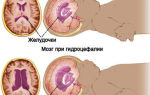
Unlike congestive discs associated with intracranial hypertension and hydrocephalus, with optic neuritis there is no pronounced protrusion (prominence) of the disc, the changes are usually unilateral.
The acute period lasts from 3 to 5 weeks. Then the hyperemia and swelling of the disc gradually disappear, the hemorrhages resolve, and the boundaries of the disc again acquire clear outlines. In more rare cases, with severe optic neuritis, atrophy of n. opticus. In this case, ophthalmoscopy reveals a pale disc with thread-like narrowed vessels and clear boundaries.
In the clinic of the retrobulbar form of optic neuritis, 3 types of inflammatory changes are distinguished: axial, peripheral and transversal.
Axial inflammation primarily affects the bundle of axons passing through the optic nerve. It is characterized by a disorder of central vision with the formation of central scotomas in the visual field and a significant decrease in functional tests.
The peripheral type of retrobulbar neuritis is associated with the occurrence of an inflammatory process in the nerve sheaths and its subsequent spread deep into the nerve trunk.
In this case, there is a significant accumulation of exudate under the optic nerve sheaths, causing the appearance in patients of so-called “shell” pain in the eye, which increases with movement of the eyeball.
Typically concentric narrowing of the visual fields while maintaining central vision. Functional test results may be within normal limits.
The most severe is the transversal type of retrobulbar neuritis, in which inflammation covers all tissues of the optic nerve. Visual acuity decreases to the point of blindness. Functional tests show extremely low results.
All types of retrobulbar neuritis are characterized by the absence of changes in the optic nerve head. Only a month after the manifestation of the disease, ophthalmoscopy can reveal disc decoloration and signs of total or partial atrophy of the optic nerve.
Since optic neuritis is an interdisciplinary pathology, its diagnosis often requires the joint participation of specialists in the field of neurology and ophthalmology. In typical cases, to verify the diagnosis, a consultation with an ophthalmologist is sufficient, during which the patient’s complaints, visual acuity test data, perimetry and ophthalmoscopy results are compared.
The most important task is to differentiate disc changes in optic neuritis from congestive disc. This is especially true for mild neuritis with minimal disturbances in visual function and when neuritis is combined with disc swelling.
In such cases, the identification of foci of exudation and minor hemorrhages in the disc tissue indicates neuritis. Fluorescein angiography of the fundus helps to distinguish between these conditions.
To exclude a stagnant disc in complex cases, a consultation with a neurologist, echo-encephalography, or lumbar puncture may be required.
In order to determine the etiology of optic neuritis, it is possible to conduct an MRI of the brain, blood culture for sterility, PCR studies, ELISA, RPR test, consultation with an infectious disease specialist, rheumatologist, immunologist, etc.
Etiotropic therapy is determined by the cause of the development of neuritis. Treatment is carried out urgently in a hospital setting. Before establishing the etiology of the disease, anti-inflammatory, dehydration, antibacterial, metabolic, desensitizing and immunocorrective treatment is usually used.
Broad-spectrum antibiotics (except for the aminoglycoside group), corticosteroids, acetazolamide with potassium preparations, intravenous glucose infusions, intramuscular magnesium sulfate, piracetam, B vitamins are prescribed.
After establishing the nature of the damage to the optic nerve, they move on to specific etiotropic therapy (for example, anti-tuberculosis treatment, surgical treatment of tonsillitis and sinusitis).
Emergency treatment for the occurrence of optic neuritis due to methyl alcohol poisoning consists of urgent gastric lavage and giving the patient 30% ethyl alcohol (vodka) orally. The latter acts as an antidote, displacing methyl alcohol from the body. A single dose is 100g and is administered every 2-3 hours.
If signs of optic nerve atrophy are detected, antispasmodics and drugs to improve microcirculation (nicergoline, pentoxifylline, nicotinamide, nicotinic acid) are additionally recommended. The outcome of both intra- and retrobulbar forms of optic neuritis depends on the type and severity of the lesion. It varies from complete restoration of visual function to the development of atrophy and amaurosis.
Source: https://www.KrasotaiMedicina.ru/diseases/zabolevanija_neurology/optic-neuritis