Concomitant strabismus is a form of oculomotor disorders, which is characterized by deviation of the eye from the general point of fixation with subsequent impairment of binocular vision. Symptoms of the disease include blurred vision, compensatory head tilt, and increased visual fatigue. For diagnosis, ophthalmological tests (Worth, Bagolini, four-point color test), synoptophore, visometry, and computer refractometry are used. Treatment includes correction of refractive errors, pleoptic methods for leveling amblyopia, orthopto-diplopic techniques and surgical correction of the vertical angle of deviation.
Concomitant strabismus is considered to be a pathology of early childhood. According to statistics, in 9% of children the first signs of the disease are detected in the neonatal period. After 3 years, the likelihood of developing the disease decreases sharply.
The prevalence of the converging type of strabismus reaches 89.9%, while the divergent type is only 10.2%. It has been proven that in 25-40% of older patients, the pathogenesis is based on a spasm of accommodation. Genetically determined forms of strabismus account for about 25%.
Strabismus occurs with equal frequency in males and females.
Concomitant strabismus
Some patients have a hereditary predisposition to the development of pathology. However, it is not strabismus itself that is inherited, but a number of factors that contribute to the occurrence of the clinical picture of the disease.
Parents often associate the symptoms of the pathology with exposure to teratogenic factors during pregnancy (infections, ionizing radiation, medications).
The main causes of the concomitant form of strabismus include:
- Monocular visual dysfunction . With unilateral visual impairment or amaurosis, the ability to simultaneously see an image with both eyes is sharply reduced or lost. The focus of the gaze on the image is impaired. This causes the eye with more severe refractive errors to deviate to the side.
- Aniseikonia . With different image parameters on the inner shell of the eye, the process of “merging” them is difficult. Even mild aniseikonia potentiates the onset of the disease. Often this cause of concomitant strabismus remains undiagnosed.
- Pathology of the central nervous system (CNS) . Organic lesions of the cortical and subcortical centers lead to dysfunction in the accommodative-refractive apparatus. In diseases of the central nervous system, adequate convergent-divergent eye movements do not develop in response to an accommodative stimulus.
Binocular vision must be considered as the gradual formation of a persistent, but at the same time dynamic stereotypy of nervous processes. Functional connections between the right and left halves of the optical analyzer are formed at 2-4 months of life.
However, the final formation of binocular vision is possible only at the age of 2-6 years. Therefore, the first symptoms of the disease are more often diagnosed in children. Due to the fact that the optomotor bifixation mechanism is not stable enough, exposure to external or internal environmental factors leads to its dysfunction.
With age, the binocular visual system becomes more stable, so violations of stereotypy are less common.
Normally, the mechanism of stereoscopic vision functions thanks to a single system consisting of several links: the receptor apparatus, cortical, cortical and subcortical centers. Strabismus occurs when there is damage at any of the levels. Cases of the development of an acquired variant of the disease with facial asymmetry and improper attachment of the extraocular muscles have been described.
The leading pathogenetic aspect is considered to be a violation of the bifixation mechanism. This leads to an inability to simultaneously direct and focus the gaze on an object. The role of aniseikonia as one of the triggering factors for the development of concomitant strabismus in childhood has been studied.
In adults, the binocular system independently adapts to the different sizes of images on the retina.
A qualitative characteristic of strabismus is deviation. It can be stable and unstable. According to the magnitude of deviation in clinical ophthalmology, minimal (up to 5°), slight (6-10°), moderate (11-20°), severe (21-35°), significantly pronounced (more than 35°) strabismus are distinguished. According to the clinical classification, the following forms of concomitant strabismus are distinguished:
- Converging (converging, esotropia) . This is one of the most common variants of strabismus. Often combined with hypermetropic refraction. The deviation of the visual axis of the eye from the point of fixation towards the medial side is characteristic.
- Divergent (divergent, exotropia) . May be transitory. Rarely combined with refractive errors. Visual dysfunction occurs secondary, against the background of previously formed concomitant strabismus. The visual axis shifts towards the temple.
- Vertical (hyper-, hypotropia) . This is the most unfavorable type of strabismus. The eye on the affected side deviates up or down. A combination of vertical strabismus with a converging or diverging variant is possible.
The leading symptom of the disease is a transient or stable deviation of the squinting eye to the side. Patients complain of progressive visual dysfunction. With the development of symptoms of strabismus after 5-6 years, double vision is observed. For children under 5 years of age, despite binocular vision disorder, diplopia is not typical.
To minimize the severity of symptoms, patients tilt their heads. Doing visual work (reading, watching a movie) leads to rapid fatigue. With high nervous tension, it is more difficult to concentrate your gaze on a specific object than in a state of complete rest.
Cases of alternating strabismus have been described, when both eyes squint alternately.
Patients whose first signs of the disease develop in early childhood suffer from torticollis (torticollis). Prolonged tilting of the head to the side leads to poor posture.
If the divergent form of the disease is not treated in a timely manner, refractive errors develop, the most common of which is myopia. Patients are at risk of developing ocular migraines.
With surgical methods for correcting concomitant strabismus in the early postoperative period, secondary reverse strabismus may occur, which is manifested by deviation of the eye in the opposite direction.
To make a diagnosis, it is necessary to analyze anamnestic information, conduct a comprehensive instrumental examination and special functional tests.
Regardless of the nature of the course, all patients undergo visometry to measure visual acuity and computer refractometry to study the type of clinical refraction.
The main methods for diagnosing the disease are represented by the following studies:
- Wars test. A sign projector is used to perform the test. In addition to studying the characteristics of simultaneous, bi- and mononocular vision, the technique allows us to identify abnormal deviation of the eyeball from the vertical axis. With stereoscopic vision, the patient can see four figures, with monocular vision - 2, and with simultaneous vision - 5.
- Belostotsky-Friedman color test. To carry out the test, the visual fields are separated using color filters. The examinee wears glasses equipped with a special light filter. The patient examines the colored holes, and by analogy with the Worth test, the binocular refractive balance is determined.
- Raster haploscopy. The division of visual fields is carried out by striped glass and Bagolini rasters, located mutually perpendicular. A person watches a point source of light. With normal stereoscopic vision, 1 light source and 2 rays are visualized at the intersection position; with monocular vision, 1 ray is visualized; with simultaneous vision, a cross-shaped figure and 2 light sources are visualized.
- Synoptophore. It is a haploscopic device that allows you to most accurately assess the parameters of visual function. The technique makes it possible not only to study the features of binocular vision, but also the magnitude of the deviation angle, the ability for bifoveal image fusion and fusion reserves.
In the treatment of strabismus, conservative and surgical techniques are used. When diagnosing ametropia in a patient, the first stage involves optical correction of visual dysfunction. In this case, lenses are selected individually, taking into account patient tolerance.
They should be 0.5-1.0 diopters lower than the degree of detected clinical refractive error. For amblyopia, the use of pleoptic therapy methods (occlusion, penalization, reflexology, limited exposure of the macula) is effective. Orthopto-diplopic treatment is represented by a system of hardware exercises.
The purpose of their use is to develop the ability of fusion and restore stereoscopy.
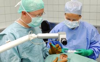
Surgical treatment is indicated for vertical deviation angles that cannot be corrected using hardware methods. The operation is not recommended for children under 4 and older than 6-7 years. Surgical treatment allows you to restore the symmetrical position of the eyes by strengthening or weakening the functions of individual muscles.
The group of surgical interventions with a weakening effect includes recession (changing the location of the posterior attachment of the muscle), tenotomy (crossing the tendon), partial myotomy (applying transverse edge notches), lengthening the muscle using plastic surgery techniques.
The action of the muscle is enhanced by resection of its section, formation of a fold of muscle or tendon tissue, and transplantation of the muscle attachment zone anteriorly.
With timely treatment, the prognosis for life and ability to work is favorable. Complete elimination of the symptoms of strabismus is possible.
To prevent concomitant strabismus, it is necessary to follow the rules of visual hygiene and correct visual dysfunction in patients with ametropia using glasses or contact lenses. Patients with a history of this pathology should undergo a follow-up examination with an ophthalmologist 2 times a year.
However, to prevent relapse of the disease after surgical treatment, a comprehensive ophthalmological examination is recommended once a year for 5 years.
Source: https://www.KrasotaiMedicina.ru/diseases/ophthalmology/friendly-strabismus
Strabismus
Strabismus is a violation of the position of the eyes, in which deviation of one or both eyes is detected alternately when looking straight. With a symmetrical position of the eyes, images of objects fall on the central areas of each eye. In the cortical sections of the visual analyzer, they merge into a single binocular image.
With strabismus, fusion does not occur and the central nervous system, in order to protect itself from double vision, excludes the image obtained by the squinting eye.
With the long-term existence of this condition, amblyopia (a functional, reversible decrease in vision, in which one of the two eyes is almost (or not at all) involved in the visual process).
Causes of strabismus
The causes of strabismus are very diverse. They can be either congenital or acquired:
- the presence of ametropia (farsightedness, myopia, astigmatism) of medium and high degrees;
- injuries;
- paralysis and paresis;
- abnormalities in the development and attachment of the extraocular muscles;
- diseases of the central nervous system;
- stress;
- infectious diseases (measles, scarlet fever, diphtheria, influenza, etc.);
- somatic diseases;
- mental trauma (fear);
- a sharp decrease in visual acuity in one eye.
Symptoms of strabismus
Normally, a person's vision should be binocular. Binocular vision is vision with two eyes with the combination of images received by each eye into a single image in the visual analyzer (cerebral cortex). Binocular vision enables stereoscopic vision - allows you to see the world around you in three dimensions, determine the distance between objects, perceive depth, and the physicality of the world around you. With strabismus, this connection does not occur in the visual analyzer and the central nervous system, in order to protect itself from double vision, excludes the image of a squinting eye.
strabismus image
normal image
Experts distinguish two forms of strabismus: friendly and paralytic.
Concomitant strabismus
With concomitant strabismus, either the left or the right eye squints, while the magnitude of the deviation from the straight position is approximately the same.
Practice shows that strabismus most often occurs in persons with ametropia and anisometropia, among whom farsightedness prevails. Moreover, farsightedness predominates in cases of convergent strabismus, and myopia is combined with a divergent type of strabismus.
The main cause of concomitant strabismus is most often ametropia, and the more pronounced it is, the greater its role in the occurrence of this pathology.
Experts also include the following reasons for concomitant strabismus:
- a state of the visual system when the visual acuity of one eye is significantly lower than the visual acuity of the other;
- a disease of the visual system leading to blindness or a sharp decrease in vision;
- uncorrected ametropia (hypermetropia, myopia, astigmatism);
- violations of the transparency of the refractive media of the eye;
- diseases of the retina, optic nerve;
- diseases and damage to the central nervous system;
- congenital differences in the anatomical structure of both eyes.
Concomitant strabismus is characterized by the following main features:
- when fixing a stationary object, one of the eyes is in a state of deviation in any direction (to the nose, to the temple, above, below);
- there may be an alternating deviation of one or the other eye;
- the angle of deviation (primary) (more often or constantly) of the squinting eye when it is included in the act of vision is almost always equal to the angle of deviation (secondary) of the fellow eye;
- eye mobility (field of vision) is fully preserved in all directions;
- there is no double vision;
- there is no binocular (volumetric, stereoscopic) vision;
- Possible decreased vision in the squinting eye;
- ametropia of various types (farsightedness, myopia, astigmatism) and various sizes (aziometropia) are often detected.
Paralytic strabismus
In paralytic strabismus, one eye squints. The main symptom of this type of strabismus is the restriction or absence of eye movements in the direction of the action of the affected muscle and, as a consequence, impaired binocular vision and double vision.
The causes of this type of strabismus may be due to damage to the corresponding nerves or a violation of the morphology and function of the muscles themselves.
These changes can be congenital or occur as a result of infectious diseases, injuries, tumors, or vascular diseases.
Signs of paralytic strabismus:
- limitation or lack of eye mobility towards the affected muscle(s);
- the primary angle of deviation (deviation) is less than the secondary one;
- lack of binocular vision, possibly double vision;
- forced deviation of the head towards the changed muscle;
- dizziness.
This type of strabismus can occur in a person at any age. It can also be caused by damage (trauma), toxicosis, poisoning, etc.
In addition, the following forms of strabismus are distinguished:
- convergent (often combined with farsightedness), when the eye is directed towards the bridge of the nose;
- divergent strabismus (often combined with myopia), when the eye is directed towards the temple;
- vertical (if the eye squints up or down).
With convergent strabismus, the visual axis of one of the eyes is deviated towards the nose. Convergent strabismus usually develops at an early age and is often intermittent at first. Most often, this type of strabismus is present with farsightedness of medium and high degrees.
With divergent strabismus, the visual axis is deviated towards the temple. Divergent strabismus is often present with congenital or early-onset myopia. The causes of divergent strabismus can be injuries, brain diseases, fear, and infectious diseases.
In addition, there are other combinations of different positions. Strabismus may be permanent or intermittent.
Atypical types of strabismus - rare, caused by anatomical developmental abnormalities (Duan syndrome, Brown syndrome, LVL syndrome, etc.)
Strabismus is distinguished according to several criteria:
By time of occurrence:
- congenital;
- acquired.
Deviation stability:
- permanent;
- fickle.
By eye involvement:
- unilateral (monolateral);
- intermittent (alternating).
By origin:
- friendly;
- paralytic.
By type of deviation:
- converging (the eye is directed towards the bridge of the nose);
- divergent (the eye is directed towards the temple);
- vertical (deviation of the eye up or down);
- mixed.
Diagnosis of strabismus
In order to confirm or refute the diagnosis of strabismus, it is necessary to undergo a thorough examination of the visual system.
At the Excimer ophthalmology clinic, diagnostics are carried out using a complex of modern computerized equipment and allow you to get a complete picture of the patient’s vision.
One of the criteria for diagnosing strabismus is examination using binocular vision tests.
Treatment of strabismus
With strabismus, usually only the eye that performs vision retains the ability to see normally. An eye deviated to the side sees worse and worse over time, its visual functions are suppressed. Therefore, treatment of strabismus should begin as early as possible.
Treatment for strabismus may include:
- optical correction (glasses, soft contact lenses);
- increasing visual acuity in both eyes (treatment of amblyopia) using hardware procedures;
- orthoptic and diploptic treatment (development of binocular vision);
- consolidation of achieved monocular and binocular functions;
- surgery.
Usually the operation is resorted to as a cosmetic remedy, since in itself it rarely restores binocular vision (when the brain combines two images received by the eyes into one).
The type of operation is determined by the surgeon directly on the operating table, since during such an operation it is necessary to take into account the particular location of the muscles in a particular person. Sometimes both eyes are operated on at once; in some types of strabismus, only one eye is operated on.
Surgery is aimed at strengthening or weakening one of the muscles that move the eyeball.
Surgical treatment of strabismus is performed in a “one-day” mode, under local drip anesthesia. The patient returns home on the same day. The final recovery takes about a week, but after such a surgical operation, doctors strongly recommend a course of hardware treatment for optimal restoration of visual functions.
- Ë
- È
- How long do you need to stay in the hospital after strabismus surgery?
Surgical treatment to correct strabismus in Excimer clinics is performed on a one-day basis, on an outpatient basis. The patient returns home on the same day.
Ë
È
If the child has strabismus, a bandage is applied to the healthy eye. Treatment takes a long time. Will he “forget” how to see with it?
Treatment using this method requires constant monitoring of visual acuity (VA) of the healthy eye, and to increase the VA of the worse eye, there is a special complex of hardware treatment (laser therapy, amblyocor, electrical stimulation, etc.)
Ë
È
I am 22 years old. Strabismus since about two years of age. Nowadays, squint is noticeable only when the eyes get tired. Is it possible to completely correct strabismus in any way: surgically or using computer programs?
Depending on the degree of strabismus and its severity, both surgical and conservative treatment are possible. To choose a treatment method and determine the state of the visual system, you need to undergo a diagnostic examination.
| Ask a Question | All questions |
Article rating: 4.8/5 (281 ratings)
Source: https://excimerclinic.ru/cross-eye/
Methods for restoring binocularity in concomitant strabismus
Strabismus involves impaired binocular vision and problems with orientation in space.
In this sense, concomitant strabismus is more easily tolerated, since when binocularity deteriorates, there is no double image and problems with visual recognition of objects do not arise.
We can say that concomitant strabismus is reversible, that is, it can be corrected and does not cause serious complications. But, like other types of strabismus, this pathology should be treated. Traditionally, eye gymnastics are used, and if it is ineffective, laser correction is used.
Specifics of the disease
Over 10 million people on the planet suffer from strabismus. Both concomitant and secondary strabismus cause significant discomfort in life and require proper correction.
The risk group includes young children whose parents have vision problems. Moreover, we are talking not only about strabismus, but also about ametropia of varying degrees.
Myopia, farsightedness and astigmatism can negatively affect refraction and lead to weakening of certain groups of eye muscles.
Timely diagnosis allows timely treatment to begin. In childhood, eye gymnastics gives good results. In some cases, it is possible to do without surgical correction and completely restore binocular vision.
Children have false strabismus from birth, which misleads parents. Sometimes it is difficult not to notice, but more often it does not attract attention and goes away on its own. Parents should not neglect such symptoms. Modern methods of studying the visual apparatus make it possible to identify pathology at an early stage and block further disorders.
Paralytic strabismus is another matter. In this case, it is almost impossible to cope with impaired binocularity without surgical intervention. But such problems are more common in adults. Infantile strabismus is easier to treat. The prognosis for early detection of pathology is positive.
Varieties of strabismus
Strabismus varies according to certain characteristics. Depending on the direction of deviation of the visual axis, the following are distinguished:
- isotropy - squinting towards the nose, also known as convergent concomitant strabismus;
- exotropia – deviation towards the ears;
- hyper- and hypotropia – vertical deviation up or down, respectively.
According to the degree of accommodation, the following forms of strabismus are distinguished:
- accommodative – occurs with refractive errors. When the difference in visual acuity reaches critical levels, the brain stops perceiving information received from the weakened eye. Subsequently, the decrease in vision progresses and the worse-seeing eye participates less and less in the process of visual perception. However, this type of strabismus is easily eliminated with the help of corrective lenses;
- non-accommodative - cannot be corrected with the help of optics, is considered incurable in the case of the use of medicinal and corrective methods of therapy. The only way to get rid of binocular impairment is surgery;
- partially accommodative - spectacle correction can improve the situation, but it is not possible to completely restore vision even with the most accurate selection of lenses. If appropriate measures are not taken, vision loss is guaranteed.
Deuteranopia - causes of distorted color perception
The peculiarity of concomitant strabismus is that the mobility of the eyeballs is preserved, and the split image is practically absent.
Symptoms of concomitant strabismus
Typically, strabismus is indicated by deviation of one or both eyes from the visual axis. These changes are visible to the naked eye, but sometimes strabismus is intermittent, and the eyes are subject to squinting alternately. More accurately, the disease is indicated by such signs as:
- deviation of the iris when fixating on an object;
- tension when concentrating;
- decreased vision in the weaker eye;
- the primary deflection angle is equal to the secondary one;
- in addition, other ophthalmological diseases may occur.
Divergent concomitant strabismus in children often occurs with progressive myopia. Hypermetropia provokes convergent concomitant strabismus. There is no direct connection between ametropia and strabismus, but a decrease in visual acuity in any direction always aggravates the situation and complicates correction.
Diagnosis of strabismus
Usually strabismus is the result of hereditary vision pathologies.
Eye diseases such as congenital cataracts, vitreous dysfunction, corneal cataract and some others can cause dysbinocular strabismus or aggravate existing strabismus.
During the diagnosis, it is possible to find out whether strabismus is accommodative or not. This allows you to select effective correction methods in the future.
The angle of strabismus is determined using the Hirshberg method. Knowing the exact angle of deviation, you can select corrective lenses or other treatment methods that will restore full binocularity.
During the diagnosis, it is possible to determine not only the degree of deviation from the axis, but also concomitant ophthalmological ailments. Visual acuity is assessed, the condition of the fundus is studied, amblyopia or the degree of ametropia, if present, is identified.
Congenital strabismus requires the study of hereditary factors, as well as the behavior of the mother during pregnancy. Once the causes of strabismus are identified, it is easier to begin the recovery process.
Without knowing what causes weakening of the eye muscles, it is difficult to prevent recurrence of strabismus even after surgical treatment.
If signs of concomitant strabismus indicate perinatal ocular microtraumas, there is no talk of restoring vision with the help of eye gymnastics and spectacle correction. In this case, the only hope is laser surgery.
Methods for restoring binocularity
If strabismus is the result of ametropia or amblyopia, then the main correction methods will be aimed at eliminating the provoking factors. In this regard, the following stages in the treatment of strabismus are distinguished:
- spectacle correction – allows you to correct ametropia and anisotropy, increases visual acuity and involves the weakened eye in the visual process;
- eye gymnastics – strengthens muscles, increases accommodative capabilities, reduces the likelihood of complications, improves blood supply to eye tissue. Traditionally, exercises are used that involve rotating the eyeballs, fixating on near and distant objects, working with light and shadow, and palming;
- pleoptics - allows you to activate the slave eye, prevents amblyopia and weakening of the eye muscles.
Treatment of concomitant strabismus is more effective the earlier it is started. Eye gymnastics in childhood gives good results. But there are no universal exercises. Depending on the type of ametropia, appropriate training is selected.
Concomitant strabismus in children can be treated with orthoptics. We are talking about special exercises on the synoptophore. Gaming programs on the computer will help normalize eye function, relieve tension, and strengthen weakened muscles. It is believed that prolonged exposure to a screen is harmful to children's vision, but a reasonable combination of work and rest gives good results.
What should everyone know about vertical strabismus?
In case of progressive ametropia, occlusion is prescribed in one eye. The dominant eye is excluded from the vision process for a short period of time. The procedures are repeated over several months. Complete occlusion is practically not used; the eye is closed for several hours every day. This method can be used from 4 years of age.
If conservative treatment does not give the desired result, surgical intervention is resorted to. Laser correction will eliminate concomitant divergent strabismus, convergent strabismus and other pathologies associated with squinting of one or both eyes. The operation involves truncation of some muscles and weakening of others. As a result, the visual axis shifts in the desired direction.
Preventive actions
To prevent strabismus, special exercises are recommended, as well as corrective glasses.
The risk group includes children with a hereditary predisposition to eye diseases, as well as people who have undergone surgery to correct strabismus in the past.
Surgical treatment does not exclude relapses, so timely and adequate prevention can prevent the pathological process.
Already at one and a half to two years old, a child can be offered corrective lenses. Available gymnastics are prescribed, which involves working with pictures and stereo images. It is useful for children to be given tasks related to finding differences in pictures or finding two identical objects among similar ones. These exercises improve concentration and help you focus faster.
It's a good idea to do accommodation training. The simplest exercise is to work with glass: place it close to the nose and mark it with a dot, and then look from the point to distant objects through the glass.
Occlusion can be considered not only as a method for correcting strabismus, but also for its prevention. If there is a decrease in visual acuity in one eye, but there is no strabismus, temporary occlusion will prevent visual impairment and weakening of the muscles of the slave eye.
To “turn off” the healthy eye from the vision process, special pads or glasses with darkened glass are used. How long to wear the patch depends on the extent of damage to the weakened eye. It is advisable to observe the dynamics and track changes.
If occlusion does not give the desired result, then other methods of vision correction and prevention of strabismus should be used.
Deterioration of visual acuity is caused by non-compliance with work and rest schedules, dark lighting in the work area, and excessive visual stress.
If you remove all provoking factors, you will be able to preserve vision for a long time and prevent binocular impairment. If there is a hereditary predisposition, observation by an ophthalmologist is mandatory.
You should visit a doctor at least once a year, and with progressive ametropia - once every six months.
Video on the topic
Source: https://MedCeh.ru/zabolevaniya-glaz/sodruzhestvennoe-kosoglazie.html
50. Strabismus: true, imaginary, hidden, methods of determination. Concomitant and paralytic strabismus. Differential diagnosis
By
the presence or absence of binocular
vision, one can distinguish real strabismus
hidden - heterophoria1.
Orthophoria2
is
the correct position of the eyeballs,
when the centers of the corneas correspond to
the middle of the palpebral fissure, and the visual axes
of both eyes are parallel when fixing
a distant object.
Ideally correct position of the eyeballs is
rare.
Most people have slight heterophoria.
Imaginary
strabismus means a displacement of the centers
of the corneas, caused by a divergence of
the visual and optical axes of the eye.
With imaginary strabismus, binocular
treatment.
Hidden
or latent
strabismus (heterophoria) means
a tendency of the eyeballs to deviate,
which manifests itself during the period when it
is turned off from the act of vision (for example,
when a person does not fixate
an object with his gaze, thinks, “goes into himself”).
Heterophoria is associated with different tone
of the extraocular muscles at such a moment.
The simplest way to identify heterophoria
is to turn off the eyeball from the act
of binocular vision, covering it with your hand
(the patient is first asked
to fix his gaze on some object).
After a few seconds, remove your hand and
look at the position of the eye.
If he
made an adjustment movement towards
the object fixed by the second eye,
then it was rejected at hand, which
means the presence of heterophoria.
Heterophoria, as a rule, does not require treatment, as
it is corrected by the act of binocular
vision.
However, if there are special
requirements for binocular vision, most
often for professional reasons,
can be performed for heterophoria
.
Pathological conditions include
obvious strabismus (heterotropia).
It is divided according to the etiology of the process into
friendly and paralytic.
A distinction is made between
monolateral strabismus, when
only
alternating), in which there is an alternating
or the other eye.
Methods
for determining the angle of strabismus
most
approximate method is
Hirshberg (Figure 16.3).
At arm's
, both eyes of the patient are illuminated with a flashlight or ophthalmoscope and asked
to fixate this object with their gaze.
On
the cornea, in the projection of the center of the pupil
of the non-squinting eye, one can see the reflection of a beam
flashlight.
In a squinting eye, the position of the reflection of light relative to the center
of the pupil will be eccentric and determines
the magnitude of the squint angle.
When the light is reflected
an average pupil width), the angle of strabismus will be
30, behind the limb - 60 degrees, or more
Determining
the angle of strabismus using a synoptophore
Synoptophore
(Figure 16.4) - a device for assessing the angle of
strabismus and quantitative assessment
of binocular vision, consists of two
tubes with a mirror located at
a right angle and a 6.5 diopter lens.
for each eye with eyepieces, through which
pictures on a slide medium are presented to each eye separately Drawing-
ki
complement each other (Figure
16.5), for example, on one - a cat with ears,
but without a tail, on the other - with a tail, but
without ears (drawings may be different).
When the visual axes of the device are parallel,
the drawings merge so that
one complete image
of the object is obtained.
If there is strabismus, in order to
obtain a single pattern, it is necessary
to change the position of the synoptophore tubes,
turning them to an angle corresponding to
the angle of strabismus.
The movements are marked on a scale, thus the synoptophore
allows you to determine the angle of strabismus.
There is
a fairly accurate method by S. S. Golovin
for determining the angle of strabismus on
the perimeter with a candle.
Currently,
to most accurately
determine the angle of strabismus, a device
ophthalmic compensator (POC).
The primary
angle of deviation is the angle
of deviation of the squinting eye.
The secondary deviation angle is the
deviation angle of the healthy eye.
They are determined by closing one eye and checking the deviation
of the second under the palm at the moment of opening.
The most important
clinical symptom of strabismus
is impaired binocular vision
The nature of vision is determined using
a four-point color test.
the right eye
with a red lens colors except red, in front of the left eye -
a green lens, which cuts off all colors
except green, and is shown a screen
with four circles: one red,
two green and one white.
If
the patient sees all four circles -
he has binocular vision, only two
red ones - monocular vision in the right
eye, two green - in the left eye, five circles
- he has simultaneous vision.
Concomitant
strabismus
Concomitant
strabismus occurs, as a rule, in
childhood and is
but also a serious functional deficiency that limits
suitability.
The prevalence of concomitant
among the childhood population.
The main
signs of concomitant strabismus are:
-
full eye movement;
-
the secondary deflection angle is equal to the primary one;
-
no doubling.
Classification of
concomitant strabismus
In
the direction of deviation of the eyeballs:
-
convergent (internal) - esophoria, esotropia;
-
divergent (external) - exophoria, exotropia;
-
vertical.
- The terms
“esophoria” and “exophoria” mean
a tendency to deviate the eyeball
deviation. - In
70% of people with convergent strabismus,
hypermetropia is observed, since in
children with hypermetropic refraction,
due to increased accommodation,
an increased impulse to
convergence occurs, and therefore the eye
deviates inward.
60% of people with divergent strabismus have
myopia , since the weakening of accommodation with
the impulse to convergence, the eye deviates outward
strabismus appears.- According to
the origin of the process, concomitant
strabismus is divided into:
accommodative (35-40%),
partially accommodative, non-accommodative.
Accommodative
strabismus is caused by the presence
of ametropia and a violation of the relationship
between accommodation and convergence and manifests itself
at the age of no earlier than 2-3 years.
If a child
a refraction that is not appropriate for his age, accommodation is disrupted:
with hypermetropia it becomes
excessive, with myopia it becomes insufficient.
Binocular vision becomes difficult,
the image of one of the eyes is suppressed
by consciousness (suppression).
Naturally, consciousness suppresses the less clear
image coming from the eye with a greater
degree of ametropia.
The eye, losing
the impulse to the correct position due to
the lack of binocular vision,
deviates in the orbit, since its
position at this moment is determined
only by the tone of the oculomotor muscles.
At first, the worse-seeing eye deviates
periodically, then the strabismus becomes
permanent
(monolateral).
With an equal or almost equal degree
of ametropia and the same visual acuity,
alternating strabismus usually occurs
, in which the cerebral
of both eyes.
a certain role in the development of strabismus
.
Anisometropia
leads to aniseikonia, and the inability
images
of unequal size into one necessary to suppress one of
them.
To
clarify whether concomitant
strabismus belongs to the accommodative variant
the correct relationship between accommodation
the accommodation of both eyes by correcting ametropia
a result of which the strabismus disappears.
a similar effect in hypermetropia
.
When wearing glasses, accommodative
binocular vision is restored.
Accommodative strabismus is
the most favorable among other
types of concomitant strabismus.
Non-accommodative
strabismus usually develops from the moment
a child is born or during the first
year of life.
Refractive errors are weakly expressed and wearing glasses does not
correct them.
non-accommodative concomitant
based on anomalies in the development of
the oculomotor muscles (changes in the place
of attachment, width of the base of the muscle
, etc.
), associated with intrauterine
or birth trauma, as well as diseases
in the postnatal period.
Often the horizontal deviation of the eyeballs
is accompanied by a vertical
Non-accommodative
strabismus should be considered as
one of the manifestations of cerebral
palsy, in the clinical picture
the incorrect position of the eyeballs comes to the fore
Partially
accommodative strabismus
occupies an intermediate position between
the two types described above.
Among
the factors leading to its occurrence
are moderate refractive errors,
anisometropia, and astigmatism.
Cycloplegics and glasses partially correct this
strabismus.
Partially accommodative
strabismus is associated both with unusual
accommodation conditions and with paresis
of the extraocular muscles.
Important
clinical manifestations of strabismus
are inhibition scotoma and abnormal
retinal correspondence.
They make it difficult to correct strabismus and, in themselves,
are difficult to treat.
In essence, the scotoma of inhibition and abnormal correspondence
a device for relieving double vision
Scotoma
of inhibition - suppression
of the image of one of the eyes by consciousness,
occurs when seeing with both eyes.
When the fixating eye is closed
, the scotoma disappears and central vision in the squinting
eye is restored.
Therefore, scotoma of
functional scotoma.
With monocular strabismus,
inhibition scotoma in most patients leads
eye.
This decrease in vision without apparent
organic causes is called
dysbinocular amblyopia.
Paralytic
strabismus
Paralytic
strabismus is caused by paresis or
paralysis of one or more extraocular
injury, tumor, infection, etc.
The following signs will allow you
to distinguish
paralytic strabismus from :
absence or limitation of mobility
of the eyeball towards the affected
muscle, inequality of the primary and secondary
angles of deviation (the second is greater than the first),
the presence of double vision (diplopia).
Diplopia
is often very painful, and patients
prefer to close one eye.
Its
presence is due to the fact that while maintaining
binocular vision, the image
of the object in question falls not
on corresponding, but on disparate
(opposite) points of the retina.
With
the long-term existence of paralytic
strabismus, the cerebral cortex suppresses
the image of the squinting eye, binocular
vision is lost and double vision stops.
Treatment of
paralytic strabismus is aimed
primarily at eliminating the cause that
caused the damage to the nerve or muscle
(elimination of the consequences of injury, removal
of a tumor, etc.).
Conservative
treatment consists of stimulating
the affected muscle with medication or
using physical therapy.
If there is no effect from conservative
treatment, surgical treatment is resorted to.
It also consists of strengthening
the affected muscle and weakening the antagonist.
Sometimes the intervention is performed on
two or more muscles.
specialists—oncologist, neurologist,
etc.—
take part in deciding
the timing of surgical treatment
Source: https://studfile.net/preview/3095912/page:18/
Concomitant strabismus: is full recovery possible?
Normally, a person has binocular vision. Strabismus, as is known, is the deviation of the position of the eyes from the central axis. This problem is specific to childhood, and therefore causes a lot of suffering to the child’s unstable psyche, causing laughter among peers.
It would be much easier if this defect was just a harmless cosmetic one. However, without appropriate treatment, the disease can eventually develop into serious visual impairment.
Ophthalmologists distinguish two forms of strabismus: concomitant and paralytic.
If we consider these two types of pathology in comparison, then the friendly one is easier to tolerate and can be treated faster.
But still, any recovery process is very long. With concomitant strabismus, although there are disturbances in binocular vision, the surrounding objects do not appear in two, which means there is no such symptom as dizziness.
Concomitant strabismus appears much more often than paralytic strabismus, usually at an early age of up to three years, and if corrected in a timely manner, it is completely corrected. But paralytic strabismus can occur at any age and the prognosis is not always favorable. According to statistics, no more than 2% of children are susceptible to strabismus, called friendly strabismus, for various reasons.
Concomitant strabismus is a disease caused by hereditary factors. The child borrows from his parents not strabismus itself, but physiological features of the structure of the body as a whole, any disturbances in the functioning of the central nervous system, or a defect in the anatomy of the eye, which can lead to such a pathology.
Moreover, the disease is often congenital. This does not mean that a visual defect is immediately noticeable in the baby. It begins to appear during the period when the eye apparatus is just adjusting to binocular vision.
However, visual impairment can also occur due to:
- infectious inflammatory eye diseases that lead to damage to the retina or optic nerve;
- myopia and astigmatism (the optical system of the eye does not have a single focus), which were not immediately identified and corrected during treatment;
- different visual acuity of the eyes, where the discrepancy is more than 0.4 diopters (it is in the squinting eye that vision is reduced);
- clouding the refractive medium of the eye.
As a rule, with various disorders of both eyes or one of them, the brain analyzer is unable to adequately put together a picture and focus the gaze on one object. In this case, the central nervous system ignores the less clear image and gradually the eye with impaired vision will lose it even more.
If treatment is not started, one of the eyes may remain completely blind. With concomitant strabismus, the work of the eye muscles is not impaired, as with paralytic strabismus, and they can move in any direction.
Depending on the direction of the squinting eye, strabismus is distinguished:
- converging (when one of the eyes or both are directed inward, towards the nose);
- divergent (direction outward, towards the temple);
- vertical (up and down);
- combined or combined (combined vertical and horizontal gaze shift).
Divergent strabismus is always accompanied by myopia, and convergent strabismus is always accompanied by farsightedness.
Depending on whether one eye or both are involved, it is customary to differentiate strabismus:
- unilateral or monolateral (when only one eye deviates);
- bilateral (both eyes are displaced);
- alternating (when both eyes shift alternately).
Before proceeding with the correction of concomitant strabismus, the doctor must determine the statute of limitations of the visual defect, whether the disease is congenital or acquired, primary or secondary.
With secondary, in addition to binocular mismatch, anatomical pathology of the visual apparatus is implied: disturbances in the functioning of nerve endings, cornea, vitreous body, etc. A complete history is taken, starting with the condition of the mother during pregnancy, childbirth, past illnesses of the parents and the child himself.
A full ophthalmological examination is then performed. The doctor needs to establish the refractive capabilities of the optical visual apparatus of each eye. It is this power that is measured in diopters.
It is mandatory, regardless of visual acuity, to check its character:
- monocular;
- monocular alternating;
- simultaneous;
- binocular.
Next, the optimal spectacle correction scheme is selected. It is worth noting that already during the examination, the ophthalmologist makes a conclusion to what extent it is possible to correct the situation with the help of optics.
In this case, a term such as “accommodation” is used. The accommodative form of the friendly type, fortunately, occurs in young children in 53% of cases.
This means that correctly selected corrective glasses can solve the problem.
If the cause of the defect lies in paresis of the eye muscles or other diseases that have caused anatomical disorders of the organ of vision, then the only solution will be surgical (non-accommodative form). Sometimes we are talking about a partial accommodative defect, when with the help of glasses it is not possible to completely correct the defect.
Treatment is a complex process that begins with wearing corrective glasses. This rule applies even to infants from ten months of age. With non-accommodative form, glasses are still required.
They will not change the position of the eyes, but will slow down the decline in vision. The selection of glasses is carried out for a wide pupil.
From the age of 3, you can use contact lenses, which not only have a positive effect on concomitant strabismus, but also correct accompanying astigmatism.
In parallel with wearing glasses, additional procedures are carried out aimed at treating and increasing visual acuity:
- occlusion (performed from the age of 4): the healthy eye is covered with a special overlay for several months to increase the load on the visually impaired one;
- irritation by the light guide of a flash lamp of the central part of the retina (from 2-3 years);
- laser pleoptics: the same area of the retina is irritated by helium-neon laser radiation;
- use of polarizing filters (Heidenberg method);
- use of a cheiroscope apparatus to combat decreased visual acuity;
- reflexology;
- electrical stimulation.
Orthoptics is the next stage, with the help of which the concomitant defect is treated. This is a game procedure on a special “Synoptophore” device with the participation of a computer, where exercises from interactive modern programs are displayed on the monitor screen.
Then follows the last, final stage - diploptics, which is designed to record and consolidate the achieved achievements, namely binocular vision. All of the above stages of treatment may take about two years.
In the event that all therapy is unsuccessful, a decision is made on surgical intervention. The operation consists of correcting the eye muscles: shortening a more developed one or increasing a less developed one. Transplantation and plastic methods are used.
When the pathology is associated with a violation of the anatomy of the eye, folk recipes, which are mostly based on herbal decoctions, are unlikely to help. However, traditional treatment can be helped to some extent by regularly drinking exactly those ingredients that help strengthen the eye muscle.
For this, calamus root, pine needle infusion, and garden currant leaf are used. True, if currant tea is pleasant to drink, then the pine needles in the concentration in which it is recommended (2 tbsp. L per 300 ml of liquid) have a very tart and bitter taste. For a small child this will be a difficult procedure.
It’s unlikely that your baby will refuse chocolate treatment. You will need a quality product with 60% cocoa in its composition. You need to eat 4 squares 3 times a day after meals. This delicacy is known to strengthen the eye muscles and nerves to some extent. There are a number of eye exercises that can be easily done at home after consulting with your doctor.
Only parents should approach this type of treatment very carefully, observing regularity. After all, for a positive result you need to do them for at least a year.
At the age when strabismus is diagnosed, the child is not yet able to understand the complexity of the disease, so all responsibility for the effectiveness of the measures falls on the parents.
The materials posted on this page are informational in nature and intended for educational purposes. Site visitors should not use them as medical advice. Determining the diagnosis and choosing a treatment method remains the exclusive prerogative of your attending physician.
Source: https://allergology.ru/glaza/sodruzhestvennoe-kosoglazie