| Belevsky Andrey Stanislavovich Doctor of Medical Sciences, Professor of the Department of Pulmonology, Russian National Research Medical University named after. N.I. Pirogova, chief pulmonologist of the Moscow Department of Health |
Pulmonology occupies an important place in the activities of the hospital and city healthcare. The clinic is consulted by the chief pulmonologist of the Moscow Department, Professor A.S. Belevsky. On the basis of the pulmonology department there is the Department of Pulmonology of the Russian National Research Medical University named after. N.I.
Pirogov. Since 1990, the hospital has been home to the Research Institute of Pulmonology, headed by Academician of the Russian Academy of Sciences A.G. Chuchalin. Thus, in the State Clinical Hospital named after D.D.
Pletnev created a powerful pulmonological and allergological school, developing and implementing the latest methods of diagnosis and treatment.
- Tel.: +7(495) 465-48-56
- The department is located in building No. 4, on the 5th floor
The Pulmonology Department of the D.D. Pletnev City Clinical Hospital is one of the main city institutions that diagnoses and treats a wide variety of diseases, including rare ones, of the respiratory system.
The department provides highly qualified medical care to patients with severe forms of bronchial asthma, chronic obstructive pulmonary diseases (physical and educational rehabilitation programs have been developed), pulmonary fibrosis, pulmonary sarcoidosis, severe and complicated pneumonia, primary and secondary pulmonary hypertension, etc. Today, the department Patients with cystic fibrosis aged 15 to 42 years from thirteen regions of Russia are observed and treated. Therapeutic and diagnostic measures carried out at the center have significantly improved the quality and life expectancy of this category of patients.
Also, one of the priority areas of the department’s work is the treatment of various respiratory dysfunctions during sleep (obstructive apnea, sleep hypopnea, etc.).
The department has been conducting long-term scientific work together with the laboratory of intensive care and respiratory failure of the Research Institute of Pulmonology.
Due to this, methods of non-invasive pulmonary ventilation (using mask devices, which is easily tolerated by patients and can reduce the number of mechanical complications arising during intubation or tracheostomy), both during intensive care and at home, have been developed and introduced into general clinical practice. ; long-term oxygen therapy (the only method that can extend the life of patients with severe forms of COPD by 6-7 years), inhalation of nitric oxide for the treatment of patients with various forms of pulmonary hypertension to reduce vascular resistance and improve gas exchange in the lungs.
The high efficiency of the methods, the absence of side effects , and ease of use have made it possible to introduce them into general clinical practice.
The algorithm for the use of many antibiotics, bronchodilators, and inhaled corticosteroids, which have become indispensable in pulmonology practice, was chosen thanks to research conducted specifically at the hospital’s pulmonology department.
In the laboratory of functional research methods, the existing pathology is confirmed, the severity of the disease is assessed, the course of the disease is monitored, and the further prognosis is determined. Patients undergo spirography, body plethysmography, computed tomography and magnetic resonance imaging of the respiratory system.
Source: https://gkb57.ru/department/17/
Research methods in pulmonology: review
To diagnose pulmonary diseases, general clinical techniques for examining the patient are used, as well as a number of special methods.
Research methods in pulmonology complement each other; without the former, the latter are often prescribed without benefit, and vice versa.
This is one of the dangers of remote contact between a doctor and a patient: the doctor does not see the patient, cannot examine him, and thereby loses a lot of useful diagnostic information, often acting at random.
When interviewing the patient, they find out what is bothering him, as well as features of the disease and life that are relevant to the diagnosis. The most common complaints are cough with sputum or hemoptysis, shortness of breath, chest pain. Palpation, percussion and auscultation data are very important: they suggest the nature and localization of the pathological process in the lungs.
Examination of the lungs using additional methods involves the use of laboratory and instrumental studies. In our article we will talk about them briefly and provide links to more detailed information.
Laboratory research
Laboratory testing requires sputum, tracheobronchial secretions, bronchoalveolar lavages and fluid obtained by pleural puncture. Less frequently, the contents of pathological cavities in the lungs, also obtained by puncture, are examined.
A microbiological examination of the obtained materials is required, which makes it possible to identify infectious agents, in particular bacteria, and find out their sensitivity to antibiotics. This allows for targeted treatment.
Sputum from pneumonia under a microscope
Cytological examination of the obtained material is always used. It allows you to detect cancer cells and other features that indicate the nature of the disease.
If necessary, a biochemical analysis of sputum or bronchoalveolar lavage fluid is performed.
Of course, all patients are prescribed a general blood and urine test. They provide non-specific information, but they can be used to determine the severity of the disease and evaluate the effectiveness of treatment. A biochemical blood test is prescribed to assess the condition of internal organs, to find out whether there are contraindications to the use of antibiotics, to clarify the severity of inflammation, and so on.
Methods of instrumental research in pulmonology
Lung examination methods include several areas:
Pulmonary function test
Functional diagnostic methods are designed to evaluate the function, but not the structure, of the lungs. The simplest of them is spirography. It gives a conclusion about the vital capacity of the lungs, inhalation capacity, exhalation capacity and other indicators. With their help, the doctor evaluates pulmonary ventilation. The method has proven itself to be effective in cases of restrictive respiratory failure: pneumonia, lung atelectasis.
In modern devices, simultaneously with spirometry (measurement of lung volumes), pneumotachometry is also performed - determining the speed of air flows in the lungs.
Pneumotachography is a continuous recording of the volumetric flow rate of inhalation and exhalation, that is, how much air passes through a person’s lungs per unit of time.
In this case, a so-called “flow-volume” curve is constructed, which allows one to assess bronchial patency. In this case, bronchospasm can be detected at a very early stage.
Pulmonary function test
Spirometry and pneumotachometry are performed at rest and after inhalation of a bronchodilator drug. This helps to judge the reversibility (impermanence) of bronchial obstruction. This is one of the main methods for diagnosing bronchial asthma.
More rarely, bronchoprovocation tests are performed, when, after determining pneumotachography parameters at rest, physical activity is given (more often in children) or the drug methacholine is administered, which significantly narrows the bronchi when they are hyperresponsive.
An important way to self-monitor asthma is peak flowmetry. This daily procedure helps assess the progress of asthma and the need for increased treatment.
Body plethysmography (whole body plethysmography) helps estimate lung volumes that are not available with spirometry. This is the residual volume, total volume, functional residual capacity of the lungs.
Body plethysmography allows one to evaluate the aerodynamic resistance of the respiratory tract, that is, it can be used to judge the effort exerted by the patient to exhale. This indicator is important for asthma.
The device is very accurate and perfectly replaces a spirometer and pneumotachograph; its disadvantage is the cost and a fairly large chamber into which a person is placed for research.
The study of elastic recoil (or distensibility) of the lungs is also used only in specialized centers. To do this, it is necessary to measure intraesophageal pressure using a special balloon. An increase in lung compliance is typical for patients with emphysema. With restrictive lung diseases (fibrosis, distress syndrome and others), the compliance of the lungs decreases.
With the dynamic measurement of extensibility indicators, it is possible to assess the work of the respiratory muscles, as well as assess the prevalence of restrictive and obstructive disorders at an early stage, which is important in the diagnosis of interstitial pneumonitis, occupational and rheumatic lung diseases.
The diffusion capacity of the lungs measures how well oxygen penetrates through the walls of the alveoli into the blood. During the study, the patient breathes a mixture of helium and carbon dioxide in a safe concentration or inhales the gas mixture and holds his breath.
Then the remainder of the gas not absorbed into the blood is determined and the diffusion of gas into the blood is calculated using a special formula.
The method is used mainly for diagnosing diseases accompanied by thickening of the alveolar walls: idiopathic fibrosing alveolitis, sarcoidosis, carcinomatosis, and the occupational disease berylliosis. Diffusion is also reduced during pneumonia.
Cardiopulmonary exercise testing helps evaluate gas exchange in the lungs during an exercise test. The load on the patient increases until he can complete it. Oxygen consumption, exhaled carbon dioxide and pulmonary ventilation parameters are determined.
Using this modern method, it is possible to assess exercise tolerance, including in athletes, evaluate the condition of a patient with heart failure, examine a patient with chronic obstructive pulmonary disease, pulmonary hypertension, interstitial diseases, and determine the patient’s condition after lung transplantation.
Blood gases and pulse oximetry are used to determine blood oxygen saturation.
The partial pressure of oxygen and carbon dioxide in the blood is determined, as well as the acidity (pH) of the blood and the oxygen saturation of hemoglobin in red blood cells.
The method is used for bronchial asthma, obstructive pulmonary diseases, sarcoidosis, severe pneumonia, tuberculosis, and occupational pulmonary diseases.
Pulse oximetry is a simple method for determining hemoglobin oxygen saturation. It does not require blood sampling or expensive tests. The sensor used is a pulse oximeter, which is put on your finger like a clothespin. Such devices are now commercially available and allow patients with severe respiratory failure to monitor their condition.
Study of the circulatory system
Very often, with lung diseases, there is a need to find out how the heart and large vessels work, because these formations are interconnected.
Therefore, all patients with pulmonary pathology undergo an electrocardiogram. It shows increased heart rate, enlargement of the right ventricle and right atrium with pulmonary hypertension, and supraventricular arrhythmias are not uncommon.
Echocardiography allows you to visualize the pulmonary artery and calculate its pressure. This indicator is very important for assessing cardiac and respiratory failure.
In addition, when conducting echocardiography, the doctor can see hypertrophy (enlargement) of the right parts of the heart due to lung diseases or, conversely, pathology of the left parts, which causes the patient to complain of shortness of breath, cough, and hemoptysis.
Echocardiography detects effusion in both the pericardial cavity and the pleural cavity.
With the introduction of modern echocardiography devices, pulmonary artery catheterization is losing its position, but it is still performed for severe circulatory disorders in the heart and lungs. In particular, this procedure is indicated in severe pulmonary hypertension to directly determine the pressure in the pulmonary trunk.
Imaging research methods in pulmonology
The most common method of examining the lungs is x-ray. Having illuminated the chest with X-rays, the doctor receives an image of the spine, ribs, and lungs and sees changes in them.
In addition to radiography, fluorography of the lungs is widespread.
Fluorography of the lungs shows structural changes in pneumonia, atelectasis and other diseases, but it is mainly used as a screening to detect tuberculosis.
Today, traditional X-ray methods are being replaced by digital radiography. It allows you to reduce radiation exposure, facilitates the work of the x-ray technician, increases the resolution of the method, and improves the quality of diagnosis.
Linear tomography of the lungs is still used. It is based on moving the X-ray tube and film in different directions and allows you to more accurately determine objects located deep in the chest. However, the diagnostic value of this study is less than that of other more modern imaging methods, in particular computed tomography.
Computed tomography of the lungs can be spiral, high-resolution, and contrast-enhanced. There are computed angiography, dynamic and expiratory computed tomography. All of these methods provide less radiation exposure to the patient, while simultaneously improving the quality of the resulting two-dimensional layer-by-layer image.
Magnetic resonance imaging
Magnetic resonance imaging has slightly different applications.
It does not recognize infiltration of lung tissue very well, but it perfectly “sees” tumors, enlarged lymph nodes, and fluid in the pleural cavity.
This is an excellent method for the early diagnosis of lung cancer and tuberculosis. Its advantage is the absence of radiation exposure to the body, that is, no human exposure occurs.
Scintigraphy is an imaging method based on the accumulation of molecules labeled with a radioactive marker in the lungs (or other organs). At the same time, the radiation dose received by the body is absolutely safe.
Scintigraphy is widely used abroad.
The main area of use of this method is the diagnosis of pathology of the pulmonary vessels, from thromboembolism of the pulmonary artery and its branches to damage to the smallest capillaries in Takayasu's disease and primary pulmonary hypertension.
In these diseases, the images reveal areas where the isotope does not accumulate, corresponding to a blood supply defect. This study is especially useful in the dynamic assessment of treatment effectiveness.
Positron emission tomography is one of the nuclear medicine methods that allows you to visualize the lungs and detect storage defects in them. The result is a three-dimensional image of the organ.
Ultrasound examination of the lungs is not very informative. The only truly valuable diagnostic information can be obtained by ultrasound examination of the pleura to detect effusion in it - pleurisy. In all other cases, lung tissue practically does not reflect ultrasound rays and does not form any significant picture.
Transesophageal and transbronchial ultrasound examination with the introduction of a sensor into the esophagus and bronze lumen, respectively, is used quite rarely, mainly for diagnostic puncture of the affected intrathoracic lymph nodes under visual control.
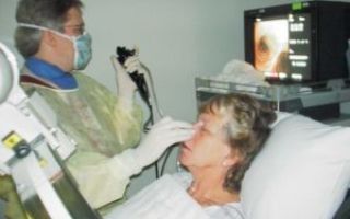
Endosonography is a combination of endoscopy and ultrasound. The doctor inserts an endoscope into the lumen of the bronchus and examines the detected disease directly at the site of the lesion using ultrasound. This technique is used to diagnose cancer of the bronchi, lungs and assess the condition of the mediastinal lymph nodes.
Contrast research techniques
As you know, X-rays pass freely through soft tissue without creating an image. Therefore, with their help, it is difficult to study blood vessels, bronchi, and hollow organs.
To eliminate this drawback, a radiopaque substance that does not transmit x-rays was introduced into the lumen of these organs.
It fills the lumen of a vessel or hollow organ, making it possible to see its internal structure.
Bronchography is one of the research methods with contrast in pulmonology. It is associated with the introduction of contrast into the bronchial cavity and is now giving way to safer and more informative methods, including endoscopic ones.
In addition, with the help of contrast, the vessels of the pulmonary circulation are studied - those in which gas exchange occurs between the lungs and blood, as well as the superior vena cava:
- angiopulmonography, which allows one to evaluate blood flow both in the entire small circle and in some part of it; this study using computed tomography is especially common;
- bronchial arteriography allows you to evaluate the vascular tree accompanying the bronchi, identify the source of pulmonary bleeding, and find abnormalities in the development of blood vessels in the lung tissue;
- superior cavagraphy – contrasting of the superior vena cava, which allows to identify compression of this vessel by a tumor of the lung or mediastinum; Due to the widespread use of tomography, the method is becoming a thing of the past.
To clarify the condition of the pleural cavity and mediastinum, methods with contrasting these formations can be used. Air often acts as a contrast. The following methods are used:
- pleurography – injection of contrast into the pleural cavity to study pleural adhesions, bursae and fistulas
- diagnostic pneumothorax: injection of air or oxygen through a puncture of the chest wall to determine the connection of a tumor in the lungs with the chest wall;
- pneumomediastinography: injection of gas into the mediastinal cavity to identify its individual organs and their tumors; due to the use of modern methods, it is used less and less;
- pneumomediastinotomography: a combination of pneumomediastinography and X-ray tomography, also rarely used; was previously used as one of the leading methods for recognizing mediastinal cysts and tumors;
- fistulography is a contrast study of fistula tracts formed between the bronchi and the chest wall, the pleural cavity, with internal fistulas, multi-chamber pleural empyema.
Endoscopic and other research methods in pulmonology
Endoscopy is gaining increasing popularity due to its informativeness, safety, reproducibility, speed, and the possibility of simultaneous diagnosis and treatment. Its use is limited only by the availability of equipment and a qualified endoscopist.
In pulmonology, the following endoscopic and other methods are used:
- bronchoscopy – examination of the bronchial mucosa if a tumor, inflammation, foreign body, polyp, bleeding and many other diseases are suspected; appropriate manipulators allow for biopsy and even treatment of detected pathology;
- biopsy - performed during bronchoscopy, as well as during lung operations, percutaneously or using video thoracoscopy - under the control of an endoscope inserted into the chest cavity; it helps to establish an accurate diagnosis of the disease at the cellular level;
- broncho-alveolar lavage: injection of therapeutic fluids into the lumen of the bronchi during bronchoscopy, followed by examination of the resulting swabs;
- pleuroscopy (thoracoscopy) – endoscopic examination of the pleural cavity;
- mediastinoscopy is a method of direct examination of the upper mediastinum, carried out during surgery and allowing one to study the lymph nodes, vessels, initial sections of the bronchi located between the lungs, and using a mediastinoscope to assess the condition of the underlying lymph nodes and perform their biopsy.
This video explains how the CT scan procedure works:
Source: https://ask-doctors.ru/metody-issledovaniya-v-pulmonologii/
Pulmonology
The branch of medicine that studies pathologies of the bronchopulmonary system is called pulmonology. A pulmonologist diagnoses and treats lung diseases. He specializes in respiratory diseases, which account for 45-50% of patient visits in different seasons of the year.
What diseases does a pulmonologist treat?
The doctor diagnoses and treats diseases of the trachea, bronchi and lung tissue of an infectious and allergic nature, caused by systemic pathology and bad habits associated with working in hazardous working conditions and heredity.
A pulmonologist treats:
- colds of the upper respiratory tract (including associated complications affecting the lungs);
- bronchitis;
- pneumonia;
- pleurisy (inflammation of the outer lining of the lungs);
- bronchial asthma;
- bronchiectasis (pathological dilatation of the bronchi);
- pulmonary emphysema (pathological expansion of the alveoli, accompanied by impaired elasticity of the lung tissue);
- pulmonary fibrosis;
- pulmonary infarction (necrosis due to circulatory disorders);
- chronic respiratory failure;
- hemothorax (bleeding into lung tissue);
- pneumothorax (compression of the lung with air and a violation of the tightness of the chest);
- pneumoconiosis (irreversible changes in the lung tissue due to the accumulation of particles in it that are present in suspension in the air, including quartz, cotton, coal, talc, iron, asbestos dust);
- pulmonary hypertension (increased pressure in the pulmonary circulation);
- pneumosclerosis (overgrowth of connective tissue);
- sarcoidosis (formation of granulomas in the lung tissue and/or in the hilar lymph nodes);
- cystic fibrosis (a hereditary disease accompanied by severe respiratory dysfunction);
- lung cancer;
- chronic pulmonary heart disease (hypertrophy and dilatation of the right ventricle in combination with pulmonary hypertension).
Symptoms of pulmonary diseases
Signs of pathologies of the bronchopulmonary system for different types of lesions are similar and include:
- cough (dry or wet);
- shortness of breath (with exertion and at rest);
- feeling of suffocation;
- chest pain when taking a deep breath;
- feeling of congestion in the chest;
- secretion of sputum mixed with blood or pus;
- increased body temperature;
- weakness;
- sweating;
- chills.
Workers in hazardous industries may be referred to undergo a pulmonary examination as part of a medical examination.
A therapist can help with colds and uncomplicated diseases, but in more complex clinical cases, consultation with a pulmonologist and therapy under his supervision is necessary.
Diagnosis of bronchopulmonary diseases
At the appointment, the pulmonologist finds out the patient’s complaints and the time of their onset. Clarifies the reasons for the development of the disease, the living and working conditions of a person. After this, the following activities are carried out:
- visual examination of the chest (with determination of its size, symmetry, as well as assessment of changes in the respiratory excursion of the lungs, participation of the organs of the shoulder girdle in breathing);
- palpation of ribs and soft tissues;
- tapping (percussion) of the chest to detect changes in percussion sound that indicate a change in the density of the lung tissue;
- listening (auscultation) to detect wheezing and other sounds unusual for healthy lungs.
Additionally, the pulmonologist can refer to:
- laboratory diagnostics (blood and urine tests, sputum examination, skin tests);
- spirography, spirometry (to assess the functional state of the lungs: methods allow you to measure lung volume, expiratory volume in one second and expiratory speed);
- peak flowmetry (determining the degree of airway obstruction);
- pneumotachometry (measurement of air velocity through the trachea and bronchi);
- radiography, computed tomography, magnetic resonance imaging (to clarify the location, size and structure of the lesion);
- endoscopy (visualization of structural changes in the bronchi);
- echocardiography (examination of the heart using ultrasound to determine pulmonary hypertension).
Treatment of pulmonary diseases
The fight against bronchopulmonary pathologies is based on an integrated approach to the disease and the development of an individual treatment regimen. Systematic treatment of pulmonary patients with pathogenetic and restorative treatment is complemented by physiotherapy.
Acute diseases of the trachea, bronchi and lungs, as well as exacerbations of chronic diseases, require bed rest.
Conservative therapy is aimed at normalizing metabolic processes in the lung tissues, stopping the inflammatory process, liquefying and improving sputum drainage, dilating the bronchi and relieving spasms. It involves the use of medications with the following effects:
- antibacterial (selected based on the sensitivity of the infectious agent to the active substance of the drug);
- antitussives;
- bronchodilators;
- expectorants;
- antipyretics;
- antihistamines;
- painkillers;
- immunomodulatory.
Local medications can be used during physiotherapy, for example, electrophoresis. Also, drugs can be administered into the bronchopulmonary system using inhalers and nebulizers. Breathing exercises are useful to restore normal lung function.
Surgical treatment of pulmonary diseases is carried out for serious indications, when the pathology threatens the patient’s life.
Sign up for a consultation with a pulmonologist at MCC CJSC. The clinic has sensitive equipment for conducting research, and consultations are conducted by highly qualified doctors.
Source: https://www.mckolomen.ru/uslugi/pulmonologiya/
Pulmonologist
A pulmonologist is a medical specialty that deals with the diagnosis, treatment and prevention of diseases of the respiratory system.
Specifics of pulmonology and its necessity
Pulmonology is a broad medical field that consists of several sections that deal with the treatment of one specific disease. For example, pulmonology includes phthisiology, which deals with the diagnosis, treatment and prevention of diseases such as tuberculosis.
This medical field is closely related to other medical specialties. In addition to diagnosing and treating emerging respiratory diseases, a pulmonologist studies the factors leading to the development of pathologies.
Deviations in the state of the respiratory system are not always associated with the penetration of pathogenic infectious microflora into the body. The following factors can provoke the development of a particular disease:
- the specifics of work activity, when a person is constantly in contact with harmful substances that settle on the lungs and mucous membrane of the larynx, causing irritation and provoking the development of diseases;
- poor environmental conditions;
- presence of bad habits, especially smoking;
- genetic predisposition. If close blood relatives had pathologies such as bronchial asthma or pulmonary embolism, the risk of developing them increases significantly.
The task of the pulmonologist is to determine which risk group his patient belongs to, which led to disruption of the functioning of the respiratory system, prescribe appropriate treatment and prepare a program of preventive measures.
Specifics of the work of a pulmonologist
Pulmonology is a medical field specializing in diseases and pathological processes occurring in the respiratory organs. A pulmonologist diagnoses and treats the following pathologies:
- pneumonia;
- pleurisy;
- bronchial asthma;
- pulmonary embolism;
- obstructive pulmonary disease of the chronic stage;
- cystic fibrosis;
- silicosis;
- histocidosis.
The overwhelming number of visits to a pulmonologist are associated with common colds - laryngitis and pharyngitis. The specialization of a pulmonologist also includes conditions such as bronchitis in heavy smokers and allergic asthma.
To treat these diseases in children, there is a separate specialty - pediatric pulmonologist. The need to identify a specialized branch of pulmonology dealing with young patients is due to the different symptomatic picture of the same diseases.
In addition, there is a need to conduct a more thorough diagnosis, since the child, due to his age, cannot always correctly describe the signs that bother him.
What are the signs that require you to see a doctor?
For most diseases of the respiratory system, the main and sometimes the very first symptom is a cough that does not go away for a long time, despite the fact that the patient takes expectorants. The following symptoms indicate the need to visit a pulmonologist:
- cough that occurs in the morning after waking up;
- periodic occurrence of cough for no apparent reason;
- dyspnea;
- labored breathing;
- temperature at 37 - 37.5 degrees;
- constant dry throat;
- refusal to eat;
- thirst that cannot be quenched by large amounts of water;
- feeling as if there is not enough air;
- chest pain;
- cough with expectoration, blood clots in the sputum.
An indication for a visit to this specialist is severe snoring during sleep. In addition to coughing, a person may often experience headaches, sneezing, and a runny nose. It is possible that signs such as itching of the mucous membrane in the sinuses and eyes may occur.
Shortness of breath, which occurs in a person in a calm physical state, may indicate the initial stage of the development of pulmonary emphysema. Although a cough does not always indicate the presence of pathologies in the organs of the respiratory system, this sign cannot be ignored, and at the first opportunity you should go to see a medical specialist.
People who have been treated for tuberculosis need to visit a pulmonologist at least 1-2 times a year. An annual examination by a specialist is also recommended for those patients with a family history of cancer of the lungs and bronchi, in order to undergo a preventive examination. This allows for timely detection of relapse and appropriate treatment.
How does a consultation with a pulmonologist proceed?
At the first appointment, the doctor conducts a detailed interview and examination of the patient. To make a diagnosis, a specialist will be interested in details regarding the nature of the symptomatic picture, what its intensity is, and how long ago the alarming signs appeared.
If a person has previously consulted other specialists and undergoes a medical examination, the test results must be provided to a pulmonologist. X-rays of the lungs are especially important for making a diagnosis.
During the examination, the doctor listens to the patient's lungs and bronchi. After the examination, the specialist asks the person to cough. The sputum that comes out when you cough is immediately sent for analysis.
Diagnostic techniques
To make an accurate diagnosis, one examination and interview of the patient is not enough. A detailed medical examination is prescribed, which includes the following tests:
- X-ray;
- general blood analysis;
- bacteriological culture of the skin;
- conducting tests of a provocative nature (these tests are carried out in the absence of a pronounced symptomatic picture);
- CT scan;
- Magnetic resonance imaging;
- echocardiography;
- blood test to determine immunoglobulin concentration;
- bronchoscopy.
If necessary, consultations with other medical specialties are carried out. If allergic reactions are suspected, an analysis is carried out to identify the irritating factor.
Pediatric pulmonologist
Diseases of the respiratory system in children are identical to the pathologies that occur in adult patients. But the symptomatic picture differs due to the age and physiological characteristics of the body, therefore the treatment of young patients is carried out by a highly specialized specialist - a pediatric pulmonologist.
Most often, children experience diseases such as pneumonia, laryngitis and pharyngitis, and bronchial asthma. The diagnostic program and treatment methods for children are selected by the doctor individually.
Methods of therapy for the respiratory system
For the treatment of diseases included in the specialization of a pulmonologist, complex therapy is selected, including the use of medications and the use of physiotherapeutic techniques. Physiotherapy includes:
- undergoing reflexology;
- use of manual therapy techniques;
- inhalation of the upper respiratory tract;
- electrophoresis;
- laser therapy;
- massage;
- magnet therapy.
Medications are selected by the doctor individually, depending on the diagnosis and intensity of the symptomatic picture.
The main medications are expectorants that help quickly remove mucus from the lungs and bronchi. To restore the body, vitamin complexes are prescribed.
If cough, shortness of breath and other symptoms are caused by allergies, appropriate medications are prescribed.
It is necessary to contact a pulmonologist as soon as the first signs of the disease appear. Diseases of the respiratory system, without timely treatment, quickly become chronic and can provoke a number of serious complications.
People who work in hazardous industries and smokers regularly need to visit a pulmonologist, since this bad habit significantly increases the risk of various pathological conditions.
Source: https://nebolet.com/vrachi/pulmonolog.html
Department of Pulmonology Integramed: Reception of pulmonologists of the highest category from the Research Institute of Pulmonology
Reception is conducted by pulmonologists with more than 25 years of experience.
Pulmonology is a section of Respiratory Medicine that studies, researches and treats lung diseases of various origins.
A pulmonologist is a doctor who diagnoses and treats diseases of the human respiratory system. Pulmonologists also practice high-tech treatment methods such as long-term oxygen therapy at home and non-invasive ventilation.
Here are just the most common diseases:
For Moscow and large cities, this specialty has long been not uncommon, there are even specialized pulmonology centers, for example ours, but still, judging by the letters we receive, it is not possible to consult a good pulmonologist everywhere and not always.
The leading scientific and practical center in the field of respiratory tract diseases is the Scientific Research Institute of Pulmonology in Moscow, under the leadership of Academician A.G. Chuchalin.
It is the specialists of this institute, spending a large amount of time with pulmonary patients, in the pulmonary hospital, constantly improving their knowledge at international congresses, who can provide a high level of care to patients suffering from respiratory diseases.
The IntegraMedservice Clinic is a highly specialized private medical clinic that deals only with respiratory diseases: the treatment and diagnosis of respiratory diseases. Our doctors are regular speakers at professional Russian and international conferences on respiratory problems. We are constantly invited as experts on television and in the media
In the diagnosis and treatment of asthma and COPD, we are guided by:
Kuleshov A.V. with the founder of the Research Institute of Pulmonology, Academician of the Russian Academy of Medical Sciences, PROFESSOR Alexander Grigorievich Chuchalin
- International clinical guidelines for the diagnosis, treatment and prevention of bronchial asthma (GINA, 2018),
- Global strategy for the diagnosis, treatment and prevention of COPD (GOLD, 2018)
- recommendations of the Russian Respiratory Society (2018).
The combination of accurate diagnosis and knowledge of treatment tactics always ensures control over diseases. Carrying out diagnostics of lung diseases with us guarantees the correctness of the diagnosis, a modern treatment and prevention regimen.
In the pulmonology department of our respiratory medicine center, only highly qualified pulmonologists are admitted: leading Moscow specialists, full members of the European Respiratory Society (ERS), students of RAMS academician A.G. Chuchalin and followers of his scientific school. This ensures high diagnostic quality of research and treatment results for respiratory diseases.
- We are consulted by pulmonologists - allergists who have worked for a long time at the Research Institute of Pulmonology ;
- Experience and expert knowledge allow us to make the correct diagnosis , as well as conduct a remote independent assessment of medical documentation;
- We carry out all breathing tests ;
- Our pulmonologists know how to manage chronic lung diseases at home ;
- The organization of work makes it possible to diagnose and prescribe treatment in the shortest possible time: for example, patients living far from Moscow can receive advice and all the necessary studies (including MRI, CT, body plethysmography, etc.) within 1-2 days of stay in the capital
NAME OF SERVICE PRICE
| Initial consultation with pulmonologist K.M.N. for adult patients | 3000 |
| Initial consultation with the head of the pulmonology department, K.M.N. Chikina S.Yu. | 3500 |
| Consultation with the head of the IntegraMed medical center K.M.N., Kuleshov A.V. | 5000 |
Source: https://integramed.info/pulmonology
Pulmonology
We work seven days a week and holidays
Doctors at the Children's Children's Center have developed medical examination and health monitoring programs for children and adults, taking into account age, lifestyle, and gender differences.
The cost of the programs is calculated at special rates with a 30% discount and remains unchanged throughout the duration of the contract. Read more...
In DCDL them. ON THE. Semashko is being treated by highly qualified doctors: consultants, KMN and DMN. The professional level of pulmonologists, technical equipment, modern equipment of the clinic, as well as extensive laboratory diagnostic capabilities create the basis for the early diagnosis of bronchopulmonary pathology.
- Diagnostics
- In the diagnosis of pulmonary diseases, in addition to traditional clinical, biochemical, and ultrasound examinations, modern laboratory and instrumental techniques are used.
- In the specialized departments of our clinic there is the possibility of a complete examination of the respiratory system:
- chest x-ray;
- study of external respiration function;
- carrying out load tests;
- fibrobronchoscopy, etc.
- study of external respiratory function with exercise and bronchodilator;
- determination of tumor markers.
- Determination of immunogram and indicators of nonspecific immunity.
Treatment
In the pulmonologist’s office you can consult and receive treatment for the following pathologies:
- cough of various etiologies;
- acute and chronic bronchitis of various etiologies;
- pneumonia of various etiologies;
- bronchiectasis;
- bronchial asthma;
- chronic obstructive pulmonary disease;
- immunodeficiency diseases;
- Malformations of the bronchopulmonary system.
Scope of diagnostic and treatment measures
- consultation of patients referred by clinic specialists;
- detection of diseases of the bronchopulmonary system;
- dynamic observation and treatment of respiratory diseases.
Dear patients! Check the cost of seeing a specific doctor at the reception desk.
- Procedures
- Diagnostics
- Analyzes
- Ultrasound
| Plain X-ray of the chest (1 image) | 800 | 900 |
| Plain X-ray of the chest + lateral projection (2 images) | 1 200 | 1 400 |
| Additional payment for second lateral chest X-ray | 570 | 735 |
| Diaskin test | 740 | 860 |
| Mantoux reaction | 560 | 635 |
Show all prices...
Source: https://cemashko.ru/pulmonology/
Pulmonologist – who is he and what diseases does he treat?
Pulmonology is a field of medicine that studies the human respiratory system, the diagnosis of pathologies and ways to treat them.
It was first identified as an independent industry only in the second half of the last century.
Until this time, science was accumulating and expanding knowledge about the respiratory system, so the diseases that are now treated by a pulmonologist were treated by different doctors - surgeons, therapists, etc.
The human respiratory system means:
- Respiratory tract (upper - nose, pharynx and lower - larynx, trachea, bronchi)
- Lungs (right and left)
- Pleura (visceral membrane lining the inside of the chest cavity and the outer surface of the lungs)
- Muscles involved in the act of breathing
- The respiratory center in the medulla oblongata, which is responsible for timely exhalation and inhalation
A pulmonologist is a doctor who studies and treats pathologies of the respiratory tract, lungs and pleura. He considers all departments of the system in close interconnection. However, not all respiratory diseases are treated by a pulmonologist:
- Surgical interventions on the chest organs are performed by a thoracic surgeon
- Threatening conditions associated with acute respiratory failure are treated by a resuscitator
- Pulmonary tuberculosis is treated by a phthisiatrician in a specialized clinic
What pathologies does it treat?
The doctor’s competence extends to a fairly wide range of diseases, from chronic rhinitis (runny nose) to lung cancer, but in some cases the help will be limited only to a consultation with a pulmonologist and diagnosis of the disease - for further treatment the patient is transferred to more specialized specialists.
A pulmonologist treats:
- Chronic and obstructive (with difficult sputum discharge) bronchitis
- Pneumonia (lung inflammation)
- Alveolitis (inflammation of the alveoli - the smallest structural units of the lung)
- Pleurisy (inflammation of the membranes of the lung)
- COPD (chronic obstructive pulmonary disease)
- Asthma
- Allergic diseases (including pneumoconiosis, silicosis)
- Hemo- and pneumothorax (effusion of blood into the pleural cavity or penetration of air)
- Emphysema (pathological dilation of bronchioles) and empyema (“ulcers”)
- Abscesses (purulent destruction of lung tissue)
- Sarcoidosis, cystic fibrosis and other fibrosis (overgrowth of connective tissue)
Can diagnose, but does not cure:
- Tuberculosis (it is treated by a phthisiatrician)
- Diseases of the upper respiratory tract (they are treated by otolaryngologists)
- Lung cancer (treated by a pulmonologist-oncologist)
In what cases should you contact this specialist?
Usually, a therapist (or general practitioner) refers to a pulmonologist, since it is quite difficult for a patient without a medical education to independently determine the disease due to the similarity of symptoms: a cough can be accompanied by influenza, pharyngitis and laryngitis (inflammation of the larynx), which do not require an appointment with a pulmonologist.
You are referred to a pulmonologist if you have the following symptoms, one way or another related to the cough reflex:
- A hacking, nonproductive cough accompanied by symptoms of respiratory failure
- Persistent cough with or without sputum production
- Changes in the nature of sputum: the appearance of pus, blood, drops of blood when coughing; discharge of pink foam
- Acute chest pain, aggravated by breathing and accompanied by increasing respiratory failure
- An increase in temperature accompanied by a cough, the appearance of shortness of breath, weakness, wet sweat and other signs of intoxication
- Choking, worsening cough in the evening/night
- The appearance of spasms, difficulty breathing under certain conditions (odors, dust pollution, changes in air temperature)
- Constant irritation of the upper respiratory tract, prolonged profuse runny nose with clear discharge, laryngospasms (“takes away” the breath)
How to make a diagnosis
An adult pulmonologist has in his arsenal a wide range of diagnostic methods, which include analysis of biological fluids (blood, sputum) for the presence of bacteria and markers of inflammation and visual hardware studies.
- Radiography and fluoroscopy are two methods of radiological diagnostics. As a result of the first, a static picture (“photograph”) of the lungs is obtained, in the second case, the organ is illuminated for some time, and the result is broadcast on the screen in real time - it is possible to evaluate the breathing function, the distribution of contrast in the vessels
- Bronchoscopy is an endoscopic visualization that allows you to examine the bronchial mucosa from the inside, take material for research, and administer a drug.
- Computed tomography – cross-sectional image of organ tissue, purpose: search for neoplasms (abscesses, fibrous cysts, cavities, foci of necrosis or purulent melting, tumors, etc.)
- Functional diagnostics: peak flowmetry (assessment of expiratory speed), spirometry (assessment of breathing volume and speed), spirography and others
Biomaterial research:
- A clinical blood test shows the presence of inflammation (with an increase in erythrocyte sedimentation rate - ESR), the acute or chronic phase of the process (according to the leukocyte formula), the presence of allergic reactions (with an increase in eosinophils), general weakness of the body (decreased hemoglobin or red blood cells)
- Sputum culture will help isolate pathogenic microflora and identify the causative agent of the disease in order to select the most effective antibiotic
- Allergy tests will establish the range of allergens that provoke deterioration
- Genetic diagnostics will help identify mutations characteristic of cystic fibrosis
Treatment methods
During a second consultation, the pulmonologist, based on the results of the study, may prescribe radical (surgical) or conservative (drug, hardware) treatment, adherence to a certain regimen and diet, and may refer for additional examination to other specialists if the problem is beyond his competence (for example, if a tumor is detected or suspected tuberculosis).
Treatment with medications can also be very varied and include, depending on the results of studies and clinical symptoms:
- Antibacterial therapy (for bronchitis and pneumonia of bacterial etiology)
- Anti-inflammatory using NSAIDs and steroids (for severe swelling)
- Antitussives (suppressing the cough reflex) or, conversely, expectorants (increasing bronchial secretion)
- Drugs that eliminate bronchospasm and dilate the bronchi - adrenomimetics, anticholinergics, antispasmodics
- Antiseptics can be administered endoscopically or using pleural puncture (for pleurisy, pneumothorax)
Hardware and instrumental treatment may include:
- Electrophoresis with various medicinal solutions
- Warming up
- Bronchoscopy with the introduction of antiseptics
- Pleural puncture (performed by a thoracic surgeon) with pumping out fluid or pus, washing the cavity with antiseptic solutions, intracavitary administration of antibiotics
Any prescription is made by a doctor after carefully studying each case of the disease. Many procedures are performed only in a hospital, and some diseases require long-term hospitalization.
Prevention
Consultation with a pulmonologist can also be preventive. Thus, chronic patients regularly go to see a pulmonologist. The purpose of these visits is to assess the current state of the body, prolong the period of remission (absence of symptoms), and mitigate the symptoms of future relapses (exacerbations).
To avoid bronchopulmonary diseases, the doctor may advise all patients without exception:
- No smoking
- Protect yourself from infections (vaccination and personal hygiene will help)
- Avoid potential allergens
- Eliminate the influence of damaging factors (when working in production with high levels of dust or toxic substances, carefully follow safety precautions and use protective equipment)
- Eat well
- Train the respiratory system (through breathing exercises, singing, playing wind instruments)
- Temper yourself and lead an active lifestyle
Advantages of carrying out the procedure at MEDSI:
- The ability to make an appointment with a pulmonologist at any time convenient for you or to see a specialist without an appointment for acute complaints
- Availability of a clinic in your area (wide network of branches)
- Availability of all hardware tests and therapeutic procedures
- Own clinical and bacteriological laboratories
- Feedback from the patient – adjustment of prescriptions and evaluation of effectiveness during treatment
- Possibility of staying in the hospital of our clinic during the entire course of treatment
- Leading pulmonologists in Moscow are world-class doctors with extensive experience and experience
It’s easy to sign up for a consultation – just call 8 (495) 7-800-500 (calls are accepted 24 hours a day).
Source: https://medsi.ru/articles/pulmonolog-kto-eto-i-kakie-bolezni-lechit/
2nd therapeutic department (pulmonology)
The Pulmonology Department of the Central Clinical Hospital of the Russian Academy of Sciences is a therapeutic department that primarily provides medical care to patients with bronchopulmonary pathology.
Treatment of diseases of the bronchopulmonary system : community-acquired pneumonia, bronchitis, chronic obstructive pulmonary disease (COPD), bronchial asthma, bronchiectasis.
To diagnose pulmonary pathology, multislice computed tomography of the chest and radiography of the lungs, bronchoscopy, bacteriological examination of sputum, examination of the immune status, and determination of the activity of the inflammatory process in the bronchopulmonary system are performed.
For daily monitoring of external respiration function in patients with airway obstruction, peak expiratory flow is monitored using a peak flow meter and pulse oximetry is monitored.
In the treatment of patients with pulmonary diseases, the department uses modern pharmacological drugs, nebulizer therapy, which allows the medicine to be delivered directly into the respiratory tract; Individual schools on bronchial asthma, adaptive medaptone therapy courses, inhalation therapy with an oxygen-helium mixture, and rehabilitation breathing exercises are conducted.
Treatment of obstructive sleep apnea syndrome (OSA) is a condition characterized by snoring, periodic collapse of the upper airways at the level of the pharynx and cessation of ventilation of the lungs with continued respiratory efforts, decreased blood oxygen levels, severe sleep fragmentation and excessive daytime sleepiness.
Diagnosis and treatment of patients with thyroid pathology : ultrasound of the thyroid gland, blood tests for thyroid hormones, ultrasound-guided fine-needle aspiration biopsy, laser therapy of thyroid nodules; diagnosis and treatment of endocrine ophthalmopathy, treatment of TDD, primary hypothyroidism.
Patients with diabetes mellitus are offered a school on diabetes mellitus, daily monitoring of glycemia with CGMS glucose monitors, selection of adequate glucose-lowering therapy, examination to clarify the presence of complications of diabetes mellitus - diabetic polyneuropathy, retinopathy, nephropathy and the choice of adequate treatment for these complications.
Diagnosis of pathology of the hypothalamic-pituitary system : MRI, CT scan of the pituitary gland, adrenal glands, hormonal profile.
Diagnosis and treatment of obesity includes the selection of individual programs for diet, physical activity, depending on gender and type of obesity, and calculation of the daily calorie content of an individually selected diet.
There are all possibilities for a complete examination of patients, clarification of the diagnosis and selection of adequate therapy for arterial hypertension, coronary heart disease - Holter ECG monitoring, 24-hour blood pressure monitoring, EchoCG, bicycle ergometry.
[ ] —
Source: https://www.ckbran.ru/hospital/therapy/pulmonologicheskoe-otdelenie