- Bone densitometry is a modern non-invasive method for determining bone density, which is performed to diagnose osteoporosis.
- With this disease, the mineral content of the bones (mainly calcium) is reduced, causing them to become more fragile.
- Osteoporosis poses the greatest danger to the spine and femoral neck, since fractures in these places are fraught with the most severe consequences.
Description of the procedure
What kind of procedure is densitometry , every postmenopausal woman should know, since during this period the likelihood of developing osteoporosis is highest. Such a study makes it possible to detect it effectively and absolutely painlessly at the earliest stages.
Densitometry refers to instrumental diagnostic methods, and allows you to determine the density of bone tissue, more precisely, to do its quantitative and qualitative analysis.
ultrasound and x-ray densitometry . The two methods are based on different operating principles and use different equipment. Sensors are used to read indicators, after which the data is transferred to a computer for calculation:
- relative bone density,
- thickness of the cortical layer,
- architectonics (spatial structure) and other parameters.
Equipment for densitometric examination can be stationary, with a table and sleeves. It is usually used to examine the spine, as well as the bones and joints of the pelvis.
Monoblock device
Stationary equipment
Monobloc equipment is also used in the form of a small device that allows scanning hands, feet and other individual joints and bones.
X-ray densitometry
X-ray bone densitometry is based on the ability of X-rays to pass through soft tissue, being retained in the dense structure of bones, which are characterized by a high concentration of calcium salts and other minerals. Based on the rate of absorption of X-rays by bone tissue, specialists calculate the degree of mineralization of its various sections.
X-ray densitometry is considered more accurate than ultrasound. It is carried out on a stationary table with a “sleeve”, where the patient is placed for 10-30 minutes.
During the procedure, the spine or its parts, hip and wrist joints, or the entire skeleton are examined. The technique is very accurate, but it cannot be used in all cases: for example, pregnancy is a contraindication for it.
The cost of the procedure ranges from 1,300 to 3,000 rubles, it is determined by the scope of the study and the type of clinic. If there is a need to conduct combined densitometry using a computed tomograph (CT densitometry), then the cost will be about 5,000 rubles.
Ultrasound densitometry
This densinometry method is similar to X-ray, but its accuracy is lower. Ultrasound densitometry is considered an indirect method for determining bone density.
Ultrasound waves travel at different speeds through areas of bone tissue that have different densities.
The process is recorded by a sensor, processed and provided in the form of data to a specialist for analysis.
The cost of this type of examination ranges from 700-2000 rubles.
Despite the lower accuracy of the results, this method is also used quite widely, due to its absolute safety, speed and ability to be performed without additional examinations. The procedure lasts from 5 to 15 minutes and can be performed for pregnant and lactating women.
Indications
Densinometry is performed when osteoporosis is suspected, as well as as a preventive examination associated with this disease.
This type of examination is used to determine:
- The amounts of minerals in any one of the bones or in the entire skeleton.
- General condition of the spine.
- The presence of osteoporosis or osteopenia (a disease characterized by a slight decrease in calcium content in bone tissue), the degree of development of the pathology.
- Fractures of bones and vertebrae.
That is, undergoing densitometry makes sense for any person who is at risk of developing osteoporosis. This is especially true for people exposed to risk factors.
The list of these factors is presented:
- Metabolic disorders.
- Pregnancy, especially multiple pregnancies.
- Diseases of the spine (spondylolisthesis, osteochondrosis), injuries.
- Endocrine diseases - hypothyroidism, diabetes, pathologies of the parathyroid gland.
- Long-term use of hormonal and other drugs that remove calcium.
- Some neurological disorders.
- Recurrent fractures.
- Rheumatism.
- Poor nutrition, frequent adherence to strict diets.
- Low body weight, alcohol abuse and smoking.
Densitometry of the lumbar spine and femoral neck can provide a 10-year prognosis for fracture and can also be used to evaluate the effectiveness of treatment.
When conducting such an examination on a child, it is possible to determine whether there is enough calcium and phosphorus in his body so that the child’s body can cope with intensive bone growth.
The amount of calcium in the bones after 30 years begins to decrease over time, so from about 40 years of age you need to control this indicator.
How often can densitometry be done? It should be carried out once every 2 years. Thanks to screening examinations, osteoporosis can be detected and treated in a timely manner. This examination regimen is recommended for women over 30 years of age who have close relatives susceptible to osteoporosis. Men should be screened for prevention starting at age 60.
How to prepare for the procedure
Preparing for densitometry involves following simple rules:
- The day before the procedure, you should stop taking medications containing calcium and phosphorus, as well as eating calcium-rich foods (cheese, cottage cheese).
- A week before the procedure, MRI or CT with contrast, as well as isotope scanning, should not be performed.
- You should not come to the examination wearing clothes with metal elements (zippers, rivets, buttons), which may affect the information content of the results.
- When starting the procedure, you need to take off your watch and hide your mobile phone in your bag.
How is densitometry done?
When performing X-ray densitometry, the patient is placed on a table equipped with a stationary device, after which the specialist leaves the room. When examining the spine, a special stand is used to support the legs.
When examining the pelvic bones, the legs are placed in a brace. After this, the arm of the device moves, during which a series of photographs are taken, and the data is transferred to a computer.
Movement during the procedure is prohibited unless such a command is given by the doctor. He may also ask the patient to hold his breath.
How is ultrasound densitometry performed? In this case, the patient lies on a medical couch, and the doctor performs an ultrasound procedure using a special attachment with a sensor. Both types of examination are absolutely painless and are performed fairly quickly.
In this way, you can examine any area: the lumbosacral spine, femoral region, heel bone, etc.
Ultrasound densitometry has no contraindications. X-rays are strictly prohibited for pregnant and lactating women, and for children it is done in case of urgent need.
How to interpret densitometry results
Of all the results that densinometry shows, the most important are:
- Bone density (T-score), compared with the norm for young people in points. A score of 1 or higher is considered normal; at 1-2.5, osteopenia is indicated; less than -2.5, osteoporosis is diagnosed.
- Bone density compared to the norm for a specific age group (Z score). This indicator must be within the specified age limits.
The list of diseases for which densitometry is indicated is presented:
- Osteopenia.
- Osteoporosis.
- Spinal fractures.
- Bone fractures
X-ray and ultrasound densitometry are effective methods for early diagnosis of such an insidious disease as osteoporosis. Thanks to their sensitivity and information content, regular examinations can promptly identify signs of the disease and prevent its progression.
Source: https://ortomir64.ru/kosti-i-sustavy/densitometriya-kostej-pokazaniya-k-diagnostike-kak-podgotovitsya-k-protsedure-osobennosti-provedeniya-obsledovaniya-rasshifrovka-rezultatov.html
Densitometry (diagnosis of osteoporosis)
Osteoporosis is an extremely common disease characterized by decreased bone density. To a certain extent, the increasing frequency of diagnosed cases of osteoporosis is explained not so much by the deterioration of the health of the population, but by an increase in life expectancy (the disease mainly affects people in the older age group).
The development of the diagnostic capabilities of modern medicine has played a certain role in the increase in the number of patients with increased bone fragility. The most informative method for diagnosing osteoporosis is bone densitometry, which allows not only to determine the percentage of bone loss, but also to identify structural disorders of bone architecture.
The mechanism of development of bone tissue pathology
Bone is a highly specific tissue that contains three structural elements:
- protein matrix, which makes up the main connective tissue that holds minerals in the bone;
- mineral component consisting of calcium and phosphorus;
- bone cells responsible for the reconstruction of bone tissue.
Contrary to popular belief, bone does not have a permanent, once formed structure. Essentially, it is a living structure whose main purpose is to provide optimal maintenance to the human body. During life, the nature of the loads on the load-bearing apparatus of the human body changes repeatedly; the reasons for the changes can be:
- weight gain;
- lifestyle changes (increasing or decreasing mobility);
- increase in external loads (systematic lifting of weights), etc.
The influence of these factors forces the bone to constantly carry out internal restructuring, allowing it to maintain stability and maximally resist changing loads.
In this case, bone tissue is destroyed in a place that does not require increased strength, and harder tissue is formed in the most “loaded” area.
The remodeling process is constant, and bone cells are responsible for it - osteoblasts, which form a new matrix and osteoclasts, which destroy it.
Regular physical activity stimulates metabolic processes in the bone structure
The age period up to 20-30 years is characterized by a high rate of metabolic processes, in which bone formation occurs under the influence of various factors (strength loads, amount of calcium consumed, hormonal changes). Once maximum bone mass is reached, the processes of loss and restoration are balanced. The main cause of osteoporosis is the predominance of resorption (destruction) processes over formation processes.
Important! If in young people the rate of metabolic processes in bones is 50% during the year, then in the age category over 50 years old it is no more than 5%, while resorption processes inevitably prevail over formation processes.
Since loss of bone mineral density (BMD) is always a consequence of some disease or condition, there are certain categories of people for whom screening for osteoporosis is indicated.
So, the indications for the examination are:
- age over 45 for women and over 55 for men;
- postmenopausal women;
- endocrine disorders (diabetes mellitus, thyroid dysfunction);
- multiple pregnancies (more than 3) or prolonged breastfeeding;
- several cases of bone fractures within 3–5 years;
- patients taking drugs from the corticosteroid group, as well as tranquilizers and anticonvulsants;
- maintaining a sedentary lifestyle (long-term bed rest, use of a wheelchair);
- sudden weight loss or constant low weight;
- presence of relatives diagnosed with osteoporosis.
Important! Insufficient intake of vitamin D in the body can cause the development of osteoporosis. Smoking and drinking alcohol are one of the causes of osteoporosis.
Diagnostics
Among the list of tests for osteoporosis, densitometry rightfully occupies a leading place, as it allows for a quantitative assessment of the condition of bone tissue.
A urine test for the amount of excreted calcium and hydroxyproline, which in patients with progressive osteoporosis are usually excreted in the urine to a greater extent than absorbed by the body, has a certain informative value, applicable to assessing the intensity of bone destruction.
- In addition, the initial examination includes testing the urine for deoxypyridonoline (DPID), which is excreted unchanged (unbound) in the urine as a result of slow or absent metabolic processes in bone tissue.
- Since the main goal of diagnosing osteoporosis is to identify a category of patients prone to low bone mass, it is advisable to carry out a comprehensive assessment of osteoblast activity, determined by the amount of osteocalcin per day, parathyroid hormone, alkaline phosphatase and deoxypyridonoline.
- Table. Normal values of biochemical markers
| Indicators | Unit change | Norm | |
| men | women | ||
| Total calcium | mmol/l | 2,2–2,65 | 2,2–2,65 |
| Phosphorus | ** | 0,80–1,5 | 0,80–1,5 |
| Parathyroid hormone | pmol/l | 0,7–5,5 | 0,7–5,5 |
| Deoxypyridonoline | DPID/mol | 2,3–5,5 | 2,9–7,5 |
| Osteocalcin | ng/ml | 12,0–52,0 | 5,5–59,0 |
Determining the concentration of female and male sex hormones has a fairly high diagnostic value, since it is endocrine disorders that often become the cause of the development of osteoporosis.
X-ray densitometry
The most commonly used method for examining bones for osteoporosis is densitometry. The term “densitometry” combines several methods of obtaining images that allow a quantitative assessment of the bone mineral density (BMD) of the patient being examined. Certain results in assessing BMD have been achieved using conventional x-rays.
However, it is not possible to obtain any significant quantitative results with its help. The determining factor that excluded radiography from the list of methods used to diagnose osteoporosis was the fact that even when assessing the image by an experienced doctor, it was not possible to detect bone loss of less than 40%.
Carrying out a dynamic assessment of the progression or regression of the disease is also quite difficult due to the low sensitivity of the equipment. Despite this, radiography is successfully used when it is necessary to assess the degree of deformation of bone structures, for example, vertebrae, since a similar phenomenon often occurs with the development of osteoporosis.
Important! It is advisable to study the degree of changes in BMD in areas of the skeleton where the proportion of trabecular tissue predominates (femoral neck, lumbar spine, wrist joint), since osteopenic changes affect it first.
Minor bone loss cannot be diagnosed using an x-ray.
The most popular methods of X-ray examination of the BMD are considered to be:
- dual-energy x-ray absorptiometry (DEXA);
- morphometric X-ray absorptiometry (MRA);
- quantitative computed tomography (QCT).
All x-ray methods for studying the degree of BMD reduction are based on the movement of ionizing radiation from a source located outside through the bone to a fixing detector. In this case, a narrow beam of X-ray radiation is directed to the object under study and the final result, that is, the intensity of the radiation transmitted through the bone is recorded by a computer system.
How often can x-rays be taken?
The main principle of the DEXA method is the use of double radiation, which allows the error to be reduced as much as possible due to the registration of two options for energy absorption (in soft tissues and bones).
The MRA method is a variant of DEXA, however, the use of a fan-shaped radiation flux has improved image quality and reduced scanning time, and accordingly reduced the radiation dose to the patient.
The QCT method allows you to obtain a three-dimensional image and not only determine BMD, but also obtain data on the layer-by-layer structure of bones, that is, assess the condition of the trabecular and cortical layers.
The negative side of using CCT is the high radiation dose, 10 times higher than DEXA, and the dependence of the accuracy of the readings on the amount of bone marrow, the percentage of which increases with age.
Ultrasound computer densitometry
The method of ultrasonic densitometric research is based on calculating the speed of movement of an ultrasonic wave through tissues of different densities. Differences in the density of the bone being examined cause differences in the speed of ultrasound transmission, that is, denser bone (well mineralized) transmits ultrasound faster than less dense bone.
The received data is recorded by the sensor and converted using computer software into quantitative indicators. A characteristic property of ultrasound densitometry is its extremely high sensitivity to the slightest changes in bone density. In this regard, it can be used to diagnose osteopenia, when the loss of mineral substances does not exceed 3–5%.
The undoubted advantages of ultrasonic computer densitometry methods include:
- sufficiently high information content;
- no negative impact on the body;
- speed of the procedure;
- affordability;
- no contraindications.
Thanks to such a large list of positive aspects, ultrasound densitometry can be used not only for diagnosing osteopenia and osteoporosis, but also for monitoring the effectiveness of therapy.
Due to significant deviations that appear when examining bones deeply embedded in soft tissue (proximal femur), ultrasound densitometry is performed exclusively on the extremities (wrist joint, calcaneus, etc.).
Long bones are the most informative when performing ultrasonic densitometry
Conduct and results
The technique of X-ray densitometry consists of performing a set of measurements using radiographs at several standard points most susceptible to osteopenic changes:
- lumbar spine;
- femoral neck;
- radius.
After taking a series of images, the software processes the results obtained by comparing them with the database included in it. The comparison is made according to two criteria:
- the result obtained with the optimal indicator for patients of the same sex (T-criterion);
- the result obtained with the average statistical indicator of patients of the same sex and age (Z-criterion).
The most informative when making a diagnosis is the T-criterion; checking the degree of its deviation from normal indicators has a significant diagnostic value:
- readings above “-1” indicate normal BMD;
- readings ranging from “-1” to “-2.5” indicate osteopenia (the initial stage of osteoporosis);
- readings below “-2.5” indicate the development of osteoporosis.
Ultrasonic densitometry is carried out by determining the density of the cortical (outer) layer of tubular bones.
To do this, using an ultrasound sensor, an ultrasonic wave is passed along the bone, determining the MIC by the speed of its propagation.
In a short period of time, the device performs thousands of measurements and calculates Z and T-criteria based on the results. Standard projections for ultrasonic computer densitometry are:
- phalanx of the middle finger;
- radius or wrist bone.
Important! The results obtained using X-ray and ultrasound methods may have some differences, but the final indicators are usually interpreted the same (normal or osteoporosis).
Structural changes in bones: on the left – normal, on the right – osteoporosis
Due to the individual characteristics of the course of the disease, there may not be obvious signs of bone tissue destruction, such as fractures. However, timely diagnosis can significantly reduce the risk of such a serious complication as a femoral neck fracture.
Despite the fact that the pathology is not fatal, a long-term decrease in motor activity and expensive treatment (prosthetics), which is also impossible to carry out in severe stages of osteoporosis, often lead to death.
Today, doctors have a large number of medications in their arsenal to treat osteoporosis, but due to the fact that the recovery process is extremely long, it is optimal to start it as early as possible.
Source: https://apkhleb.ru/prochee/densitometriya-diagnostika-osteoporoza
Densitometry for osteoporosis: indications, implementation, results
Osteoporosis is a systemic disease characterized by metabolic disorders in bone tissue. Bones lose an important macronutrient - calcium, which is responsible for their hardness and strength.
The loss of the leading link from the metabolic cycle leads to serious complications in all organs and systems. Therefore, densitometry for osteoporosis is an important diagnostic method that allows one to detect the disease “at the crime scene.”
Screening for osteoporosis
Often, patients do not suspect the existence of an insidious pathology, seeking qualified traumatological care to exclude a fracture (a frequent actual manifestation of osteoporosis).
Meanwhile, the disease slowly but surely weakens the protection of the bones and makes them vulnerable (fragile) to external environmental factors.
Sometimes it is enough for a person to receive a minor injury (bruise), which turns into a serious problem if the bone is incompetent (crack or fracture).
Therefore, it is important to detect the disease at an early stage of development so as not to waste precious time. Based on the results of densitometry, the degree of osteoporosis is indicated by displaying digital values on the screen. The outcome of the disease directly depends on the neglect of the pathological process.
Patients who may have osteoprosis due to age or other risk criteria undergo not only diagnostic, but also screening and preventive examinations.
As you know, science does not stand still, which is why medicine is developing at a rapid pace. To help specialists in various fields, a “miracle” was invented - a device (densitometer) that quickly and accurately determines even minor deviations from the norm in calcium levels in bone structures.
Densitometry against osteoporosis
Densitometry scan
The uniqueness of the method is that surgical manipulation is not required to determine the level of calcium in bone tissue. Thus, the examinee does not experience additional inconvenience during the medical procedure, except for touching the sensor in the examination area. The designated area serves as a subject for studying the amount of calcium depending on the patient’s gender and age.
The sensitive device is ready to immediately display the obtained indicators on a computer monitor and compare them with normal values, taking into account possible deviations (errors).
The miracle technique allows you to determine the very first signs of osteoporosis, which is very important for a favorable outcome of the disease.
It is better to prevent the disease than to face serious complications (frequent bone fractures with and without displacement).
Densitometry indicators for osteoporosis reflect how much the mineral composition of bone tissue and bone density in general are affected. In addition, it will not be difficult for an experienced specialist to determine the stage of osteoporosis using densitometry.
Who needs examination
The following categories of patients are at risk:
- representatives of the fair sex during the critical period before and after menopause;
- men over 60 years of age;
- men and women over 40 years of age due to a fracture in the absence of serious trauma (fracture out of the blue);
- representatives of both sexes with a height below 150 cm, who do not have a genetic predisposition to short stature;
- patients with a history of endocrine pathology.
It is important to note that all patients are recommended to undergo densitometry in the presence of osteochondrosis, spinal deformities (lordosis, scoliosis, kyphosis), protrusions (bulging discs) and intervertebral hernias.
In addition, a long course of treatment with hormones implies a mandatory examination of bones for osteoporosis. If the patient has been treated for a detected calcium deficiency in the bones, then it is necessary to undergo an examination to monitor the effectiveness of the therapy received.
Bone Densitometry (DEXA)
The test is also called a dual-energy X-ray absorptiometry or (DEXA) scan. The DEXA scan is the established standard for measuring bone mineral density (BMD).
This is a simple, quick and non-invasive medical test that involves exposing specific parts of the body to very small amounts of ionizing radiation and is used to produce images of the inside of the body.
DEXA scans are usually performed on the lower spine and hip bones.
Scanner Features
The scanner itself is different in that it produces two x-ray beams, each beam with a different energy level. One beam is high energy while the other is low energy.
The amount of energy that can pass through the bone is measured separately for each ray, and depends on the thickness of the bone.
Based on the difference in results regarding the amount of energy capable of passing through the bone between the two beams, bone density is calculated.
Density is directly related to bone calcium content. Therefore, thick bones contain more calcium than thin bones.
Conventional X-ray scanners cannot accurately measure the calcium content of bone tissue, and therefore a patient with osteoporosis may have completely normal X-rays.
Densitometry is used to obtain accurate measurements of bone density. A regular x-ray is much more reliable when it comes to detecting a fresh bone fracture.
How is scanning done?
Information that is usually of interest to patients:
- The examination is carried out on an outpatient basis. The patient comes to the hospital and, after examination, goes home the same day.
- The scan takes from 10 to 30 minutes, depending on the equipment used and the area being examined.
- It is important that the patient remains still during the procedure and may be asked to hold their breath for a few seconds while the image is projected.
- Patients can eat immediately before the test and remain fully clothed as long as the clothing does not have metal snaps or fasteners.
If a doctor is checking for osteoporosis in the lumbar spine and hip, the forearms will usually be looked at as well, as this will give an idea of the difference in calcium levels between parts of the body.
Recently, portable scanners have become popular, which can even be used in a general practitioner's office and check the mineral density of the heel bone or phalanges.
results
- After the DEXA scan has been performed, the radiologist (the doctor who monitors the examination process and interprets the image) gives the result of the examination.
- The results will be in the form of 2 points or criteria, T-criterion and Z-criterion.
- T-score : This is a comparison of bone mass with a younger person of the same sex at peak bone mass density.
Any score up to -1 is considered normal. A score of -1 to -2.5 is considered decreased bone density and is classified as osteopenia. Below -2.5 is classified as osteoporosis.
The T-score provides an indication of the risk of fractures and the need to begin treatment, and is accepted by WHO to assess the condition of bone tissue and is used by practitioners.
Z-score : Reflects the amount of bone mass compared to other people within the same age group, weight and gender.
Why are different bones considered for diagnosis?
The density of bone tissue is generally determined by the density of the two rather different layers or substances of which it is composed: trabecular and cortical. The damage to each type of bone tissue depends on the causes of osteoporosis. For example, in osteoporosis caused by postmenopause and long-term hormone therapy, the trabecular substance is more likely to suffer.
The cortical substance of the bone suffers in senile, diabetic osteoporosis, or dysfunction of the thyroid and parathyroid glands.
The approximate ratio of trabecular and cortical substance in different bones of the skeleton:
| Skeleton section | Trabecular substance | Cortical substance |
| Middle third of long bones | 5% | 95% |
| Distal third of long bones | 25% | 75% |
| Femoral neck | 25% | 75% |
| Greater trochanter of the femur | 50% | 50% |
| Lumbar vertebrae | 66% | 34% |
| Thoracic vertebrae | 75% | 25% |
| Ultradistal part of the forearm bones | 75% | 25% |
| Calcaneus | 90% | 10% |
- More information can be obtained during the examination by determining the mineral density of different bones (one element with a predominance of trabecular substance, and the other - cortical substance), and also draw conclusions about the causes of osteoporosis in a particular patient (depending on in which bones the process is detected ).
- Video about densitometry from Elena Malysheva’s program:
Methods of conducting the examination
Quite often, patients ask themselves the question: which densitometry best detects osteoporosis? Of course, all modern methods for detecting problems with calcium metabolism in bone structures are effective.
But when screening, experts still prefer ultrasound densitometry for osteoporosis as the safest method of examination.
For a more accurate diagnosis, X-ray densitometry or DEXA is performed.
In addition, it is important for patients to know how often can densitometry be done for osteoporosis? Only the attending doctor is ready to answer this question, because the diagnosis of osteoporosis by densimetry is carried out in each individual case according to an individual scheme.
DEXA scans are generally recommended at yearly intervals, although more frequent monitoring, approximately every 6 months, is possible.
It is important to consider that calcium in the human body is necessary not only for the proper growth and development of bone tissue. The health of internal organs and systems directly depends on the optimal level of this macronutrient.
Muscle tissue reacts acutely to calcium deficiency. The heart muscle is especially sensitive to metabolic disorders. The main engine of the human body must work without interruption, so calcium is vital for it.
Source: http://spinaisustav.ru/densitometria-pri-osteoporoze/
What will densitometry show in osteoporosis?
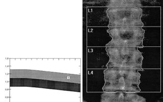
Densitometry for osteoporosis is a method of measuring bone density. Detects the disease at the earliest stage. Since destroyed bone tissue transmits x-rays more strongly and reflects ultrasound less, the device accurately detects these changes. The lumbar spine, femoral neck, forearm, and less often the entire skeleton are scanned.
X-ray densitometry is recognized as the most informative; ultrasound is used in selecting patients for further examination. Quantitative computed tomography helps make a diagnosis in doubtful cases. The recommended diagnostic frequency is once a year.
Why is densitometry prescribed for osteoporosis?
Densitometry for osteoporosis is needed to identify the initial signs of decreased bone density and leaching of calcium from them. Pathology at an early stage responds well to treatment. This helps protect the patient from fractures of the spine and femoral neck, which often lead to disability and even death.
The disease is difficult to detect before a fracture; it has no typical manifestations, and in half of the patients it is completely asymptomatic. Without densitometry, a diagnosis can only be made using radiography, but it provides information when at least 30% of the bone tissue is destroyed.
Densitometers (devices for studying bones) show osteoporosis already with a decrease in mineral density of 2.5%. Therefore, the method is considered the “gold standard” for identifying the disease, as well as subsequent monitoring of the patient during therapy.
We recommend reading the article about tests for osteoporosis. From it you will learn about who is recommended to take a blood test for osteoporosis, a basic comprehensive blood test and other research methods for women and men.
And here is more information about diffuse osteoporosis.
Indications for the diagnosis of densitometry for osteoporosis in women and men
A diagnosis using densitometry to identify osteoporosis in women and men is necessary if at least two risk factors for bone destruction are present:
- hereditary predisposition (fractures in blood relatives due to minor trauma);
- smoking;
- lack of physical activity – sedentary lifestyle, bed rest;
- rare exposure to the sun;
- lack of dairy products, fish in the diet, an abundance of protein and fatty foods, sweets, alcohol, coffee;
- thin build;
- short stature;
- multiple births (from 3), prolonged breastfeeding;
- infertility;
- late puberty, menstruation after 15 years;
- early onset of menopause;
- age from 65 years for women and from 70 for men;
- previous fractures, especially with minor trauma (for example, a fall from your own height);
- diseases - renal failure, diabetes mellitus, Itsenko-Cushing syndrome, increased function of the thyroid gland (thyrotoxicosis), disorders of the parathyroid glands, intestinal inflammation, rheumatoid arthritis;
- lack of sex hormones (estradiol in women, testosterone in men);
- long-term use of hormones from the group of corticosteroids (for example, Dexamethasone), anticonvulsants, Heparin.
Factors for turning to densitometry
The examination is carried out before prescribing drugs to strengthen bones, as well as during their use to assess their effectiveness.
Contraindications
Ultrasound bone scanning is absolutely safe and has no contraindications. X-ray densitometry and computed tomography involve radiation exposure to the body, so they are not prescribed to pregnant women. A relative limitation is the inability of the patient to lie in a supine position when it is necessary to examine the spine.
Types of densitometry examination for osteoporosis
Examination for osteoporosis using densitometry takes the form of ultrasound diagnostics, double adsorption radiography and tomography.
X-ray
It is called dual-energy, or double, since the bone is affected by a stream of rays of low and high strength. This is necessary to accurately distinguish bone tissue from surrounding tissue. The adsorption method is called because the bone absorbs (adsorbs) radiation, and the remaining flux is recorded by the apparatus.
X-ray densitometry
The obtained data are compared with the average norm of indicators (T and Z criteria for osteoporosis). Based on them, a conclusion is made about normal or reduced mineral density. The research is carried out in the following areas:
- lumbar vertebrae;
- neck of the femur;
- forearm (middle third of the radius in right-handed people).
Modern devices have the function of scanning the entire spine. Advantages of the method:
- high accuracy;
- relative affordability;
- radiation exposure is comparable to standard fluorography.
The densitometry result must be deciphered by the doctor prescribing the procedure.
Disadvantage : with bone deformation, calcium deposits in the aortic wall or intervertebral discs, and scoliosis, erroneous results may occur.
Ultrasonic
Dense and thin bone tissue reflect ultrasonic waves differently.
The conclusion about the condition of the bones is made by scanning the forearm, lower leg, heels, and hands, since they are closest to the surface. Two parameters are studied - the speed of ultrasound propagation and its attenuation.
The first depends on the elasticity and density of the bone, and the second shows its mass, the number and location of bone partitions.
Advantages: low cost, complete harmlessness. Disadvantage: Bone ultrasound does not show changes accurately enough and is used only for selecting patients for further diagnosis.
Ultrasound densitometry
Quantitative computed tomography
Bone density is determined and compared with a reference sample. It is carried out on a computer tomograph, which has a special “Osteo” program. Most often used to study the spine. Advantages:
- the condition of the porous and dense parts of the bone (cancellous substance and cortical layer) can be assessed separately;
- three-dimensional volumetric image;
- there is no overlap of adjacent tissues.
Flaws:
- expensive equipment;
- not carried out in all diagnostic centers;
- high dose of radiation.
Quantitative computed tomography of the pelvic joint
There is an option to examine only the radius or only the femur. This method is called peripheral CT. It is carried out using a portable device, but the reliability of its data is still questioned.
Which densitometry best detects osteoporosis?
Double X-ray densitometry is recognized as the optimal option for diagnosis, since it best identifies osteoporosis, provides all the necessary information for prescribing treatment and allows you to monitor its results.
The ultrasound method is recommended during pregnancy and as an initial stage of examination. Computed tomography is used in difficult diagnostic cases when symptoms of osteoporosis contradict X-ray densitometry data.
How to prepare for the procedure
Densitometry does not require any special restrictions and can be performed at any time. It is important to exclude pregnancy before X-ray diagnostics and tomography in women of childbearing age. The study is not recommended if, 2 weeks before, the patient underwent any other type of radiography or radioisotope scanning.
During the day you need to exclude increased intake of calcium. Drugs, vitamins and dietary supplements containing it are discontinued. Immediately before densitometry, all metal objects are removed from the body; there should also be no elements with metal on clothing, as they create interference.
Watch the video on how to prepare for the densitometry procedure:
Conducting a survey
During an ultrasound scan, the patient sits on a couch, and the doctor examines the selected anatomical area with a sensor. Diagnostic time is about 5 minutes.
X-ray densitometry takes place on a table under a special device in the form of an L-shaped scanner. It is installed over the area being examined. The X-ray source is located below, and their passage through the bones is read by a scanner. The information enters the program for processing.
The procedure lasts no more than 15 minutes. To ensure that the pelvic bones and spine are located on the same plane, a cube is placed under the patient’s bent legs.
Carrying out densitometry
Quantitative computed tomography is performed on a tomograph. It is a ring into which a couch with the subject is driven. To determine the structure of the spine, 10-15 minutes are enough.
Indicators of osteoporosis by densitometry
This diagnostic method is comparative. It is important for him not only to obtain absolute indicators, but also to compare them with normal values.
Densitometry criteria for osteoporosis
Two criteria for assessing osteoporosis by densitometry are used:
- T-criterion – the bone density of a woman aged 30 years is taken as a sample;
- The Z-score is determined for each age period, taking into account the weight and gender of the patient.
For diagnosis in women, the first criterion is suitable if their age is over 35 years. In all other cases, children and men are recommended to use the second criterion.
Degree of osteoporosis densitometry
Using the T-criterion, you can assess the degree of osteoporosis during densitometry:
- from 1 to -1 – normal bone tissue;
- up to -2.5 – bones are less dense (osteopenia), osteoporosis has not yet begun, preventive measures will be sufficient;
- -2.5 – osteoporosis, intensive therapy is required;
- from -2.5, there was a fracture - an advanced form of the disease, long-term treatment will be required, it is important to prevent repeated falls.
Graphic display of bone mineral density test results
Ultrasound compares the bone density of the subject with those of healthy people. The results are evaluated as follows:
- 87-105% correspond to the norm;
- 68-86% means osteopenia;
- below 67% occurs in osteoporosis.
When performing quantitative computed tomography, the following threshold (critical) indicators are taken into account:
- bone density below 100 mg/cm3 is a sign of its destruction;
- decrease to 50 mg/cm3 – the risk of fracture increases sharply.
Densitometry results for osteoporosis
The final conclusion of the functional diagnostics doctor will reflect the following densitometry results:
- patient diagnosis;
- absolute values obtained;
- comparison criterion;
- skeletal area where densitometry was performed;
- level of radiation received (for x-ray diagnostics).
How often to do densitometry for osteoporosis
There is no point in doing densitometry frequently, since long-term treatment will be required to restore bone density. It is carried out once a year.
After diagnosis, re-examination is necessary to assess the results of therapy.
It should be borne in mind that it is best to undergo all subsequent procedures in the same diagnostic center as the initial examination. This is due to the unequal sensitivity of the devices.
We recommend reading the article about osteoporosis of the hip joint. From it you will learn about the degrees of osteoporosis of the hip joint, diffuse and local osteoporosis, symptoms, as well as methods for diagnosing and treating osteoporosis of the hip joint.
Read more about osteoporosis of the foot here.
Densitometry helps identify osteoporosis at the earliest stages. Dual absorption radiography is best suited for this purpose. Its results are assessed using T and Z-criteria. During pregnancy and for initial examination, ultrasound scanning of bone tissue is used.
The quantitative method of computed tomography is needed to determine the structure of the spine in doubtful cases. After treatment is prescribed, repeat densitometry is necessary a year later to assess the effectiveness of the drugs.
Source: https://endokrinolog.online/densitometrija-pri-osteoporoze/