Prostatitis is a common male disease. Its insidiousness lies in its extensive, vaguely defined symptoms. It is almost impossible to recognize prostatitis at the initial stage, so it is important to get tested on time. Their results will allow you to diagnose the disease and determine the stage of the pathology. Correct diagnosis is the key to success. The basis of effective treatment is accurate diagnosis. The results of clinical studies allow us to select a treatment method that will ensure recovery in the shortest possible time. Analyzes will allow you to select medications that are most effective in a particular case. In addition, the examination reveals what caused the inflammatory process in the gland. Getting tested for prostatitis is very important. Analysis for prostatitis includes a whole range of clinical studies. They make it possible to determine the general condition of the organ, the presence of an inflammatory process in it, the stage of tissue damage, as well as the presence of microorganisms that caused the development of the disease. Clinical examinations give the specialist a complete picture of the patient’s health status, the presence of bacteria in the body and the immune system’s response to them. Frequent urge to urinate. The following symptoms are present:
- Urination is difficult.
- Sexual dysfunction.
- Fast fatiguability.
- Long night erections.
- Burning in the perineum and urethra.
- Pain during bowel movements.
- Frequent urge to urinate.
- Threads are observed in the urine.
- Urination is intermittent.
Additional research.
- Main study.
- Additional research.
- General blood analysis. Blood is taken from a finger or a vein. The study determines the leukocyte sedimentation rate, which helps identify infections and inflammation in the body. An elevated ESR threshold indicates the presence of prostate inflammation.
- Three-glass urine sample. The collection of material for research is carried out in stages. First, urine is collected from one urination into one container, then into a second, and finally into a third. Before collecting urine into the third glass, a prostate massage is used. This method is used if chronic prostatitis is suspected. The urologist performs a rectal massage of the prostate, stimulating the release of prostate secretions into the urethra. An increased level of leukocytes in the third container indicates the presence of a disease.
- General check of prostate secretions. The collection of material is carried out only by a health care worker. The gland is massaged rectally. The secretion released from the external opening of urine is collected in a test tube or applied to a glass slide. Transparent or yellow color of the material may indicate the presence of a disease. A large number of red blood cells, as a rule, indicates the presence of a malignant tumor in the gland or prostate stones.
- Examination of urethral discharge. The material is collected using a special probe with a swab. It is carefully inserted and a swab is taken for further laboratory testing. The results of the study provide information about possible microflora disturbances, as well as detect inflammation of the urethra.
- Study of ejaculate. The most informative and accurate test for assessing reproductive function. Submit the material in the laboratory or at home. However, in the second case, he must be taken to a medical facility no later than an hour later. The study allows us to identify the presence of infectious pathologies that are a possible cause of the development of prostatitis. Such infections can be transmitted through unprotected sex.
- PCR diagnostics. It stands for polymer chain reaction to various hidden infections. Allows you to determine the DNA of a pathogenic agent. A positive result indicates the bacterial nature of the pathology. Using the analysis, acute, sluggish and latent infections are determined. Thanks to diagnostics, effective drugs are selected against a specific pathogen.
- Bacterial culture of urine. Collecting material for testing for prostatitis is performed in different ways. The main condition is to exclude the entry of foreign microorganisms. After urination, a clean cotton pad is pressed to the outer opening of the canal, which absorbs enough liquid.
- Magnetic resonance and computed tomography. An MRI scan allows you to obtain three-dimensional images of an organ. The most accurate method for examining the pelvic organs. It can be used to identify a tumor or other organ pathologies. The specialist prescribes an MRI if prostate cancer is suspected. The method helps determine the extent of spread of the disease to nearby organs.
- PSA blood test. PSA is a specific antigen produced by the prostate. The amount of this substance in the blood makes it possible to detect prostate cancer at an early stage. Malignant cells produce antigen in conjunction with certain serum proteins. PSA is considered a tumor marker. An elevated PSA level does not only indicate the presence of prostate cancer. It can increase in the presence of any inflammation or infection. The likelihood of developing cancer is determined by the ratio of total PSA to bound protein.
- Spermogram. Prostatitis and spermogram are interrelated concepts. The analysis is carried out in the presence of symptoms of prostatitis. The study makes it possible to detect pathology and the cause of its development already at the initial stage, which significantly speeds up the recovery process during treatment. Using a spermogram, the qualitative and quantitative composition of sperm is determined, which can change with prostatitis. Before collecting material, certain preparation rules must be followed. Otherwise, the results may not be reliable.
- Ultrasound of the bladder. The study is carried out using the transabdominal method, and the bladder must be full. It is performed through the abdominal wall. The method allows you to diagnose all pelvic organs. The last meal should be six hours before the procedure. Used when an inflammatory process or prostate adenoma is suspected. Helps confirm or exclude the presence of cystitis or other inflammations.
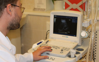
- TRUS of the prostate gland. TRUS – transrectal ultrasound examination. The most informative and safe diagnostic method. A special sensor is inserted into the rectum. It displays an image of the organ on the screen. Ultrasound waves act directly on the organ, resulting in a clear image of the gland being displayed on the screen. By examining the organ, the doctor can confirm or deny the presence of pathological changes.
Three-glass urine sample.
General check of prostate secretions.
Examination of urethral discharge.
To obtain correct test results, the study is repeated, especially if there is even a slight deviation from the norm. Perform no earlier than a week later.
The presence of inflammation can be determined by the type of reaction. With prostatitis, it is alkaline, which leads to the death of sperm.
Bacterial culture of urine.
Before collecting the material, the area around the external opening of the urethra is disinfected.
The sampling is also performed in the laboratory using a sterile catheter. The liquid is placed in a sterile container, then it is placed on special bacterial media. The culture is grown and diagnostics are performed. The analysis reveals the presence of pathogenic microflora in the genitourinary system and determines the type of infection that caused the development of the pathology. With prostatitis, cultures may reveal colonies of bacteria, fungi and viruses.
Magnetic resonance imaging.
A CT scan of the prostate gland is performed to determine an effective treatment method and clarify the nature and type of tumors. Using X-rays penetrating the body, a three-dimensional image of the gland is created.
It clearly shows not only the prostate, but also the vessels, lymph nodes and surrounding soft tissue. During the examination, the specialist examines the density of the organ, its contour, the nature of the angle, as well as the ratio of sizes with other organs.
The condition of the seminal vesicles and canals and fatty tissue is determined.
The method provides information about the internal state of the organ and the presence of tumors in it.
PSA is a specific antigen.
This test is performed on all men after the age of forty to monitor antigen levels.
Prostatitis negatively affects the quantity and quality of sperm. Disturbances in the functioning of the gland gradually lead to irreversible processes leading to infertility.
TRUS of the prostate gland.
An important part of diagnosis is deciphering the tests.
- Urine - leukocyte level - no higher than ten. The presence of ketone bodies indicates a risk of developing diabetes. Blood should not be present in the urine.
- Blood test - an increased level of leukocytes indicates the presence of an inflammatory process. With prostatitis, several units of band leukocytes may be observed. Hemoglobin during inflammation is usually reduced.
- PCR analysis - if the quantity and quality of red blood cells is low, it means the gland is injured. Clumped red blood cells indicate the likelihood of prostate cancer.
- Analysis of prostate secretions - the level of leukocytes is not higher than ten. A higher value indicates the presence of inflammation in the prostate.
- Examination of the ejaculate - a small number of leukocytes is an indicator of a normal prostate. The number of live sperm is at least 50%. Mobility must be at least 32%. Morphology – at least 14% of normal forms.
- Ultrasound and TRUS - shows the density and volume of the prostate. Normally, the size of the seminal vesicles should not be more than 5 cm. The volume of the prostate is up to 25 cm. Cubic.
Source: https://oprostatite.guru/prostatit/lechenie-i-analizy/analizy-na-prostatit/
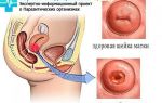
Features of a rectal examination of the prostate
A rectal examination of the prostate gland is a way of feeling (palpating) it through the patient's anus. It is part of a direct examination of the genitourinary system by a doctor to determine the direction of a diagnostic search or during preventive examinations. This is a routine procedure that does not carry anything humiliating for the patient.
general characteristics
Modern means of self-defense are an impressive list of items that differ in their operating principles. The most popular are those that do not require a license or permission to purchase and use. In the online store Tesakov.com, you can buy self-defense products without a license.
Doctors recommend such studies for all men over 50 years of age, regardless of the presence or absence of signs of pathology of the genitourinary organs. This significantly increases the likelihood of diagnosing the causes of not only pronounced changes, but also poorly manifested, but serious and dangerous diseases in the early stages. Which sometimes saves not only the health, but the life of the patient.
The high information content of this study is due to the close anatomical relationship of the lower intestine and the prostate gland. It is directly adjacent to the rectum in its anterior section, where you can palpate the posterior surface of the prostate.
Before palpation, the anal area is also examined, cracks, signs of inflammation and the presence of hemorrhoids are identified. This makes it possible to exclude concomitant intestinal pathology and identify the spread of the process beyond the gland tissue in certain diseases (neoplasms, abscesses).
Technique
Technique for performing a rectal examination of the prostate
The procedure is carried out with the patient lying on his side with his legs tucked to the chest (with the hip and knee joints bent) or in the knee-elbow position. The examination is performed by a urologist or andrologist.
Sequencing:
- The doctor puts on sterile gloves.
- To facilitate the procedure, lubricate the index finger of one hand with Vaseline and insert it into the rectum.
- At a distance of up to 5 cm from the anus along the anterior inner surface of the rectum, the doctor feels the prostate, which is shaped like a chestnut.
- Examines not only the tissue of the prostate gland, but also the inner surface of the rectal mucosa.
- To obtain gland secretions (if inflammatory or oncological processes are suspected), the doctor performs a gland massage.
- The resulting secretion is sent to the laboratory for analysis.
If it is necessary to palpate the seminal vesicles (most often when an inflammatory process is suspected), then the procedure is carried out in the Picker position, when the patient takes a position as if sitting on the doctor’s index finger. This allows you to palpate the deep parts of the rectum.
Parameters and their assessment
To form a conclusion, the doctor evaluates a number of characteristics of the prostate gland and surrounding tissues.
The prostate normally has a tight-elastic consistency, uniform throughout the organ; in the middle part, the median groove between its lobes is palpable; in the lateral areas of the gland, seminal vesicles are detected.
The boundaries of the prostate are clear, the transverse size of the gland is 2.5-4.5 cm, and the longitudinal size is from 2.5 to 3.5 cm. All structures are painless, and the rectal mucosa above the prostate is displaceable, the tissues are not fused together.
Criteria for evaluation:
- size of the gland (if enlarged, then evenly or in a certain area);
- shape of the prostate (in pathology – irregular, asymmetrical);
- boundaries of gland tissue and seminal vesicles (clear or unclear);
- density (during diseases, areas of sharp compaction or softening may appear);
- characteristics of the interlobar groove (during inflammatory processes of various natures it is smoothed out until it disappears);
- degree of sensitivity and pain (individual areas and the entire organ);
- mobility of the intestinal mucosa at the site of contact with the prostate (if the pathological process spreads beyond the gland, intestinal tissue may be involved and fixed);
- characteristics of the tissues around the gland, seminal vesicles (pain, consistency, size).
Changes in various pathologies
Changes in the prostate due to inflammation
A rectal examination of the prostate can suggest the inflammatory, tumor nature of changes in the organ, and reveal the extent of the spread of the pathological process and the involvement of surrounding structures.
With acute inflammatory changes (prostatitis), an increase in size is observed, with sharp pain, the consistency becomes denser, and the boundaries are somewhat smoothed out.
With a complicated course and the formation of an abscess, foci of a sharp decrease in density are added, up to situations where fluid is palpated in a limited cavity (focus of fluctuation).
Chronic prostatitis is characterized by a slight enlargement of the prostate and its uneven consistency; slight pain is possible.
Adenoma (benign hyperplasia) is characterized by an increase in the size of the prostate gland and a significant smoothing of the median groove while maintaining normal density. There is no pain.
The likelihood of a malignant neoplasm (prostate cancer) is indicated by:
- pronounced and uneven density (even rocky);
- increase in size;
- soreness when palpated;
- adhesion of the tissues of the gland and rectum, the inability to displace the intestinal mucosa at the point of contact with the prostate.
In healthy people, the seminal vesicles are not palpable. They enlarge and become painful only with pathology, usually with inflammatory changes.
Thus, digital rectal examination of the prostate gland is a traditional method of direct examination of the patient, which allows one to determine the feasibility of further research during preventive examinations, as well as prescribe instrumental methods to confirm the diagnosis.
- Is your beard not growing? Or is it not as thick and chic as you would like? All is not lost.
- Cosmetics and accessories for proper care of your beard and mustache. Log in now!
Source: http://MenQuestions.ru/urologiya/prostatit/rektalnoe-obsledovanie-prostaty.html
Ultrasound of the prostate gland: indications and types of studies, procedure and preparation for each type
Examination using ultrasound or echography is an important preventive measure for male diseases. The doctor prescribes an appointment for examination based on the patient’s complaints. In addition, preparation for the study is carried out annually by a man after 40 years of age to exclude diseases of the genitourinary system. Among the indications for prostate ultrasound, the following problems stand out:
- Inability to conceive a child. (If the wife does not have problems with reproductive function).
- Pain in the perineum and lower abdomen.
- Pain or discomfort when urinating.
- Weak urine stream or difficulty urinating.
- Problems with potency.
- Tests of urine, blood and semen reveal problems in the body.
- There is an inflammatory process in the epididymis and seminal vesicles.
When any diseases of the male genitourinary system , for example, prostatitis in acute or chronic form, its complications, adenoma, malignant tumor and other ailments, an ultrasound scan is required.
Types of prostate ultrasound
Most often, only two types of ultrasound are used - transabdominal and transrectal ultrasound of the prostate gland. How is it carried out?
- In the first case, echography of the gland is performed through the lower outer part of the abdomen. This study does not bring any discomfort to the man, but the information content is low. External ultrasound shows only a general image of the prostate without nuances. This type of examination is used only in advanced cases when access to the prostate through the anus is impossible. It is necessary to prepare for the transabdominal ultrasound method.
- The transrectal ultrasound method of the gland is performed by inserting a sensor into the rectum. This procedure is not particularly painful, but psychological and physiological discomfort is still present. Each man has his own reaction to this ultrasound method. However, this type of study gives a complete picture of the condition of the prostate, since the sensor is close to it.
Transrectal ultrasound of the prostate gland has no contraindications . Therefore, it can be performed even with hemorrhoids; in this case, special preparation and ultrasound techniques will be required. The attending doctor will definitely tell you about this.
Ultrasound of the prostate gland: preparation
To complete the procedure, you need to take some steps in advance. To prepare for a prostate ultrasound, you need to know the type of ultrasound that was prescribed to the man by the doctor.
Preparing for a transabdominal examination
If an external procedure is to be performed to diagnose the prostate gland, then preparation for it consists of the following measures:
- 4 days before the ultrasound examination, you need to make adjustments to your diet to eliminate bloating and gases in the intestines. Otherwise, the image of the prostate gland will be distorted. Among the forbidden foods: peas and beans, brown bread, baked goods, cabbage, fruits. You should also avoid store-bought juices, carbonated drinks, and alcoholic beverages.
- External examination of the gland is done on an empty stomach. If the analysis is carried out in the morning, then breakfast is prohibited; if in the afternoon, you can allow yourself a small portion of food in the morning.
- External scanning is performed on a full bladder.
There are two options for taking water , each depending on the disease. So, for infertility, sexual disorders and as a preventive measure, you need to drink 3-4 glasses of still water 1-2 hours before the ultrasound procedure.
If an ultrasound examination is carried out to identify problems with urination, you should come to the procedure 30 minutes before the start and start drinking a liter of still water right in the clinic. As soon as you feel the urge to urinate, you need to notify the doctor about this - he will perform an ultrasound procedure without waiting.
Preparation for transrectal examination
If a man has been referred for this type of procedure, the preparation will be completely different.
To conduct an internal examination of the prostate gland, an important condition must be met - to cleanse the intestines of feces. This determines the receipt of complete information regarding the health of the prostate, as well as the absence of discomfort during the procedure. Preparation for transrectal echography can be done in several ways:
- With the help of a special laxative “Fortrans”. This drug is used to prepare for medical procedures. It allows you not to use cleansing enemas, since it is able to remove all impurities. The drug "Fortrans" consists of 3-4 sachets, each of which is diluted in a liter of water and taken a day before the procedure. If the ultrasound is scheduled in the afternoon, two sachets are left for the morning.
- The second way to cleanse the intestines is an enema. It is done 2-3 hours before the ultrasound procedure. There are two options for colon cleansing using this method. The first is based on taking 200-300 ml of warm water, the second - 1.5 cool. Consult your doctor about which enema cleansing method to choose.
- To cleanse the intestines in the third way, you need to purchase Microlax microenemas at the pharmacy. Glycerin suppositories are also suitable.
Preparation for a transrectal type of prostate examination for some specialists involves a full bladder . Opinions on this matter vary - some believe that a full bladder is necessary, as in an external examination, others are sure that it is empty. In any case, you need to ask your doctor about everything in advance.
Preparation for ultrasound examination of the prostate in men is performed during routine examinations. However, in emergency cases, the procedure can be carried out without preparatory measures.
Source: https://urolog.guru/lechenie/vidy-obsledovaniy/podgotovka-k-issledovaniyu-predstatelnoy-zhelezy-uzi.html
Laboratory methods for examining the prostate gland
1794
Of great importance in the diagnosis of prostate is laboratory research methods, including a general blood test, serological methods and PCR diagnostics to exclude atypical intracellular infection ; general urine analysis, culture of the middle portion of urine, microscopic examination of pancreatic secretion, bacteriological examination (culture) of three portions of urine and pancreatic secretion (Meaxes-Stamey test), as well as bacterioscopy of urethral smears to detect gonococci. The most important indicators of a general urine test are protein, leukocytes, and bacteria. Normal urine does not contain protein. In the urine of a healthy person, 1-2 (up to 5) leukocytes are detected in the field of view of the microscope. Leukocyturia and pyuria (>60-100 leukocytes in the field of view) indicate an inflammatory process in the organs of the genitourinary system.
Bacteriuria in a general urine test does not have significant diagnostic value. More informative are the calculation of the number of microbial bodies per unit volume (degree of bacteriuria) and bacteriological examination of urine.
Normally, human urine obtained non-invasively can be contaminated with microflora from the distal urinary tract and perineal skin. The detection of one or more bacterial cells in the field of view of the microscope indicates the presence of 105 microorganisms or more in 1 ml of urine. Determination of one or more leukocytes in the field of view of the microscope is an indicator of infection. In a urine smear of a healthy person, the presence of only a few bacterial cells and leukocytes is allowed. Urine collection for bacteriological examination is carried out after a thorough toilet of the external genitalia with a 0.5% solution of potassium permanganate or boiled water and soap. Urine is collected in an amount of 10-15 ml in a sterile container with a lid. Samples are delivered to the laboratory within 2 hours after collection or stored in a refrigerator at a temperature of +4 ° C until delivery to the laboratory (no more than 18 hours). To detect hidden leukocyturia, erythrocyturia and cylindruria (with single leukocytes, erythrocytes and cylinders in the field of view), a urine test is performed according to Nechiporenko, in which their content in 1 ml of urine is calculated (normally 4000, 1000 and 500, respectively). Urine analysis in three portions with examination of prostate secretions (Fig. 1-5) (three-glass sample, Meares-Stamey test, four-glass sample) is a key laboratory method for diagnosing and determining the category of prostatitis.
Figure 1-5. Meares-Stamey sample
Before the study, the patient is recommended not to have sexual intercourse for 2-3 days and immediately before taking samples for analysis not to urinate for several hours (the patient’s bladder should be well filled). After toileting the external genitalia and retracting the foreskin, the patient begins to urinate (10-15 ml) into the first container (urethral test), then 100-200 ml into the toilet, and then, without interrupting the flow, 20-30 ml of urine is collected into the second container (bubble test); after this, the patient should stop urinating. To obtain prostate secretions, massage the right and then the left lobe of the prostate gland from the periphery to the center for one minute. The whitish-yellow fluid released from the urethra is collected on a glass slide or in a Lippendorff tube for analysis. If it is not possible to obtain prostate secretion, then examine the urine sediment obtained immediately after prostate massage (no later than 5 minutes). Then the very first urine (10-15 ml) excreted by the patient after massage of the prostate gland is collected into the third container (prostatic sample). The resulting three portions of urine and/or prostate secretion in its pure form are subjected to microscopy and cultural examination.
Laboratory signs of prostatitis under microscopy are an increase in the number of leukocytes (more than 10-15 in the field of view of the microscope), a decrease in the number of lecithin grains and the presence of macrophages containing fat (oval fat bodies) in the secretion of the prostate gland or in the third portion of urine.
In case of bacterial prostatitis, bacteriological examination reveals uropathogenic flora in the secretion of the prostate gland or in the third portion of urine: most often Escherichia coli, less often Klebsiella, Proteus, Enterobacter, Staphylococcus and Pseudomonas aeruginosa.
It should be borne in mind that negative results of a single study of prostate secretion do not mean the absence of an inflammatory process in the prostate gland.
At the same time, examination of prostate secretion alone does not reveal inflammation in almost 50% of patients with inflammatory syndrome of chronic pelvic pain.
The detection of more than 15 leukocytes when examining the sediment of the first portion of urine confirms the diagnosis of urethritis.
When examining urine sediment for any histological form of a tumor, two types of epithelial cells are found: some are dark, degeneratively changed, often elongated, scattered, others are large, located in complexes or in the form of papillary structures. Nuclei with coarse chromatin, some with nucleoli. Both types of cells can be found in the same patient in different portions of urine. As a rule, the background of the drug consists of detritus, elements of inflammation, and erythrocytes. In most cases, such cytograms can only be used to judge the presence or absence of malignant neoplasm cells. A tampon with a narrow applicator is inserted into the urethra to a depth of 3-4 cm, carefully turned and removed. For the study, material obtained from both the tip and the rod of the tampon applicator is used. To collect material for the purpose of microbiological research, it is also possible to use a sterile bacteriological loop.
A reliable sign of urethritis is four polymorphonuclear leukocytes (or more) in one field of view of the microscope (oil immersion system). In addition to determining the number of leukocytes, microscopy of a smear from the urethra determines the presence of microflora (including gonococci, trichomonas, fungi).
Scraping from the mucous membrane of the urethra to determine sexually transmitted infections is done using a probe to take material from the urethra.
Clinically significant pathogens - Chlamydia trachomatis, Ureaplasma wealyticum, Mycoplasma hominis, Mycoplasma genitalium, Herpes simplex, Cytomegalovirus, Trichomonas vaginalis, Gardnerella vaginalis, Neisseria gonorrhoeae, Candida spp.
It should be noted that the most widely used methods for detecting pathogens ( immunofluorescence reaction - RIF , polymerase chain reaction - PCR ) do not have 100% sensitivity and specificity.
Therefore, to increase the reliability of the research results, as well as to exclude false-positive and false-negative results, it is desirable to determine sexually transmitted infections using two different methods.
Urine testing for tumor markers is a relatively new diagnostic method, used for early detection of bladder cancer (Bard-BTA stat and NMP22 tests) and prostate cancer (PCA3 test - urine is examined after prostate massage).
The main indication for the study of ejaculate is the inability of a patient who has been sexually active for a year and has not used protection to conceive a child. Material for research is usually collected through masturbation. On the eve of donating the ejaculate, the patient is advised to abstain from intimate life, alcohol and taking hot baths for 3-4 days. It is considered optimal to conduct the study within an hour from the moment of receiving the ejaculate, since when the sample stands for a long time, its physicochemical properties change, which affects the activity of sperm and, accordingly, the results of the analysis.
The scope of laboratory research includes the study of microscopic characteristics of sperm (spermogram), as well as a number of physical and biochemical indicators of the ejaculate. The ejaculate is assessed in accordance with the standards of the World Health Organization ( WHO) (Table 1-4).
Table 1-4. Evaluation of ejaculate according to WHO standards
The study of ejaculate allows, in some cases, to carry out a differential diagnosis with inflammatory and non-inflammatory chronic pelvic pain syndrome, to identify the involvement of the reproductive system organs in the inflammatory process and to determine the quality of the seminal fluid.
The most frequently detected abnormalities in semen analysis are oligospermia (For leukocytospermia (the number of leukocytes in seminal fluid >1.0x10⁶/ml), a microbiological examination of the ejaculate is recommended.
The concentration of pathogenic bacteria in the ejaculate >103 CFU/ml is clinically significant bacteriospermia. Since the 90s
Since the last century, the determination of a tumor marker in the blood serum - prostate specific antigen (PSA) - . PSA is a serine protease produced almost exclusively by the prostate epithelium.
This marker is organ-specific, since its increase can be caused not only by cancer, but also by hyperplasia, prostatitis and other causes (digital rectal examination, prostate massage, bladder catheterization, ejaculation).
However, PSA is superior in diagnostic value to digital rectal examination and transrectal ultrasound ( US) . The study is carried out for suspected prostate cancer, as well as for all men over 45 years old, once a year for its early detection. The normal level of prostate specific antigen is 0-4 ng/ml.
However, there is currently a trend towards a decrease in the upper limit of normal PSA from 4 to 2.5 ng/ml, since some studies have found that in this range of values the risk of detecting prostate cancer is more than 20%, which leads to the diagnosis of clinically insignificant tumors which could lead to death. Thus, according to various authors, the probability of developing prostate cancer within 7 years is: with prostate-specific antigen 3-6 ng/ml - 34%; 6-10 ng/ml - 44%; >10 ng/ml – 71%. A false increase in PSA lasting about 10 days can occur after a digital rectal examination or prostate massage performed the day before, after ejaculation, rectal ultrasound examination, and in patients suffering from constipation. It is also known that the PSA level directly correlates with the volume of the prostate gland and the degree of activity of the inflammatory process, and therefore PSA can be increased with a large prostate size and with an active inflammatory process.
To optimize the diagnostic value of prostate-specific antigen, clinical algorithms have been developed, including a number of indicators:
- PSA density (PSA D);
- PSA density in the transition zone (PSA TZ);
- age-specific PSA norms;
- free PSA (PSA F);
- PSA growth rate (PSA V);
- PSA doubling time.
For a more specific diagnosis, especially when prostate-specific antigen levels are in the “gray zone” (from 2.5 to 10 ng/ml), the ratio of free (unbound) and total PSA is determined (0.75 ng/ml per year - for non-benign changes in the prostate). All of the above indicators are designed to distinguish cancer from benign processes, especially with an intermediate PSA level (2.5-10 ng/ml). There is no consensus yet on their combined use. SOUTH. Alyaev
Published by Konstantin Mokanov
Source: https://medbe.ru/materials/predstatelnaya-zheleza/laboratornye-metody-issledovaniya-predstatelnoy-zhelezy/
Tests for prostatitis, PSA, blood, urine, noma, examinations
When visiting a urologist regarding suspicion of prostatitis, the doctor’s algorithm for diagnosing the disease begins with collecting data, for which a survey is carried out about the patient’s complaints and rectal palpation.
But without laboratory and instrumental diagnostic methods, it is impossible to accurately diagnose the disease.
In what cases is examination necessary?
- Prostatitis in the initial stage of development occurs with mild symptoms, which may indicate other, often more dangerous, diseases for men.
- For this reason, a complete clinical picture can only be obtained by using a set of diagnostic methods.
- The following complaints are grounds for prescribing a comprehensive diagnostic technique:
- impaired bladder emptying;
- decreased sexual activity;
- repeated burning sensations in the perineal area;
- mental depression associated with dissatisfaction with sex life;
- excessive fatigue in the absence of an increase in normal load;
- erectile dysfunction;
- repeated pain during bowel movements;
- increased frequency of urination;
- “rate of fire” - rapid ejaculation;
- prolonged erections at night.
What can tests show?
Comprehensive diagnostics for prostatitis allows one to assess the condition of the prostate gland, the presence of infectious agents in it, the presence (absence) of an inflammatory reaction, and the degree of the pathogenic process.
Auxiliary examination techniques enable the doctor to identify:
- the general health of the man;
- the degree of involvement of the prostate in the development of neoplasms;
- the ability of existing infectious agents to activate;
- the degree of resistance of the body to the effects of pathogenic organisms.
What tests are done for prostatitis?
Tests for suspected prostatitis are conditionally classified into 2 groups:
- The main ones are bacteriological examination of urine, molecular genetic diagnostics (polymerase chain reaction analysis), computed tomography and magnetic resonance imaging, general analysis of prostatic fluid and urine.
- Auxiliary – semen analysis, after which a spermogram is deciphered, ultrasound examination of the bladder, detection of prostate tumor markers in the blood, sonography in the form of transrectal ultrasound examination of the prostate gland.
Manual examination - concept and procedure
- Functional testing is the main examination technique among manual methods.
- It is appropriate to carry out it in the absence of symptomatic signs of acute prostatitis.
- The technique shows reliable results in chronic forms of inflammatory reactions in the prostate gland.
- One of the test methods for assessing the functioning of the prostate is massage of the gland, performed transrectally.
- A urologist has the opportunity to assess the degree of organ damage, the size and shape of the gland, the density and consistency of the follicular tissue.
- With acute damage to the prostate, there is a risk of damage to purulent-necrotic areas and their migration into the blood, which threatens a systemic inflammatory reaction known as “sepsis”.
- When massaging the prostate during an acute course, the pain threshold for the procedure is significantly reduced, which increases the patient’s suffering during the procedure.
POPULAR WITH READERS: Spermogram interpretation, normal
PSA analysis for prostatitis - concept and norm
- The prostate specific antigen (PSA) indicator is a significant diagnostic tool used in the diagnosis of urological pathologies in blood tests.
- By identifying the number of tumor markers in the blood, the analysis shows the presence or absence of cancer in the prostate gland, and also allows you to monitor the level of tumor markers during therapeutic methods for treating cancer.
- A slight increase in prostate specific antigen in the blood means any damage to the gland or the presence of inflammatory processes in it, accompanied by organ hyperplasia.
- For this reason, before donating blood for a PSA test, a man should take utmost care to avoid even minimal trauma to the prostate, so as not to distort the PSA test.
- Microtraumas can occur during sex, masturbation, transrectal massage, TRUS and cystoscopy.
- The optimal period for refraining from analysis after these procedures is 7-11 days.
- For prostate biopsy, the interval between procedures is about 1 month.
The week before taking the PSA test should pass without active physical training, eating foods with high fat content, spicy seasonings and salt. During this time, do not drink alcohol or smoke.
On the day of testing for a specific antigen, the drinking regime is only non-carbonated water.
During the development of a malignant tumor, the number of tumor markers in the blood exceeds the norm many times.
The normal concentration of prostatic antigen is considered to be a concentration in 1 ml from 1.5 ng to 6.5. The upper limit of normal is considered for men aged 70 years.
Each decade below this age reduces the rate:
- in 60-year-old patients it is 3.5 ng/ml;
- 50-year-old men have a normal PSA concentration of 2.5 ng/ml;
- 40-year-olds - 2.0 ng/ml;
- 35-year-olds – no more than 1.5 ng/ml.
- Age over 70 years allows any increase in PSA levels, provided there is no malignancy in healthy prostate cells.
- An increase in the concentration of prostate antigen over 10 ng/ml indicates a possible benign change in the prostate (adenoma) or hyperplasia of the gland, and the level of bound PSA is 15% relative to the free antigen.
- Normally, the concentration of free PSA in the blood is 90% of the total concentration of antiproteases.
- In bound form, in an amount of 10% of the total specific antigen, PSA is associated with blood serum proteins.
At a PSA concentration of up to 30 ng/ml, the possible transformation of a benign tumor into a malignant form is considered. Exceeding the level of prostate antigen above 1000 ng/ml clearly indicates extensive damage to the prostate gland by a cancerous tumor.
- On the paper form after the analysis, the PSA level should be looked at in the column where the antiprotease is designated by the Latin symbols PSA.
- Treatment of prostatitis, in which the value of the indicator does not exceed 7 ng/ml, occurs according to a simplified scheme.
- Men over 55 years of age are required to undergo testing for PSA levels in the blood to prevent the development of prostatitis.
General blood test for prostatitis and its norm
For a general blood test, the sampling procedure takes place from a vein. Indicators for the study are the concentration of total hemoglobin, the presence (absence) of helminths, and the presence of an inflammatory process.
The latter indicator is the basis for the assumption of the possibility of developing prostatitis.
A general blood test cannot suggest a diagnosis of prostatitis if the hemoglobin level in the blood does not drop below 110 g/l. The leukocyte concentration should not exceed 9x10⁹.
- When deciphering the leukocyte form of blood, pay attention to the number of band leukocytes, the norm of which is no more than 4.
- With the development of inflammation in the prostate, the ESR in the blood increases above 5 mm/h.
- Exceeding the norm in the general blood test indicators changes proportionally with the degree of development of the inflammatory process in the prostate gland: the more the indicators exceed the norm, the more severe the inflammation occurs.
- A referral for a general blood test is made by a urologist after examination with other diagnostic techniques.
- A general blood test is not the only and independent basis for making a diagnosis.
Urinalysis for prostatitis
- Indicators for urine analysis for prostatitis are the quantitative parameters of protein, salts, ketone bodies and acetone, blood cells and pathogenic bacteria.
- The analysis is divided into bacteriological and cytological components, and general analysis.
- The patient prepares especially carefully for submitting urine for analysis, monitoring the cleanliness of the body, diet and urine collection procedure.
Analysis of prostate secretions and urine culture
- Prostate secretion is subject to collection and subsequent determination of pathology using a microscopic method.
- The presence of pathological contents and the consistency of the secretion are examined under a microscope.
- The results of the analysis allow us to judge the degree of damage to the prostate and the nature of the inflammatory reaction.
- Analysis of prostate secretions is not carried out during acute prostatitis.
- Prostate secretion is obtained by isolating a drop of clear mucous fluid from the prostate through transrectal stimulation of the organ.
- The secretion released through the urethral opening is placed on a glass slide in the form of a smear and subsequently examined through the eyepiece of a microscope.
After a few minutes, the prostatic secretion begins to resemble a distant image of a fern. If there is pathology, fragments of different shapes appear in the picture.
- In cases where there is no secretion, even after transrectal stimulation, the patient is asked to collect the first portion of the stream during urination, which contains prostate secretion.
- Chemical and bacteriological analysis of urine shows sharp deviations in indicators with the aggressive development of prostatitis and pronounced inflammatory processes.
- The process of collecting urine during bacteriological analysis is characterized by a high degree of sterility of the collection procedure, which should prevent the entry of infectious agents into laboratory glassware during discharge from the urethra.
- There are 2 methods of sterile urine collection:
- Using a sterile urinary catheter, collection is made by inserting an instrument through the urethra towards the bladder. This method is the most sterile, since, provided the metal tube is sterile, it eliminates other conditions for urine contamination.
- Using a sterile swab placed over the opening of the urethra, after which urination begins. The first portion of the jet abundantly wets the swab, which is subject to subsequent release of moisture and placing it on a glass slide. The disadvantage of the method is the high probability of penetration of pathogenic microorganisms from the skin of the penis to the tampon and by other means. In the absence of a urinary catheter, the skin of the penis adjacent to the head should be treated with disinfectant materials.
The results of bacteriological urine culture are revealed several days after an increase in the number of individuals of pathogenic microorganisms or colony growth.
Analysis of urethral secretion
To determine the level of leukocytes and the content of pathogenic microorganisms in the urethra, a smear technique is used using a special probe with a tip in the form of a cotton swab.
After probing, the cotton swab is wiped in a circular motion on the glass slide.
Semen analysis
- Seminal fluid is a complex of secretions from the bulbourethral, prostate glands, testicles and seminal vesicles.
- By the nature and composition of the secretion, necessarily represented by the prostate, the presence and degree of development of the inflammatory process is judged, based on the leukocyte and possible erythrocyte presence.
- Fragments of weakened and dead male germ cells arise during pathogenic metabolic processes and immune reactions.
The inflammatory process in the prostate can also be judged by the acid-base balance of the fluid. The normal pH of ejaculate has a slight shift towards the acidic side.
- When the pH of the environment deviates from neutral towards alkaline, the suspicion of an inflammatory process in the gland increases.
- Prostatitis is indicated by 8 out of ten sperm being dead or inactivated.
- A larger percentage of gametes that are unable to fertilize an egg reveals cancerous conditions of the prostate or pathologies that affect not only the prostate, but also the testicles of a man.
- Transformed sperm show pathologies of endocrine function, the development of malignant prostate conditions or genetic abnormalities.
- If difficulties arise with the bacteriological analysis of the contents of the prostate, the ejaculate is cultured.
- Semen analysis is considered one of the reliable laboratory methods for detecting inflammation in the prostate gland.
- For this reason and due to the high reliability of the results, analysis of seminal fluid by urologists is often used when diagnosing prostatitis.
- IMPORTANT TO KNOW: In acute forms of prostatitis, the cystography procedure is contraindicated.
Additional research methods
- The main methods for detecting prostatitis listed above do not fully guarantee a reliable positive result.
- In order to increase the reliability of the results, several additional techniques are used, indicated above in the classification, if infection of the prostate and clinical signs of inflammation do not stop even after the start of therapeutic methods.
- Additional examination is required if symptoms of prostatitis are detected, resulting from the development of a malignant tumor in the prostate or pelvic organs.
- An auxiliary technique is inevitable when complications arise after prostatitis.
Summary of the rating of diagnostic techniques
Despite the large number of main and additional sets of methods for detecting prostatitis, the effectiveness of one of them exceeds the others in terms of rating both in terms of frequency of use and degree of reliability of the results.
This technique is TRUS, when a sensor inserted into the ampulla of the rectum shows the consistency, size of the prostate gland, the presence of stones and calcifications in it.
Source: https://ProstatitMedic.ru/analizy-obsledovanie.html