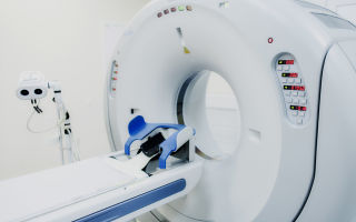
Computed tomography is one of the modern research methods that is successfully used in gynecology. CT scan of the pelvic organs allows timely detection of disturbances in the functioning of internal organs, diagnosis of neoplasms, and obtain images of organs in all projections.
CT is often used to study the pelvic organs
In what cases is the procedure used?
A CT scan of the pelvis shows pathological changes in the uterus and also examines the condition of the ovaries. In addition, the procedure allows you to assess the condition of the genitourinary system and the condition of the rectum. Using tomography, you can determine which organs are healthy and which are not. Identify their size and location in the body of a woman and a man.
If the results of a gynecological examination and ultrasound do not satisfy the doctor, then he may prescribe a study called computed tomography of the pelvis. Most often, to exclude or confirm the presence of malignant tumors. If the patient complains of:
- pain in the lumbar region;
- pain in the pelvic area;
- long menstruation;
- irregular cycle;
- infertility.
CT scan is performed for pain in the pelvic area
This means that the internal organs need careful examination. A CT scan of the pelvis in women allows timely detection of pathologies and prescribing a course of drug treatment.
How is the preparation carried out?
Advice may be different for each patient. It depends on the type of disease that the gynecologist suggests. Preparation for a pelvic CT scan includes the following:
- take a blood test for creatinine, if this indicator is within acceptable values, then a CT scan can be performed; when creatinine is elevated, the procedure is rescheduled;
- adhere to a special diet - this recommendation is for individual cases;
- take a laxative or do an enema;
- Do not eat or drink several hours before the test.
A diet is needed to prevent gases from forming in the intestines. Therefore, a few days before the CT scan, you need to stop eating foods that contribute to gas formation. Since gases can distort the results of the study.
The basic diet is:
- eat pureed, liquid food for three days;
Before the contrast procedure, you should not eat for 6 hours.
- if contrast will be used, then you should not eat for six hours before the procedure;
- in other cases, a light breakfast is allowed;
- a couple of hours before the CT scan you should take gas-removing drugs (Espumizan);
- if the bladder is examined, it should be full. Therefore, you cannot go to the toilet until the end of the examination.
How is a CT scan performed?
CT scans are expensive procedures. They are held in government institutions, but there is a long queue for them. Therefore, if the study is needed quickly, you will have to contact private medical centers.
In addition to CT, MRI, there is a method of MSCT of the pelvis. Its difference is that it produces better images of soft tissues.
The examination is carried out by appointment. You need to be at the clinic 15-20 minutes before the procedure. The patient is given clothes that should not contain metal (buttons, zippers).
The procedure itself is carried out on a horizontal surface, the patient is located on the table of the device. Do not be afraid of the fact that a person is restrained with belts.
After all, any movement during the procedure will worsen the image of the organs and the image will be unclear.
All metal jewelry must be removed before CT scanning.
Metal jewelry must be removed. They may interfere with research.
What are the features of CT with contrast?
A CT scan using contrast is done so that the images show pathological tissues and healthy ones. With the help of contrast, diseased tissue will stand out in white.
Since malignant neoplasms have a better blood supply, the contrast will accumulate in them. The contrast agent allows you to assess the condition of the patient’s veins and arteries.
Contrast is administered intravenously using a catheter. Sometimes this happens once per procedure, but sometimes there are several injections. After the patient is prepared and moved to the scanner, the medical staff goes to the next room. A loudspeaker is used to maintain communication with the patient.
Examination of organs takes different times. As a rule, this is 10-30 minutes. If the obtained images of the organs are clear, then the procedure is stopped.
In addition, there are several ways to administer a contrast agent:
- Using a syringe-injector. This method is called bolus.
Contrast can be administered through intravenous injections
- Intravenously.
- Orally, that is, you need to drink the contrast.
- Rectally. The substance is administered through the rectum.
What is the result
As a result, the doctor receives images of the internal organs in different projections. The radiologist deciphers them, describes them and issues a conclusion. A pelvic CT scan may reveal:
- inflammation of the female organs;
- injuries, internal hematomas, fractures of the pelvic bones;
- uterine polyps, benign formations;
- malignant formations in the pelvic organs;
- hollow tumors on the ovaries;
- frozen pregnancies;
- pathological changes.
- This video will tell you how to properly prepare and undergo a CT scan:
- If a patient is planning an operation on the pelvic organs, then the CT procedure will help in visualizing the organs; the surgeon will have more information on how to carry out the intervention.
What are the risks of the procedure?
These X-ray examinations can be performed every six months. This applies to the procedure with and without contrast. The main danger lies in the dose of radiation that the patient receives. Although doctors say that it is minimal. In any case, procedures can be performed more frequently if the patient's condition requires it.
When contrast is used, a small group of people (about 3%) experience negative reactions in the body. If such a picture is observed, then the study can be carried out only if absolutely necessary. Main pathological reactions:
- the appearance of swelling on the face;
- changes in the frequency and rhythm of breathing;
Some people are allergic to contrast and may experience bronchospasm
- rashes on the body that are accompanied by itching;
- bronchospasm effect;
- a sharp decrease in blood pressure;
- the appearance of nausea and vomiting.
If at least one of these reactions occurs, this indicates that the body perceives the contrast agent as an allergen. Such conditions require medical attention.
Pregnant women are strictly prohibited from conducting the study due to the risk of x-ray radiation. It can negatively affect the development of the fetus. Also, nursing mothers should approach the procedure with caution.
If you cannot do without a CT scan, then you need to limit yourself to artificial mixtures for several days after the examination.
When CT cannot be performed
All procedures have their contraindications, CT is no exception. With CT and MSCT, the radiation dose is minimal, but for some groups of patients it is contraindicated:
- research should not be carried out on pregnant women and nursing mothers;
- patients with renal and heart failure;
CT machines are not suitable for overweight people
- women who are overweight (the tomograph is designed for 130 kg);
- people with neurological disorders who suffer from claustrophobia (in this case, patients can be put into medicated sleep);
- children under 12 years old;
- patients diagnosed with myeloma, diabetes mellitus, thyroid problems;
- allergy sufferers if there is a reaction to contrast.
Did the article help? Rate it (No Ratings Yet) Loading…
Source: https://infouzi.ru/kt/brjushnaja-polost-i-malyj-taz/osobennosti-tomografii.html
CT scan of the pelvis in men and women: what it shows, preparation, examination and price
Computed tomography (CT) of the pelvis is a modern type of x-ray examination. Using a special apparatus, photographs are taken in different projections.
Most often, the procedure is prescribed for suspected oncology, acute inflammatory processes, the presence of cystic formations, or anomalies. The results make it possible to assess the condition of the blood vessels, lymphatic system, internal organs, and tissues in this area.
In order for the diagnosis to be as reliable and accurate as possible, it is necessary to prepare for the tomography in advance and study the list of possible complications.
Benefits of pelvic CT in men and women
The computed tomography method is a layer-by-layer scan of all organs and tissues of the pelvis. Its purpose is to study the structure and various pathologies using x-rays.
The main advantage of CT for the female body is a comprehensive study of the reproductive and genitourinary systems. Subject to diagnosis:
- ovaries;
- uterus;
- fallopian tubes;
- vagina;
- space behind the uterus.
In men, other areas are covered during the procedure. These include:
- prostate;
- vas deferens;
- testicles.
In both cases, pictures of the bladder and rectum are taken.
Like ultrasound and MRI, computed tomography is a non-invasive diagnostic method. The only difference between the procedure is that it can be performed even if there are metal elements, valves, implants, prostheses, or pacemakers in the body.
Indications and contraindications
A mandatory stage of preparation before surgery on the pelvic area is computed tomography. The study allows you to clarify the diagnosis and assess the prevalence of the disease. It can also be used to monitor the effectiveness of treatment or prevent relapses during rehabilitation.
Main indications for hardware diagnostics:
- injuries of the pelvis, lower back;
- sharp pain, discomfort when walking or other activities;
- inflammatory processes in the uterus, fallopian tubes, ovaries;
- inflammation of the bladder or rectum;
- suspicion of the presence of cystic formations;
- abscesses;
- suspicion of oncology, presence of metastases.
Tomography is routinely performed on people with birth defects and anomalies.
CT scanning is recommended for women with inability to become pregnant and irregular menstrual cycles. Detailed data analysis will help determine the cause of the problem.
Among the contraindications of the tomographic method:
- pregnancy;
- lactation;
- weight over 150 kg;
- unconsciousness, coma;
- mental disorders;
- allergy to iodine;
- chronic diseases of the heart, kidneys, liver;
- severe forms of diabetes mellitus;
- abnormalities in the functioning of the thyroid gland.
CT scans are not recommended for children under 12 years of age. However, if necessary, the procedure is performed even on newborns. It is important to understand here that the harm from late detection of the disease can be much greater than from X-ray radiation.
If the child has no contraindications, computed tomography is replaced by a safer diagnostic method - magnetic resonance imaging (MRI).
Radiation dose during examination
During a CT scan, a person receives a certain dose of radiation. The value varies from 1 to 10 millisieverts (mSv). Factors that influence the perception of X-rays:
- equipment quality;
- time of the procedure;
- scanning area.
When examining the pelvis and abdominal cavity, the radiation dose is 5.8 mSv, the head - 0.4 mSv, the chest - 2.9 mSv, and other internal organs - 7-9 mSv.
Radiation from CT is much higher than from other x-ray diagnostic methods. That is why the doctor must take into account the number of previously performed procedures to avoid exceeding the norm.
How often can a pelvic CT scan be done?
The optimal interval between x-ray sessions is 6 months. If there is an urgent need, it is allowed to do a CT scan once every 1-2 months. In this case, the doctor must strictly control the received radiation dose. Otherwise, the growth of malignant cells or the development of pathological diseases can be provoked.
When multiple anatomical areas need to be scanned, it is safer to do a full-body CT scan. In this case, the radiation load will be distributed evenly and the risk of complications will be reduced.
Preparing for the study
You need to start preparing for a pelvic CT scan with contrast 2-3 days before the procedure. First of all, you should balance your diet. Basic diet rules:
- Eliminate brown bread, potatoes, mushrooms and legumes from your diet. These products cause increased gas formation and pollute the body.
- Reduce the amount of fresh fruits, vegetables, berries and herbs you consume. They contain coarse fiber, which causes bloating.
- Carbonated drinks are prohibited.
- Allowed dishes include liquid soups, broths, semolina porridge, boiled meat and fish.
- You can eat boiled sausages, cheeses, butter, and fermented milk products (except cottage cheese) in small quantities.
On the day of the study itself, it is recommended to limit food intake to a minimum. Give preference to teas, still mineral water, natural juices, and low-fat broths. Otherwise, the doctor will see spots and darkening in the pictures that are not related to the disease. This will distort the clinical picture and complicate the diagnosis.
To completely cleanse the intestines, an enema is performed in the evening and morning before the procedure. If this option is not suitable, the clinic will select a laxative. The action time of the product is 16-21 hours.
All patients must be prescribed special medications. They act as contrast agents during CT scanning, blocking X-rays. 1 ampoule of urografin is dissolved in 500 ml of water and drunk in two doses (morning and evening). The morning portion is drunk in the clinic before the diagnosis.
Computed tomography is performed on an empty stomach, with a full bladder. Women additionally need to insert a dry gauze swab into the vagina.
Carrying out the procedure
Computer diagnostics of the pelvis is carried out in the first half of the day and takes 15-20 minutes. How the procedure goes:
- The patient is placed on a movable table.
- The table slides into the tomograph ring.
- The doctor observes the examination process and gives instructions to the patient using a speakerphone.
You must remain still during the CT scan. At the request of the specialist, you need to hold your breath for a short time while the device takes pictures. The ring and table can move to scan the area from all angles.
After completing the procedure, all food restrictions are lifted. It is recommended to drink more fluids and rest for the first few days. This will speed up the process of removing the contrast agent from the body.
Modern tomographs have reduced radiation exposure. If you follow all the specialist’s recommendations and carefully prepare for the procedure, the risk of complications is minimized.
Interpretation of examination results
The patient receives a doctor’s conclusion and completed images within 30 minutes after undergoing a CT scan. Many clinics also provide data in electronic format. The results can be sent by email or recorded on any storage medium (disk, flash drive). Several factors affect image quality:
- tomograph model;
- immobility during diagnosis;
- compliance by the patient with the rules of preparation and completion of the procedure.
In women, a CT scan of the pelvic organs shows the condition of the vagina, body and cervix, ovaries and bladder. Using images, it is easy to identify a tumor, its stage of development and nature, and the presence of metastases. In men, the examination helps identify cancers of the prostate, rectum, soft and bone tissues.
Possible complications after CT scan
Radiation exposure in any quantity has a negative impact on health. Additional harm during computed tomography is caused by contrast agents. They come in low and high osmolarity. The latter are of lower quality, are cheap and most often cause complications.
The excretory system is primarily affected. The patient may develop nephropathy or acute renal failure (ARF). Circumstances in which the risk of complications is greatest:
- elderly age;
- chronic renal failure;
- diabetes;
- dehydration;
- high dose of administered drug.
It is also impossible to assume an immune reaction to a CT scan with contrast. This depends on many factors, including the individual characteristics of the body.
Symptoms for which you should consult a doctor:
- heat;
- pain in the pelvic area;
- nausea;
- vomit;
- severe dizziness;
- loss of consciousness;
- hives;
- bronchospasm.
Full recovery after diagnosis takes 1-2 weeks. During this time, the contrast agent is completely eliminated from the body, and painful symptoms disappear. For this period, antihistamines, plenty of fluids, and bed rest may be prescribed.
approximate cost
The quality of equipment affects the cost of the procedure
The cost of computed tomography is calculated based on several indicators:
- Clinic. The reputation of the institution, its location and equipment determine the price of the service. For example, in Moscow, an examination will cost 20-30% more than in a small provincial town or regional center.
- Field of study. The more pictures taken during the procedure, the higher the final cost will be.
- Contrasting. When performing a CT scan with contrast, an additional fee for the substance is charged. The price of the drug varies from 4 to 7 thousand rubles, which is about 40% of the total cost of the service.
- Equipment. The more modern and high-quality the tomograph, the more expensive scanning with it costs. However, this has its advantages. The procedure using the latest generation devices is much safer, and the result is more accurate.
When choosing a suitable center for CT scanning, do not be guided solely by the institution’s favorable pricing policy. Carefully study patient reviews on thematic forums and websites to get not only high-quality images, but also their qualified interpretation.
Computed tomography has been trusted by Western doctors for many years. Scanning equipment is being improved and poses almost no harm to health. Don't be afraid of the procedure. To prevent complications, consult a specialist in advance and donate blood for a biochemical test. This will help eliminate most of the risks and conduct a CT scan safely.
Source: https://NogoStop.ru/taz/kt-malogo-taza.html
Multislice CT for pelvic diseases: what diagnoses, preparation, scanning process
A pelvic CT scan is a method of X-ray imaging of organs, soft tissues and bone structures. In clinical practice, it is used to determine the condition, functional abilities of anatomical structures, identify complications and evaluate the effectiveness of treatment.
MSCT data provide an accurate description of the changes that led to the development of the disease, reflect the dynamics and predict the outcome of the pathological process.
Pelvic CT scan in female patients
- The multislice CT technique evaluates the condition of the reproductive system, hollow organs of the urinary system, and the final part of the digestive tract in women.
- Diagnostics provides layer-by-layer detailed images of the uterus with appendages, fallopian tubes, bladder, and rectum.
- Examination of the pouch of Douglas and regional lymph nodes reveals purulent-destructive complications of the underlying disease and the spread of oncopathology from the primary focus.
- Pelvic scan shows:
- benign and malignant formations;
- metastases to bone tissue, lymph nodes;
- determining the stage of spread of malignant neoplasms;
- rupture of the bladder, urethra;
- hematoma, phlegmon of the pelvic tissue, retroperitoneal space;
- traumatic damage to the pelvic bones, joints, acetabulum;
- pathology of intrapelvic vessels;
- varicose veins;
- foreign bodies in the pelvic cavity - catheters, intrauterine devices;
- postoperative complications: vesicovaginal, ureterovaginal fistulas, hemorrhages, ureteral strictures;
- chronic pain of unknown etiology in the pelvic area.
Pelvic CT scan in male patients
In men, the study is carried out for similar indications as in women. The scanning area includes the prostate gland, seminal vesicles, distal colon, soft tissues, and bone structures.
Multislice CT detects the following diseases with 97-98% confidence:
- androgenital form of varicose veins;
- causes of erectile dysfunction;
- malignant tumors of the rectum;
- benign hyperplasia, fibrosis, prostate calcifications;
- prostate cancer.
In what cases is it prohibited to conduct research?
Relative contraindications to performing CT of the pelvic organs are:
- inappropriate behavior;
- alcohol or drug intoxication;
- pregnancy;
- hypersensitivity to iodine substances;
- body weight more than 150 kg;
- waist circumference more than 130-140 cm;
- diseases of the kidneys, thyroid gland, cardiopulmonary system in the stage of decompensation.
The study is not prescribed during menstruation. To clearly differentiate the anatomical structures, it is recommended to schedule an appointment in the second phase of the cycle, a week after the end of discharge from the genital tract.
If there are contraindications, the doctor considers alternative diagnostic methods. The restriction on the study can be lifted subject to the elimination of negative factors, correction of the general condition, premedication and the use of anesthesia.
How to prepare for scanning
During a routine examination, before being referred for MSCT, the patient undergoes ultrasound and MRI diagnostics of the area of interest.
To an appointment with a radiologist, the patient brings the conclusion of consultations with specialized specialists and previous research methods. The referral from the attending physician indicates the diagnosis, goals, and objectives.
If the administration of iodine-containing drugs is required, then the functional abilities of the liver and kidneys are additionally assessed.
Pathology of the hematopoietic system is excluded. 3 days before the tomography, blood is donated to determine creatinine levels. On the day of the test, a test is taken to confirm the absence of an allergy to iodine.
To obtain high-quality tomograms, scanning is performed on a full bladder and cleansed intestines. Preparing the patient for the study on the day of the procedure includes:
- self-administration of a cleansing enema;
- to fill the bladder within two hours before diagnosis, ingest 2 liters of clean water at room temperature;
- in patients with a urinary catheter, the bladder is filled by clamping the tube or retrograde fluid injection.
Diet
Within two days before the diagnosis of OMT, a diet is established for the patient, which allows eliminating excess gas formation and stagnation of feces. To do this, exclude gas-forming and hard-to-digest foods from the diet:
- fresh fruits and vegetables;
- baked goods;
- beans, peas, beans;
- black bread;
- animal meat;
- milk;
- canned food;
- smoked meats, pickles, marinades;
- beer, kvass, sparkling water.
It is recommended to consume liquid, non-rich soups, cereal porridges, compote, and sweet, weak tea. 6 hours before the study, food intake is completely stopped.
How the research works
In the CT room, the doctor conducts an explanatory conversation with the patient, then takes informed consent for scanning and administration of contrast agents. In order to avoid the appearance of artifacts and reduce the information content of the procedure, the patient changes into clothes without metal accessories.
The x-ray technician places the patient on the couch in a horizontal position on his back. The arms are placed behind the head, and the lower limbs and torso are secured with belts. After determining the study area, the scanning process starts.
During the procedure, the radiologist gives commands through an intercom and monitors the patient through an observation window. Upon completion of the diagnosis, the medical staff helps the patient off the couch and escorts him to a separate room to await the results.
Contrast agents in diagnostics
Contrast enhancement in computed tomography is carried out to detail the detected changes if the tumor nature of the disease is suspected.
The tomography technique with contrast is performed by intravenous and intracavitary administration of drugs:
- For bolus delivery of contrast agents into the bloodstream, a catheter is installed in the vein of the elbow, to which a syringe injector is attached. The flasks of the automatic system are filled with iodine substance in a volume of 90-120 ml and physiological solution 50 ml. The rate of drug administration is determined by the radiologist;
- to contrast the bladder on the day of the study, 1.5 hours before the procedure, the patient takes orally 1200 ml of liquid diluted with 40 ml of a radiopaque iodine-containing substance (Urografin);
- to study the distal parts of the large intestine, the patient drinks 0.5-1 liters of a contrast agent solution on the eve of the procedure and 0.5 liters in the morning on the day of scanning. The rectum is filled with contrast before the examination using an enema.
Alternative options
- The first-line method in diagnosing pathological changes in the pelvic organs is ultrasound.
- Ultrasound with a sensitivity of 98-99% determines inflammatory processes in the reproductive organs, bladder: thickening of the walls, fluid accumulation, cystic inclusions, signs of peritoneal irritation, adhesions.
- In oncological diseases, it detects tumor infiltrates, the spread of formations beyond the organs, and damage to the lymph nodes.
- The use of Doppler sonography diagnoses disturbances of venous hemodynamics and pathology of the vascular bed.
- MRI is highly accurate in visualizing organs, soft tissue structures of the pelvic floor, bones, and lymph nodes.
- The technique reveals the localization of pathological foci, determines the structure, size, and presence of metastases in the lymph nodes and bone tissue.
Conducts differential diagnosis of the nature of tumors, assesses the dynamics of tumors, and the effectiveness of therapy. Provides detailed information on defects and anomalies of the genitourinary system.
Cost of the procedure
The final price for the study is formed by adding the cost of scanning without contrast enhancement, the use of X-ray contrast agents and general anesthesia.
Some clinics provide discounts on repeated examinations, overnight diagnostics, and simultaneous scanning of two anatomical areas.
To get a CT scan, on average, a patient will need 5,000 rubles. Diagnostics with iodine contrast agent will cost 8,000-10,000 rubles.
Video
- Timely diagnosis of diseases using multispiral scanning technology increases the effectiveness of treatment and the patient’s chances of a full recovery.
- Availability, high speed of research and a minimum of contraindications allow the use of MSCT in emergency cases and routinely, to clarify previously identified changes.
Source: https://osnimke.ru/tazovye-organy/kt-malogo-taza.html
CT scan of the pelvic organs
Computed tomography (CT) of the pelvis is used in the diagnosis of diseases of the following organs and systems:
- organs of the reproductive system,
- Bladder,
- urinary tract.
In addition, computer scanning is used to monitor the results of treatment, preliminary assessment of the condition of the pelvic organs before surgical procedures, as well as after surgery and during the rehabilitation period.
In some cases, to obtain the most complete information about the organ being studied, a CT scan with contrast is performed. An iodine-based dye is used as a contrast agent, which is injected into a vein.
The drug is impenetrable to X-rays, so vascular pathologies and tumor formations are visualized much better than during examination without staining. The contrast agent is completely harmless to humans, with the exception of people suffering from various forms of allergies.
The drug is excreted through the human excretory system within 1-2 days.
Indications
A CT scan of the pelvic organs is usually prescribed by the attending physician to clarify the results of other diagnostic methods, for example, ultrasound, or if the patient has complaints of a certain nature:
- traumatic damage to pelvic organs and tissues;
- pain in the lumbosacral region without identified causes;
- pain when urinating and/or the presence of blood in the urine;
- suspicion of oncological pathology or the presence of a benign tumor;
- study of the structure of regional lymph nodes and determination of metastases in them;
- inflammatory diseases of the urinary tract, uterus and appendages (in women), prostate gland (in men);
- urolithiasis, ureteral obstruction;
- abnormal development of organs and/or bones of the pelvis;
- various vascular diseases;
- clarification of the causes of infertility.
Contraindications
A CT scan of the pelvic organs is a safe procedure, even though the scanning technology uses X-rays. The radiation dose is so small that it makes it possible to examine the same person repeatedly, at short intervals.
However, there are a number of conditions in which CT is strictly prohibited or some additional measures must be taken to perform it:
- pregnancy and breastfeeding – expectant mothers are not recommended to have a CT scan at all, since X-ray radiation, even in a minimal dosage, can negatively affect the development of the fetus. And for nursing mothers - it is possible, but on the condition that during the period of contrast removal (if the study was conducted with it) the woman will not breastfeed;
- the patient’s serious condition – the patient is connected to life support resuscitation equipment or cannot do without constant medical manipulations;
- obesity – weight more than 120 kg and the patient’s waist circumference exceeding the diameter of the tomograph makes scanning difficult or impossible;
- an allergic reaction to iodine is an absolute contraindication for CT with contrast;
- chronic renal failure - the patient should not be administered a contrast agent, which is excreted in the urine and puts a large burden on the kidneys;
- childhood (up to 14 years) – the child’s body is actively growing and developing during this period, therefore CT scans are not recommended for children under 14 years of age, except in emergency cases;
- mental disorders or fear of closed spaces (claustrophobia) - patients with such disorders are given medication (administration of sedatives);
- hyperkinesis (inability to control the motor activity of one’s body) – makes the examination very difficult, since during the scan it is necessary to remain completely still. In this case, the patient can be put into medicated sleep (light anesthesia).
Preparing for the study
2 days before a CT scan of the pelvic organs or urinary tract, the patient is advised to follow a diet that excludes foods that increase gas formation. Otherwise, a swollen intestine can affect the quality of the image and blur the picture.
On the day of the study, 2 hours before the procedure, you can take anti-flatulence medications (Espumizan) and any antispasmodic (No-shpa).
A pelvic CT scan is performed with a full bladder, so before the scan you will need to drink 1-2 glasses and then not urinate. If a CT scan is performed with contrast enhancement, then you cannot eat before the procedure - the dye is administered strictly on an empty stomach.
Methodology for performing CT scans of the pelvic organs and urinary tract
During the examination, the patient lies on the diagnostic table of the tomograph, and a scanner ring that emits X-rays rotates around him. Each rotation lasting 1 second produces a series of slice-by-slice images of the area under study. Subsequently, using a computer program, the obtained images are simulated, from which the specialist makes a conclusion about the presence or absence of any pathology.
Contrast-enhanced CT requires prior drug administration. When the drug enters the blood, the patient may feel a slight heat or cold, as if spreading along the vein. This is a normal reaction of the body. After the injection, itching and redness may occur at the site where the needle was inserted, which go away on their own after some time.
In scanning mode, the tomograph makes specific sounds: clicks, hum, crackling. For those to whom these noises cause psychological discomfort, the nurse provides headphones or earplugs. For children this condition is mandatory.
During the procedure, the patient must wear a special uniform suggested by the nurse. These clothes are body-neutral, do not have rough seams that irritate the skin, and do not cause any discomfort. Or the patient can use his own things, preferably made of cotton fabric.
Modern tomographs have a built-in feedback system with medical staff. Therefore, if health deteriorates or panic occurs, the patient can always inform the diagnostic room staff about this.
Important! If a biopsy or other study using contrast was performed the day before, you must warn your doctor about this.
- Results of CT scan of the pelvic organs
- After the examination, which lasts 15-30 minutes, the patient is given a conclusion, or it can be transferred directly to the doctor who referred the patient for diagnosis.
- Side effects after CT
No complications were observed after CT without contrast. An allergic reaction may occur only to the administration of a coloring agent. But since before the procedure the doctor conducts a thorough examination and questioning of the patient, such risks are eliminated even at the preparation stage.
If allergy symptoms appear (hives, itching, redness, skin rashes, swelling, etc.), you should immediately inform your doctor so that he can take appropriate measures to eliminate them.
Alternative methods for diagnosing the pelvic organs
Computed tomography, as mentioned above, is usually prescribed to clarify the results of other research methods.
Ultrasound does not provide a complete picture of the disease, and the image on the ultrasound specialist’s computer monitor is only in one plane. With CT, a whole series of layer-by-layer images is obtained in different projections;
MRI in some cases provides more information than CT because CT and MRI images are from different angles. Doctors prefer to combine these diagnostic methods, which complement each other perfectly.
Source: https://www.diagnos.ru/procedures/manipulation/kt/kt-taz
CT scan of the pelvic organs: methodology, indications and preparation for the procedure
Computed tomography is a modern diagnostic method for layer-by-layer structural scanning of organs and entire body systems. The implementation of this research occurs thanks to a computed tomograph, the work of which is based on X-ray radiation.
Features of CT of the pelvic organs
Carrying out a CT scan of the pelvic cavity is the main method that helps to visualize the anatomy of the organs located in it in the form of layer-by-layer images in three planes. This technique is possible through the use of X-rays that pass through the medium under study.
The intensity of the rays depends on the area being examined, since the density of the organ affects the clarity of the image, its contrast and the reliability of visualization of the smallest structures.
In some situations, for example, to conduct a CT scan of the pelvic organs in women, a method of introducing a contrast agent, the main component of which, as a rule, is iodine, can be used.
Computed tomography is used to detect various pathologies, injuries and neoplasms located in both soft tissue and bone structures of the area under study. In addition to the above, this diagnostic method allows you to identify various pathological changes in the human lymphatic system.
Carrying out CT scans of the pelvis in women and men
Computed tomography of the pelvic organs in women is performed to study the anatomy of the reproductive and urinary systems. The examination is carried out:
- Bladder;
- rectum;
- “Douglas space” (rectumuterine cavity);
- ovaries;
- fallopian (uterine) tubes;
- uterus
A CT scan of the pelvic organs in men is performed to examine:
- Bladder;
- prostate gland;
- rectum;
- vas deferens.
Purpose of the study
Often, diagnosis of the pelvic organs using CT acts as both a primary examination and to confirm the diagnosis and clarify the data of a previously performed CT scan. Computed tomography is repeatedly used to monitor the treatment of cancer and evaluate the dynamics of tumors.
Indications and contraindications for CT of the pelvic organs
Indications:
- Traumatic damage to the pelvic area or sacral spine;
- Pain or stiffness in this area that gets worse with movement;
- To identify inflammatory processes in the uterus or appendages;
- If there is an inflammatory process in the lower intestines or bladder;
- If you suspect the development of cancer or cystic diseases;
- To conduct preoperative examinations of this area;
- To control the tumor and the extent of its metastases (if any) to soft tissue and bone structures;
- Women with an unclear etiology (cause) of irregular menstrual cycles or infertility.
Contraindications:
The effect of x-rays on the body is contraindicated during pregnancy and lactation, as this can lead to the development of pathologies in the formation of organs and systems of the fetus.
CT scans may not be performed on patients who are overweight because they cannot be placed in a CT scanner (however, there are now CT scanners that can examine patients weighing more than 120 kg).
CT scanning cannot be performed on patients who are unconscious or in a coma.
Conducting CT with contrast has its limitations in patients with allergies, which were detected during the administration of iodine-containing substances, chronic diseases of the cardiovascular system, liver or kidneys. In case of disruption of the endocrine system, namely: diabetes mellitus and thyroid pathologies.
Preparing to Scan
- 2-3 days before the CT scan, you need to follow a certain diet (vetoed on bread products and products that contribute to increased gas formation);
- Two hours before the procedure, it is advisable to refuse solid food;
- Do not drink excessive amounts of fluid on the day of the diagnosis, as the bladder should be moderately full;
- When visiting a tomography room, the patient is recommended to put on comfortable and loose clothing in advance that will not hinder his movements or change into a medical gown;
- Before the procedure, it is recommended to leave all metal-containing objects outside the tomography room (jewelry, glasses, hairpins, etc.).
The specialist will provide detailed information about this procedure to the patient before it is performed.
Preparation for a pelvic CT scan: when examining the pelvis with contrast, it is necessary to undergo an appropriate allergy test in advance to identify the body's hypersensitivity to iodine (if suspected). On the eve of the examination, you should stop drinking alcoholic beverages and iodine-containing medications, which can indirectly affect the CT tests obtained.
CT technique
Diagnostics begins with the patient being placed on his back on a special automated table-couch, which is moved into the tomograph tunnel using a control panel.
Throughout the entire procedure, the X-ray tube, which is located in the body of the device, rotates around the patient.
The sensors, in turn, transmit signals to the doctor’s monitor in the form of an image of the area of the human body being examined.
A CT scan without contrast lasts a maximum of half an hour.
The pelvic tomography itself has a minor effect on the patient in the form of x-ray radiation, but there is no need to worry about this due to the fact that the radiation dose is extremely small.
During the examination itself, the patient does not feel anything. At the end of the study, the patient receives a detailed series of images of organs and bone structures, which are subsequently used for diagnosis.
A CT scan of the pelvis with contrast, compared to a standard study, takes longer (diagnosis takes about an hour). As a rule, a computed tomography scan with a contrast agent is prescribed to study blood circulation in regional vessels or if the development of a malignant neoplasm is suspected.
An iodine-containing contrast agent is injected into the human body (the dosage of the drug does not exceed 20 mg, the parenteral (intravenous) route of administration is used).
After some time, the active component of the contrast agent spreads throughout the circulatory system, thereby coloring it and demonstrating the places of greatest accumulation. Carrying out this technique is of fundamental importance if the presence of a tumor is suspected.
The fact is that they are more actively supplied with blood, compared to other parts of the body. Due to this, the contrast agent accumulates in them and distinguishes the neoplasm from other areas of the pelvis.
- This technique is contraindicated in patients with disorders of the urinary system, in particular with kidney diseases of various etiologies.
- Side effects of using contrast agent
- Despite all the positive properties of using contrast, it has a number of negative effects on organs and tissues. The most common side effects of contrast media use include:
- The occurrence of hyperemia (redness), skin itching and minor urticaria, even with negative results of an allergic test for iodine-containing substances;
- Nausea and dizziness during the procedure itself;
- The appearance of a salty or metallic taste in the mouth after the procedure;
- Increased blood pressure levels.
It is worth noting that all the above symptoms, as a rule, disappear without a trace shortly after the procedure. Often their intensity is so insignificant that the patient may not even feel any discomfort. Metabolites of the contrast agent are excreted through the kidneys over time.
Source: https://CheckUpAdviser.ru/kt-scan/malogo-taza/kt-organov-malogo-taza