Intestinal amebiasis is an infectious protozoal disease caused by protozoan microorganisms (amoeba) that affects the intestines, causing ulcers. The disease is capable of generalization. The human disease is transmitted through unwashed hands, the oral-fecal route, through dirty fruits, through a handshake with a sick person if both did not comply with sanitary standards.
You can also become infected through someone else's bedding and underwear, while swimming in sewage or stagnant water. Amoebas can also be found in the soil.
Amebiasis is an intestinal infection, the course of which can be acute or chronic. The disease can cause abscesses in the lungs and liver, intestines. Symptoms of the onset of the disease resemble an intestinal infection.
Pathogen
The disease is caused by histolytic and dysenteric species of protozoa in active form and in the form of cysts. Cyst amoeba develops when treatment has not been sufficient, there are no symptoms, but amoeba cysts are present. Then we can talk about carriage, or chronic amoebiasis.
- The problem is that such a patient, without knowing it, can infect healthy people.
Diagnostics
- General blood test, thymol test, bilirubin, protein, AST, ALT,
- Analysis of stool or intestinal lavage.
- Additional methods: ultrasound of internal organs, X-ray, sigmoidoscopy with smears, CT scan of the abdominal cavity.
Symptoms
The incubation period ranges from 2 weeks to several months, most often two to three.
Acute amoebiasis
Manifested by fever, malaise, impaired bowel movements - up to 6 times a day, abdominal pain, nausea and vomiting. As the disease progresses, stool may increase - 20 times a day. If you have such symptoms of amoebiasis, you should consult a doctor; those infected are treated in infectious diseases institutions.
If you do not undergo specific treatment, the disease can develop into cyst carriage, so a person can infect others. After some time, acute, untreated amoebiasis can become chronic.
- The chronic form is expressed by the following symptoms:
- Decreased appetite.
- Fatigue.
- The liver becomes enlarged and affected.
- Anemia.
- Malfunctions of the cardiovascular system.
Complications may occur, such as appendicitis, peritonitis, tumor and gangrene of the intestine, bleeding. The brain, liver, lungs, genitourinary system, and skin are also affected.
Liver damage
If the liver is damaged, amoebic hepatitis and abscess may develop. The course is acute and chronic, the prognosis is not favorable. Through the bloodstream, tissue forms of protozoa enter the liver and begin to multiply there. They clog blood vessels and cause inflammation.
Hepatitis develops with diffuse liver damage, and an abscess with focal damage. There may be several lesions.
Skin lesions
Develops in patients with insufficient immunity. It appears as ulcers between the legs, near the anus, and buttocks.
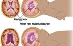
Brain damage
Cerebral amebiasis can develop when amoebae enter the brain through the bloodstream. Abscesses develop, which are fatal. The development is immediate.
Damage to the genitourinary system
It is observed when amoebas enter through the rectum into the bladder and internal genital organs, or rather through wounds in the intestines. Characterized by inflammation of these organs.
At the first symptoms of amebiasis in children, you should call an ambulance and do not delay treatment. The disease can develop much faster than in adults and affect all of the above organs and systems.
Treatment
Medicines for the treatment of amebiasis in adults are divided into 3 groups:
Agents that act on protozoa located directly in the intestines. These include the antibiotics tetracycline and oxytetracycline; diiodoquine and quiniophone. For the skin form, use ointment with chemofon. The latter is used in the form of enemas. The drugs destroy amoebas during carriage and chronic course.
Preparations for the treatment of amoebiasis in acute and extraintestinal forms. Protozoa can be found in the intestinal mucosa, so medications are used that tend to accumulate in the mucous membranes and destroy tissue forms of the pathogen.
The most commonly used dosage forms are emetine hydrochloride; chloroquine, hingamine. They treat amoebic lesions of the liver and kidneys, pneumonia, skin form and are ineffective against luminal forms of amebiasis.
Drugs of the third group enter the microorganism and disrupt the proteins of their cells, as a result of which the amoeba dies. These include metronidazole, ornidazole, tinidazole. They treat all types of amoebic lesions, including carriage.
Treatment of amebiasis can be effective if you consult a specialist in a timely manner.
Prevention
It consists, first of all, of observing sanitary standards and frequent hand washing. People at risk include people living in poor conditions, sewer workers, greenhouse workers, those who work with food, and people who have been in a subtropical climate, such as Mexico or India. In these countries, the water supply system is poorly established, and amoebiasis is widespread.
Photo of amoebiasis
- We also recommend viewing:
Help the project, tell your friends:
Source: https://doktoradvice.ru/amebiaz/
Amebiasis - Diagnosis, symptoms and treatment of amebiasis
Last updated June 30, 2017 at 05:06 pm
Reading time: 6 min
Amebiasis is an infectious disease caused by protozoan parasites.
With it, ulcers appear in the large intestine and on the walls of other internal organs.
Amoeba is the simplest single-celled creature that causes this disease. More than 10% of the world's inhabitants are infected with amoebiasis, mainly in warm and hot climates.
Low levels of sanitation and environmental pollution also provoke the appearance of amoebiasis.
Today, this disease is one of the serious medical problems, although there is treatment and doctors use all their knowledge and medicines to treat people even from poor countries and cities.
What causes amoebiasis?
The causative agent of this disease is dysenteric or histological amoeba. This bacterium can be active or passive. In its usual form it is not dangerous, but in a state of rest it provokes ulcers and ulcers.
If treatment is carried out incorrectly or incompletely, the ulcers turn into cysts, and the symptoms of amoebiasis disappear. In this case, the person still continues to be a carrier of this disease and release substances during bowel movements that can infect other people.
How is this infection transmitted?
This single-celled bacterium is transmitted from one person to another.
Amebiasis is also called the “disease of dirty hands” - as are many similar infectious diseases. The carrier of the infection must carry out all hygiene procedures. You need to wash your hands after going to the toilet, otherwise it will transfer bacteria to personal items, clothing, food, and even to another person upon contact.
Other people should also follow these simple rules to avoid getting infected. Wash your hands, vegetables and fruits before eating them, and do not drink dirty water.
How does the disease develop?
Once in the large intestine, amoeba bacteria begin to transform into an active form. However, infection does not always occur.
These protozoa can simply live in the large intestine and quietly feed on its contents. In this form they will not cause you any harm. This form of amebiasis is called asymptomatic carriage.
At the same time, you still release bacteria that can infect other people.
If you have a reduced immune system, then most likely you will get amoebiasis.
A person with impaired intestinal function, subject to frequent stress, or fasting can also become infected with this disease. In this case, the amoebas begin to show aggression, because they do not have enough of the substances that are in the intestines of such people.
Then they stick to the walls of the organs and begin to suck out their nutrients. The intestinal wall reacts to this by the appearance of ulcers.
At the same time, amoebas throw their own waste into the human body, gradually polluting it and poisoning it.
Most often, ulcers are located on the intestinal walls, but they can also be on the appendix. Their depth can be quite large; some large ulcers eat away the entire intestinal wall, forming a perforation in it. As a result, peritonitis develops - inflammation of the abdominal cavity, because all waste from the intestines enters it.
If there is a blood vessel at the site of such an ulcer, then there is a high risk of internal bleeding. The entry of amoebas into the blood will contribute to their spread throughout the body, and this will lead to the formation of abscesses - ulcers throughout all organs. This is very dangerous for the patient!
What forms of amoebiasis exist?
Intestinal amebiasis - main symptoms
There is a separate form in which the disease is asymptomatic. It poses no threat to the carrier, but other types of amoebiasis can be very dangerous and in some forms are fatal for the patient.
This form is accompanied by certain symptoms, in contrast to asymptomatic amoebiasis. It is also called amoebic dysentery or amoebic dysenteric colitis.
It can be acute and clinical
The symptoms of intestinal amoebiasis are very similar to ordinary dysentery.
The incubation period usually takes from a week to 4 months. After this, symptoms such as frequent stools, fever, pain in the lower abdomen and a false, frequent urge to defecate begin to appear. At the beginning of the development of the disease, the carrier can go to the toilet from 4 to 6 times a day, and at its peak - 10-20 times.
The temperature at the beginning of the disease may be normal or slightly elevated, and then fever begins - from 38.5 or more.
In a more severe version, the disease is accompanied by symptoms of vomiting and decreased (lack of) appetite. Intestinal amebiasis lasts for 3-5 weeks and can be completely cured during this period. After this, a period of remission begins and all symptoms disappear. The disease moves to the chronic stage. You can also try to cure it, but it will be much more difficult and longer.
During the chronic stage of this form of amebiasis, the following ailments are observed: general weakness of the body, loss of appetite, enlarged liver, exhaustion, pale skin, rapid heartbeat.
Deadly complications include intestinal tumors and colon gangrene.
Extraintestinal amoebiasis
Symptoms of amoebiasis in this form depend on where the complication appears.
When amoebas enter the liver, it begins to enlarge and thicken. In this case, the body temperature remains up to 38 degrees, does not fall, but does not become higher. However, with the development of an amoebic liver abscess, the temperature will begin to rise by more than 39 degrees. And the liver will increase sharply, begin to fester and become very painful. The patient's skin will become yellowish.
If the amoebas enter the lungs, then purulent pleurisy begins to develop. This inflammation of the lungs is accompanied by a cough with phlegm, chest pain, less often a cough with blood, and fever.
When bacteria enter the brain, cerebral amebiasis occurs.
The consequences are sad, often doctors cannot make a diagnosis and the patient dies. In the genitourinary system, when amoebas enter, genitourinary amebiasis develops. It is accompanied by inflammation of the genital organs in both men and women, equally.
Cutaneous amebiasis: what are the symptoms?
This form of amebiasis can develop as a complication. It is often experienced by patients with reduced immunity. Body ulcers mainly appear in areas where the skin comes into most contact with the toilet seat and human waste.
They look like erosions with dark edges and smell unpleasant.
Diagnosis of amoebiasis
The diagnosis can only be made by a doctor after a complete examination of the person. At the same time, reports on the sanitary and epidemiological situation of the area or region where the person lives are also taken into account.
After a long study, the doctor will prescribe the correct treatment and necessary medications, their dosage and frequency of use.
Amebiasis in adults and children is recommended to be treated only with medications, and in severe cases - with surgery.
How is amoebiasis treated in the hospital?
Patients with severe disease or in later stages are sent for treatment to an infectious diseases hospital.
For effectiveness and speed of recovery, doctors advise using medications. They can be recognized when diagnosing amoebiasis. The doctor will prescribe the necessary medications for you and select the correct dosage.
Also, if there are ulcers or cysts on the internal organs, surgery may be used. For skin amebiasis, special ointments are often prescribed.
How to treat amoebiasis using folk remedies?
Treatment of amebiasis with folk remedies at home is possible only in mild or initial stages of the disease.
Even in ancient times, when there were no medicines, patients with amoebiasis were treated with decoctions of sea buckthorn, hawthorn or bird cherry.
They also used garlic - they mixed one hundred milliliters of vodka and forty grams of crushed and dried garlic, infused for two weeks and took ten drops before meals with a dairy or fermented milk product.
When using folk remedies for treatment, remember that medications are much more effective and cope better with amebiasis. However, you can use them together to achieve the best effect.
Is it possible to completely cure amoebiasis?
This disease can be completely cured in a few months. If there was no treatment for the disease at all or the disease was detected too late, it could even be fatal.
Now you know what amebiasis is, and if you notice the symptoms of the disease listed above, consult a doctor immediately. In this case, it is better not to self-medicate, but to trust a professional.
Disease prevention
Firstly , always remember that the causative agent of amoebiasis is an amoeba that lives in a polluted environment. Therefore, it is very important to follow all the rules of normal hygiene.
And we are taught this from childhood - to wash our hands after using the toilet and before each meal, not to drink dirty water, not to touch insects and animals that mainly live in landfills.
They can also carry this contagious disease.
Secondly , there are certain risk groups - people who need to be checked more often for the presence of such bacteria. These are those who suffer from chronic diseases of the intestines and excretory system, as well as workers in treatment facilities and sewerage systems. Also homosexuals and people who have visited a country with warm, hot temperatures and low levels of sanitation.
Also, all persons who get a job in preschool and school institutions, food industry factories, and sanatorium-resort complexes are subject to a similar examination. If the result is positive for the presence of bacteria that causes amebiasis, they are not hired until they have completely completed the course of treatment.
To prevent amebiasis, water is purified with filters at wastewater treatment plants, and the cleanliness of public toilets and places where sewerage is not provided is monitored.
The rules of personal hygiene are promoted among the population and absolutely everyone is taught this from an early age. After all, no one has any questions about washing their hands after using the toilet or before eating.
And all buildings where food is served must follow sanitary rules - the presence of a toilet and a sink where you can wash your hands with soap after use is a must!
conclusions
So, if you find any symptoms of the disease, you should go to the doctor and get examined.
Prevention is just as important as annual inspections. It is important to carry them out even if you are not at risk. Self-diagnosis and attempts to treat amoebiasis should not be carried out. This disease is quite insidious and can be fatal for you if it is not treated at all.
Source: https://GemoParazit.ru/parazity/zabolevanie-kishechnyj-amebiaz-simptomy-i-diagnostika
Amebiasis - symptoms, treatment, causes of the disease, first signs
In order to diagnose amoebiasis correctly, it is necessary to conduct microscopy in physiological solution from freshly isolated feces of native smears, as well as smears that are pre-stained in Lugol's solution.
If the disease is at an acute stage or at a pre-acute stage, then as a result of the study, specialists should detect the vegetative tissue form of the amoeba. If the patient’s carriers are asymptomatic, then a cyst and a small luminal form are detected in the laboratory.
But the detection of luminal forms and cysts in feces is not a sufficient indication to make a diagnosis.
During laboratory diagnosis of amoebiasis, feces are used no later than fifteen minutes after defecation. This is a very important factor that must be taken into account, otherwise an incorrect diagnosis may be made.
There are also cases when specialists are not one hundred percent sure of the presence of effective signs of amoebiasis in a patient. And then they use the trichrome staining method for long-term storage of the preparations, and then send them for special examination to the reference laboratory.
Diagnosing intestinal amebiasis by examining freshly excreted feces under a microscope is the simplest and most convenient for doctors. In order to carry out such diagnostics, the clinic must be equipped with the latest modern equipment.
But even the most experienced laboratory technician is not always able to assess the patient’s condition. And in such cases, it is precisely necessary to send the patient’s tests to more trained laboratories for further research and obtain a final assessment.
Differential diagnosis of amebiasis
In some cases, colonoscopy and rectoscopy are recommended. This is done when other tests indicate intestinal damage.
If the doctor has ordered a colonoscopy and rectoscopy, a biopsy of the infected area of the intestine is taken from the patient, examined to identify amoebas, and then a diagnosis is made.
Differential diagnosis of amebiasis helps to identify the presence of all kinds of ulcers and amoeba in the intestines. If, nevertheless, the patient has amebiasis, then the type of lesion will not be diffuse, but focal.
In order to diagnose extraintestinal amebiasis, as a rule, doctors prescribe computed tomography and ultrasonography to patients. Thanks to this, the number and quantity of abscesses in the patient’s body, their location are determined, and most importantly, the results of treatment are monitored.
It is also recommended to take an x-ray. It will help determine the number of abscesses in the lungs, whether there is effusion into the pleural cavity, as well as the condition of the diaphragm.
It should be noted that differential diagnosis is carried out in the case of diseases that are accompanied by hemocolitis.
Dysenteric amoebiasis is characterized by a fairly short incubation period, very acute symptoms at the onset of the disease and minor clinical manifestations over a fairly short time period.
And in this case, pathological changes in the blood progress very quickly. In addition, this disease tends to recur and is accompanied by manifestations of hemocolitis already in severe stages of the disease.
But the most important thing to remember in this case is the fact that the correct diagnosis can only be made by an experienced doctor who has a fairly high level of qualifications. You should never listen to the advice of ordinary people (friends, acquaintances, relatives).
As soon as you experience the first signs and symptoms of a disease such as amoebiasis, you should immediately seek help from a specialist. And try to choose a doctor with a very good reputation. This is necessary to avoid misdiagnosis of the disease.
Treatment
Treatment of amoebiasis takes place in infectious diseases departments of hospitals. As a rule, it is aimed at completely eliminating any manifestations of the disease, as well as replacing the loss of blood, electrolytes and fluid that occurred during the illness. Even as a result of the treatment of amebiasis, all pathogens of this disease must be completely destroyed.
Amoebas that occur during amoebiasis in the human body can choose the intestinal wall, the intestinal lumen, or be outside this area as their habitat.
And because of this, not all drugs are able to eliminate these microorganisms. That is why doctors recommend using different medications during treatment. That is, combine them.
And then it will be possible to talk about some positive result.
Groups of drugs for the treatment of amoebiasis
Based on the symptoms of amoebiasis, treatment can be prescribed using different drugs. They are divided into three main groups. Namely:
- Contact drugs. They have a very destructive effect on those microorganisms that cause this disease in the patient.
- Drugs that act on tissue forms of amoeba. They are considered quite effective if the patient suffers from extraintestinal or intestinal amebiasis.
- Universal drugs. They are used in the treatment of any form of amoebiasis.
Each group of drugs is very effective at one or another stage of the disease. Basically, treatment is prescribed under the strict guidance of a specialist. That is, you should never self-medicate. The doctor himself knows very well which medications you need to prescribe.
The choice of drugs for the treatment of amebiasis in adults, as well as for the treatment of amebiasis in children, directly depends on the stage of the disease.
As soon as you experience the first symptoms, you should immediately seek help from a specialist; he will examine you, diagnose the disease and prescribe treatment, based on the state of your tests.
Medicines for amoebiasis of the first group
The first group of drugs for the treatment of amoebiasis includes drugs such as diiodoquine and yatrene. They must be taken in doses of half a gram three times a day for a week and a half.
After use, a certain period of time must pass, and then it is recommended to repeat the course. Namely, again, you need to take diiodoquine for one and a half weeks.
Only in this case it’s 0.25-0.3 grams and about four times a week.
Medicines for amoebiasis of the second group
The second group includes emetine hydrochloride, dihydroemetine, ambilgar and delagil. The first of the above drugs is taken in doses of 1 gram per kilogram per day. It is injected under the skin intramuscularly for a week. If this does not help, then, as a rule, doctors prescribe a repetition of the course after a month and a half.
The second is also taken intramuscularly for one and a half weeks at one and a half milligrams per kilogram per day. This drug is considered better than the previous one, since it contains much fewer toxins and is much more effective.
The third of the above drugs may outperform the first two combined. It is taken for a week at 25 milligrams per kilogram per day. But it has one drawback. Ambilgar can contribute to the manifestation of neuropsychic disorders and headaches.
And finally, the fourth drug. It has a fairly pronounced effect. It is able to fully concentrate in the liver and intestines. Therefore, delagil is considered very effective for amebiasis of both organs. It must be taken within three weeks. Moreover, in the first week - 0.75 grams per day, in the second - 0.5 grams, and in the third - 0.25 grams.
Medicines for amoebiasis of the third group
The third group of drugs for amoebiasis includes metronidazole. This medicine is prescribed for intestinal and extraintestinal amebiasis.
In the first case, it is taken three times a day for five days, 0.4 grams. In the second - 0.8 grams three times a day for one day, then 0.4 grams three times a day for five days.
Some doctors suggest taking this drug for one and a half weeks.
Another medicine that belongs to the third group of drugs is furamide. It is used within five days. Take two tablets three times a day. It can also be used for prevention. Namely, two tablets during the time the patient is sick.
Antibiotics can also be used as an aid. As a rule, doctors prescribe monomycin, tetracycline, metacycline and other drugs. Antibiotics are sometimes combined with antimoebic drugs. This treatment is prescribed for abscesses of the lungs, liver, brain, and so on.
But in any case, treatment should be prescribed by an experienced specialist who is a professional in his field and knows a lot about it. Never self-medicate. All antibiotics and other anti-inflammatory drugs should be prescribed to you by professionals.
It should also be noted that some patients prefer treating this disease with folk remedies. Here I would immediately like to emphasize that when treating serious diseases you should always use common sense. And again, treatment by any means should only be prescribed to you by an experienced specialist.
In addition, in our time, amebiasis is almost completely curable.
Medicines
cdn.thedailybeast.com
To treat amebiasis, various drugs can be prescribed depending on the symptoms of the disease. Namely:
- Luminal drugs.
- Contact drugs.
The former are usually used to treat non-invasive amoebiasis. That is, in the case of asymptomatic carriers. Specialists can also prescribe treatment with luminal drugs in case of prophylaxis after treatment with systemic tissue amoebicides. With their help, they eliminate amoebas that could remain inside the intestines. And thus, doctors minimize the possibility of relapses.
If the clinical case is quite complex, and doctors realize that it is impossible to prevent a relapse, then luminal drugs are not prescribed, since this is impractical.
In such cases, luminal amoebicides should only be prescribed if there is a possibility that others may be infected with the disease. For example, if the patient works in a certain structure with a large crowd of people.
Then luminal drugs for amoebiasis are prescribed.
Dosage of drugs in the treatment of amoebiasis
The use of special drugs for the treatment of amoebiasis gives very good results. But only the patient needs to seek help from a specialist when the first symptoms appear. This should be done in order to prevent the spread of the disease at the initial stage. As a rule, doctors prescribe drugs for patients in the following dosages:
Intestinal amoebiasis:
- metronidazole – 30 mg/kg in three doses per day for one and a half weeks;
- ornidazole – for children 40 mg/kg in two doses per day for three days, and for adults 2 g in two doses for three days;
- tinidazole - children 50 mg/kg once a day for three days, and adults 2 g per day for three days in one dose;
- secnidazole - children 30 mg/kg per day in one dose for three days, adults 2 g per day in one dose for three days.
Source: https://yellmed.ru/bolezni/amebiaz
Amoebiasis
Amebiasis is a parasitic disease caused by histolytic amoeba and occurring with intestinal and extraintestinal manifestations. Intestinal amebiasis is characterized by copious mucous stools mixed with blood, abdominal pain, tenesmus, weight loss, anemia; extraintestinal - the formation of abscesses of the liver, lungs, brain, etc. The diagnosis of amebiasis is based on clinical data, sigmoidoscopy, colonoscopy, microscopy of smears of abscess contents, serological examination, and radiography. In the treatment of amebiasis, medications are used (luminal and systemic tissue amoebicides, antibiotics), surgical methods (opening and draining abscesses, intestinal resection).
Amebiasis is a protozoal infection, manifested by an ulcerative process in the large intestine and damage to internal organs with the formation of abscesses.
Amebiasis is most widespread in regions with tropical and subtropical climates; In terms of mortality among parasitic infections, it ranks second in the world after malaria.
In recent years, due to a significant increase in migration and foreign tourism, the number of imported cases of amoebiasis in Russia has increased. Amebiasis is recorded in sporadic cases, epidemic outbreaks are rare. Amoebiasis predominantly affects middle-aged patients.
Amoebiasis
The causative agent of amebiasis, the histolytic amoeba (Entamoeba histolytica), is a pathogenic protozoan and has two stages of the life cycle: a resting stage (cyst) and a vegetative stage (trophozoite), which replace each other depending on living conditions.
Vegetative forms of amoeba (precystic, luminal, large vegetative and tissue) are very sensitive to changes in temperature, humidity, pH, and therefore quickly die in the external environment.
Cysts exhibit significant stability outside the human body (they persist in soil for up to 1 month, in water for up to 8 months).
Mature cysts, once in the lower gastrointestinal tract, are transformed into a non-pathogenic luminal form that lives in the lumen of the colon, feeding on detritus and bacteria. This is the stage of asymptomatic carriage of amoebas.
Subsequently, the luminal form either encysts or turns into a large vegetative form, which, due to the presence of proteolytic enzymes and specific proteins, is introduced into the epithelium of the intestinal wall, turning into a tissue form. Large vegetative and tissue forms are pathogenic and are found in acute amoebiasis.
The tissue form parasitizes in the mucous and submucosal layers of the colon wall, causing destruction of the epithelium, disruption of microcirculation, formation of microabscesses with further tissue necrosis and multiple ulcerative lesions.
The pathological process in the intestine with amebiasis most often extends to the cecum and ascending parts of the colon, less often to the sigmoid and rectum. As a result of hematogenous dissemination, histolytic amoebas are capable of entering the liver, lungs, brain, kidneys, and pancreas with the formation of abscesses in them.
The main source of amoeba infection is patients with a chronic form of amoebiasis in remission, as well as convalescents and carriers of cysts. Flies can be carriers of amoeba cysts.
Patients with an acute form or with a relapse of chronic amoebiasis do not pose an epidemic danger, since they secrete vegetative forms of amoebas that are unstable in the external environment.
Infection occurs through the fecal-oral route when food and water infected with mature cysts enter the gastrointestinal tract of a healthy person, as well as through household contact through contaminated hands. In addition, transmission of amebiasis is possible during anal intercourse, mainly among homosexuals.
Risk factors for contracting amebiasis include poor personal hygiene, low socioeconomic status, and living in areas with a hot climate. The development of amebiasis can be triggered by an immunodeficiency state, dysbiosis, unbalanced diet, and stress.
The incubation period of amoebiasis lasts from 1 week to 3 months (usually 3-6 weeks).
According to the severity of symptoms, amebiasis can be asymptomatic (up to 90% of cases) or manifest; according to the duration of the disease - acute and chronic (continuous or recurrent); according to the severity of the course - mild, moderate, severe.
Depending on the clinical picture, there are 2 forms of amebiasis: intestinal and extraintestinal (amoebic abscesses of the liver, lungs, brain; genitourinary and cutaneous amebiasis). Amebiasis can manifest itself as a mixed infection with other protozoal or bacterial intestinal infections (for example, dysentery), helminthiasis.
Intestinal amebiasis is the main, most common form of the disease. The leading symptom of intestinal amebiasis is diarrhea. The stool is copious, liquid, initially fecal in nature with an admixture of mucus up to 5-6 times a day; then the stool takes on the appearance of a jelly-like mass mixed with blood, and the frequency of bowel movements increases to 10-20 times a day.
Characterized by constant increasing pain in the abdomen, in the iliac region, more on the right. When the rectum is damaged, painful tenesmus occurs; when the appendix is damaged, symptoms of appendicitis occur. There may be a moderate increase in temperature and asthenovegetative syndrome.
The severity of the process in intestinal amoebiasis subsides after 4-6 weeks, after which a long remission occurs (several weeks or months).
Spontaneous recovery is extremely rare. Without treatment, an exacerbation develops again, and intestinal amebiasis acquires a chronic recurrent or continuous course (lasting up to 10 years or more).
Chronic intestinal amebiasis is accompanied by disorders of all types of metabolism: hypovitaminosis, exhaustion, even cachexia, edema, hypochromic anemia, endocrinopathies.
In weakened patients, young children and pregnant women, a fulminant form of intestinal amebiasis may develop with extensive ulcerations of the colon, toxic syndrome and death.
Of the extraintestinal manifestations of amebiasis, the most common is amoebic liver abscess. It is characterized by single or multiple abscesses without a pyogenic membrane, most often localized in the right lobe of the liver.
The disease begins acutely - with chills, hectic fever, profuse sweating, pain in the right hypochondrium, aggravated by coughing and changes in body position. The condition of the patients is serious, the liver is sharply enlarged and painful, the skin is sallow in color, and sometimes jaundice develops.
Pulmonary amebiasis occurs in the form of pleuropneumonia or lung abscess with fever, chest pain, cough, and hemoptysis. With amoebic brain abscess (amebic meningoencephalitis), focal and cerebral neurological symptoms and severe intoxication are observed.
Cutaneous amebiasis occurs secondarily in weakened patients, manifested by the formation of mildly painful erosions and ulcers with an unpleasant odor in the perianal area, on the buttocks, in the perineal area, on the abdomen, around fistulous openings and postoperative wounds.
Intestinal amebiasis can occur with various complications: perforation of an intestinal ulcer, bleeding, necrotizing colitis, amoebic appendicitis, purulent peritonitis, intestinal stricture.
In case of extraintestinal localization, abscess breakthrough into surrounding tissues with the development of purulent peritonitis, pleural empyema, pericarditis or the formation of fistulas cannot be ruled out.
In chronic amebiasis, a specific tumor-like formation from granulation tissue is formed in the intestinal wall around the ulcer - an amoeba, leading to obstructive intestinal obstruction.
When diagnosing intestinal amebiasis, clinical signs, epidemiological data, results of serological studies (RNGA, RIF, ELISA), sigmoidoscopy and colonoscopy are taken into account.
Endoscopically, with amebiasis, characteristic ulcers of the intestinal mucosa are detected at different stages of development; in chronic forms, cicatricial strictures of the colon are detected. Laboratory confirmation of intestinal amoebiasis is the identification of tissue and large vegetative forms of amoeba in the patient’s feces and discharge from the bottom of ulcers.
The presence of cysts, luminal and precystic forms of the pathogen indicates amoebic carriage. Serological reactions show the presence of specific antibodies in the blood serum of patients with amebiasis.
Extraintestinal amoebic abscesses can be visualized by a comprehensive instrumental examination, including ultrasound of the abdominal organs, radioisotope scanning, plain chest radiography, CT scan of the brain, and laparoscopy. The detection of pathogenic forms of the pathogen in the contents of abscesses is evidence of its amoebic origin.
Differential diagnosis of amebiasis is carried out with dysentery, campylobacteriosis, balantidiasis, schistosomiasis, Crohn's disease, ulcerative colitis, pseudomembranous colitis, colon neoplasms; in women – with endometriosis of the colon.
Amoebic abscesses of extraintestinal localization are differentiated from abscesses of other etiologies (echinococcosis, leishmaniasis, tuberculosis).
Treatment of amebiasis is carried out on an outpatient basis; hospitalization is necessary for severe cases and extraintestinal manifestations. To treat asymptomatic carriage and prevent relapses, direct-acting luminal amoebicides (etofamide, diloxanide furoate, iodine preparations, monomycin) are used.
Systemic tissue amoebicides (metronidazole, tinidazole, ornidazole) are effective in the treatment of intestinal amebiasis and abscesses of various locations. To relieve colitis syndrome, accelerate reparative processes and eliminate pathogenic forms of amoebas, iodochlorooxyquinoline is prescribed. If metronidazole is intolerant, the use of antibiotics (doxycycline, erythromycin) is indicated.
The combination of drugs, their doses and duration of therapy are determined by the form and severity of the disease.
In the absence of effect from conservative tactics and the threat of abscess breakthrough, surgical intervention may be required.
For small amoebic abscesses, it is possible to perform a puncture under ultrasound control with aspiration of the contents or an opening with drainage of the abscess and subsequent introduction of antibacterial and amoebicidal drugs into its cavity.
In case of pronounced necrotic changes around an amoebic ulcer or intestinal obstruction, intestinal resection with colostomy is performed.
Forecast and prevention of amebiasis
With timely specific treatment, in most cases the prognosis for intestinal amebiasis is favorable. In case of late diagnosis of amebic abscesses of other organs, there is a risk of death.
Prevention of amebiasis includes early detection and full treatment of patients and amebia carriers, compliance with a sanitary and hygienic regime at home, ensuring high-quality water supply and wastewater treatment, food safety control, and health education.
Source: https://www.KrasotaiMedicina.ru/diseases/infectious/amebiasis
Amoebiasis
The first signs of the disease appear approximately 7-10 days after infection or within several months (incubation period). Depending on the symptoms of the disease, intestinal and extraintestinal amebiasis are distinguished.
With intestinal amebiasis (intestines are affected), several symptoms come to the fore.
- Stool disorder: the appearance of diarrhea (frequent loose stools), its frequency can reach 20 times a day. The stool is first mixed with feces, formed, then (on days 4-7) with an admixture of mucus, blood, pus (the feces look like raspberry jelly); in severe cases, bloody diarrhea occurs.
- Pain in the lower abdomen: cramping, intensifying after defecation (emptying the rectum).
- Painful abdominal cramps (tenesmus).
- An increase in body temperature (usually slight), but in some cases the body temperature may be within normal limits.
- When the appendix is damaged (amebic typhlitis), symptoms of acute appendicitis (inflammation of the appendix of the cecum) occur, for example, severe pain in the right hypochondrium, increased body temperature, tension in the abdominal muscles, etc.
- Dehydration (dehydration).
- Weakness, drowsiness.
- Acute manifestations of amebiasis last from 4 to 6 weeks, then the condition normalizes, and a period of remission begins (from several weeks to several months).
Symptoms of extraintestinal amebiasis (not associated with intestinal damage, localized (located) in other organs).
- Abscess (ulcer) of the liver. The disease usually begins acutely and the following are observed:
- increase in body temperature to 39°C and above;
- chills, increased sweating;
- pain in the right hypochondrium, which intensifies when coughing, changing body position, or palpating the liver;
- jaundice (yellow discoloration of the skin of the sclera (outer membrane) of the eyes).
When an abscess occurs inside the liver, symptoms may be absent or mild.
- Amoebic pneumonia (pneumonia), in which the following symptoms are observed:
- increase in body temperature to high numbers;
- chills;
- pain, discomfort in the chest;
- cough;
- dyspnea;
- hemoptysis.
- Damage to the pericardium (outer lining of the heart).
- Encephalitis (inflammation of the brain). Symptoms depend on the location (location) of the abscess in the brain. For example, convulsive seizures when the abscess is localized in the frontal lobe, impaired coordination of movements when localized in the cerebellum, etc.
- Cutaneous amebiasis - usually occurs in weakened patients, with erosions (superficial skin defects) and/or ulcers (deep skin defects) on the skin of the abdomen, buttocks, and perineum.
If untreated, amebiasis can last for years (sometimes even decades) with periods of exacerbation and remission (chronic recurrent (renewed) course), sometimes a continuous course is observed. There are:
- asthenia (weakness, drowsiness, decreased performance);
- anemia (anemia, decreased levels of erythrocytes (red blood cells) and hemoglobin (oxygen carrier protein));
- weight loss, up to cachexia (extreme exhaustion);
- hypovitaminosis (vitamin deficiency in the body).
The incubation period lasts from 1-2 weeks to 3 months.
Based on the severity of the symptoms of amebiasis, the disease is divided into asymptomatic and manifest .
- Asymptomatic (hidden) course: the disease does not manifest itself for a long time.
- Manifest course: There is an increase in body temperature, diarrhea (frequent loose stools), weakness, and abdominal pain. There are several types of manifest amoebiasis:
- intestinal amebiasis (intestines are affected);
- extraintestinal amebiasis (damage to other organs is detected);
- cutaneous amebiasis (with predominant skin lesions, most often occurs in weakened patients and/or is a complication of the intestinal form of the disease).
According to the stages acute and chronic are distinguished .
- Acute stage: symptoms develop very quickly, patients feel seriously ill.
- Chronic stage:
- chronic continuous course;
- chronic relapsing (renewing) course.
Extraintestinal amebiasis, in turn, is divided into:
- amoebic hepatitis (liver inflammation);
- liver abscess (ulcer in the liver);
- pulmonary amebiasis (lung damage);
- cerebral amebiasis (brain damage);
- cutaneous amebiasis (skin lesions).
There are also invasive (symptoms of the disease are observed) and non-invasive amebiasis (carriage of amoebic cysts is a temporary form of existence of microorganisms that forms in unfavorable conditions and protects the microorganism), which is characterized by:
- asymptomatic course of the disease;
- absence of hematophagous trophozoites (an active form of the microorganism found in tissues and organs);
- absence of pathological (abnormal) changes during endoscopic examination (using a special optical instrument (endoscope));
- absence of specific antibodies (proteins that are produced in the body when antigens (foreign proteins) enter).
- The causative agent of the disease is a single-celled parasite, the histolytic amoeba (Entamoeba histolytica). It exists in 2 states: in the dormant stage (cyst) and in the vegetative form.
- In the external environment, the amoeba is in the cyst stage, this ensures its spread and protection from various unfavorable environmental factors.
- The vegetative form (trophozoite) is formed from cysts in the human body; outside the body, trophozoites (the active form of the microorganism) die. The source of infection is a sick person who releases cysts into the external environment along with feces (stool).
- Transmission mechanisms - fecal-oral (that is, feces (infected, containing amoeba cysts) of a sick person enter the environment (water, soil), and then they enter the body of another person (did not wash hands, drank dirty water, etc.)) .
- Routes of transmission: alimentary (food), water, household. The disease is transmitted through contaminated water, food, soil, and household items. Amoebas can be transmitted sexually (among homosexuals).
- When amoebas reproduce and parasitize (exist at the expense of humans) in the intestine, microabscesses (small ulcers) are formed in its wall, which open into the intestinal lumen, forming ulcers (deep defects in the organ wall), which can merge and even lead to perforation (rupture) intestines.
- As a result of the penetration of amoebas into the vessels of the mesentery (the fold of the peritoneum through which the abdominal organs are attached to the posterior wall of the abdomen), the parasite is carried into other organs, where abscesses develop.
LookMedBook reminds you that this material is posted for informational purposes only and does not replace medical advice!
-
The therapist will help in treating the disease
Make an appointment with a therapist
- Analysis of the medical history and complaints (when (how long ago) did the symptoms of the disease appear, what is the frequency and nature of the stool (mixed with blood, pus, mucus), whether the patient visited areas endemic (areas with a high prevalence of the disease) for the development of amebiasis (hot, tropical and subtropical countries), whether symptoms of the disease appeared after the trip, and after some time).
- Physical examination. Pain on palpation (palpation) of the abdomen (with caution), symptoms of extraintestinal manifestations (for example, skin ulcers, liver abscess, etc.) of amoebiasis are determined.
- Blood test (eosinophilia is often observed - a sharp increase in eosinophils (a subtype of white blood cells) in the blood, which is an indirect sign (since an increase in eosinophils in the blood can also occur due to allergies or as a manifestation of blood tumors, etc.) of the presence of a parasitic (caused parasites) diseases), anemia (anemia) may be detected.
- Biochemical blood test to detect changes in enzymes (substances that accelerate metabolic processes in the body) of the liver, determination of total protein, etc.
- Stool analysis. This test can detect living amoebas. It is very important to conduct research immediately after defecation - emptying the rectum (to detect live parasites).
- Examination of the intestine (colonoscopy, sigmoidoscopy) is an endoscopic method (the examination is carried out using a special optical instrument (endoscope)), which allows you to identify ulcers (deep defects in the organ wall) in the intestinal wall and immediately perform an analysis to detect live amoebae. For research, unformed feces (feces) with mucus and blood are taken, since it is in them that vegetative forms (temporary forms of existence of microorganisms that form in unfavorable conditions and protect microorganisms)) of amoebas are found.
- Methods of immunological diagnostics - detect antibodies (proteins that are produced in the body when antigens (foreign proteins) enter) and antigens (foreign proteins) of amoeba: enzyme-linked immunosorbent assay (ELISA) - a laboratory method.
- Molecular diagnostic methods - allow you to detect amoeba DNA in stool using PCR (polymerase chain reaction).
- Instrumental research methods for extraintestinal forms (for example, damage to the liver, lungs, brain, etc.):
- ultrasound examination (ultrasound) of the abdominal organs to identify possible changes in the liver;
- X-ray examination of the chest (if lung damage is suspected);
- Computed tomography (CT) and magnetic resonance imaging (MRI) of the abdominal organs can detect changes in the liver, brain, and lungs.
- A consultation with a gastroenterologist is also possible.
Treatment of amebiasis can be conservative (non-surgical) and surgical.
Conservative treatment.
Specific drugs are used to treat amebiasis.
- Antiprotozoal drugs (to kill amoebas).
- Antibacterial drugs (antibiotics for the treatment of infections), if there is intolerance to antiprotozoal drugs or if complications develop).
- Symptomatic treatment:
- multivitamin preparations;
- restoration of disturbed water-salt balance;
- antispasmodics to eliminate pain;
- hepatoprotectors (drugs that protect the liver).
Surgical treatment if conservative treatment is ineffective or if there is a threat of rupture of an abscess (abscess). In the intestinal form (intestinal damage):
- perforation (rupture) of the intestinal wall;
- peritonitis (inflammation of the peritoneum);
- intestinal bleeding;
- specific appendicitis (inflammation of the appendix - the vermiform appendix of the cecum);
- rectal prolapse;
- detachment of the intestinal mucosa and its gangrene (tissue death);
- stricture (narrowing) of the intestine;
- infectious diseases (secondary infections, that is, they join an existing amoebiasis).
For extraintestinal forms (for example, damage to the liver, lungs, brain, etc.):
- an abscess (ulcer) of the liver can be complicated by a breakthrough into the abdominal cavity with the development of peritonitis or into the pericardium (heart sac) with the development of pericarditis (inflammation of the outer lining of the heart - the heart sac (pericardium)). Mortality (mortality) in this course of the disease without specific treatment reaches 25% or higher;
- lung abscesses (purulent pleurisy (purulent inflammation of the pleura (the membrane lining the inner surface of the chest wall and the outer surface of the lungs)), empyema (accumulation of pus between the layers of the pleura), pneumothorax (accumulation of air in the pleural cavity), hepatopulmonary fistulas (pathological (abnormal) ) channels between the bile ducts and bronchi)).
- Refusal to drink raw water (you need to boil it).
- Eating only thermally processed foods.
- After suffering from amebiasis, follow-up with a doctor at your place of residence for at least 1 year.
- Individual prevention of the development of amebiasis for persons planning to visit endemic areas (hot, tropical and subtropical countries).
- Identification and treatment of cyst carriers (people who are found to have amoebic cysts (a temporary form of existence of microorganisms that forms in unfavorable conditions and protects the microorganism)).
Source: https://lookmedbook.ru/disease/amebiaz