There are a huge number of diseases in the world that can affect any person, and in modern conditions it can be very easy to get seriously injured, but many people, even if they experience ailments or any unusual manifestations, do not turn to a specialist, sparing money and believing that everything will go away on its own. Unfortunately, this happens very rarely, and most often the disease continues to progress and irreversible consequences appear, often even leading to death. If you consult a doctor in a timely manner, then using diagnostic methods he will help make a correct diagnosis and prescribe the necessary therapy for a full recovery, if possible. Let's look at how a CT scan of the spine is done, one of the most common procedures.
How does computed tomography work?
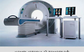
To conduct a CT or MRI scan of the spine, a special device is used - a tomograph, consisting of a moving table and a special block equipped with a round hole into which the table slides.
When such an examination of the spine is performed, an X-ray source located in the ring part of the equipment is activated. On the opposite part of this ring there is a receiving grid that captures X-rays passing through the patient’s body.
Computed tomography is a painless, non-invasive examination technique.
During a study on a computed tomograph, namely, when rays pass through tissue, their physical characteristics may change, and the degree of change depends on the density of these tissues.
The already mentioned grid registers such changes in characteristics and transmits them to a specialist’s computer, where the data is processed and, therefore, full-fledged images are created. We have been asked many times about how x-rays of any vertebra or intervertebral discs differ from SCT.
In fact, the difference between these examinations is significant, because with computed tomography it is not just possible to achieve a reduction in radiation exposure. An image, or rather a series of them, turns out to be much more accurate and informative, and modern technologies even make it possible to make a 3D model of the spine.
This happens due to the fact that the radiation source, as well as its receiver, are constantly moving around the patient, this allows you to carefully study an organ or part of the body from different angles, and there are a lot of them, but it all depends on the capabilities of specific equipment. Here are the main advantages of computed tomography:
- excellent clarity of images, allowing you to carefully study them;
- absence of any pain in the patient during the study;
- speed of the procedure;
- minimal X-ray radiation (compared to conventional radiography);
- the ability to carry out diagnostics in the presence of certain implants in the body;
- the ability to take pictures when there is slight movement of the patient (this does not mean that you can lie as you want, but this factor improves quality and reduces error).
Important! The question is often asked: “MRI or CT of the spine: which is better?” In fact, there is no definite answer to this question, since it all depends on your condition and the opinion of specialists.
Despite the fact that diagnosis of the spine can be carried out using both methods, there is far more than one difference between MRI and CT. Magnetic tomography of a resonant nature is safer, but much more expensive.
The fact is that it is carried out without the use of X-ray radiation, which is harmful to any organism, but magnetic resonance imaging also has unique contraindications, for example, the presence of metal elements or even electrical devices in the body.
Now, the difference between a CT scan and an MRI of the spine will not be a mystery to you. Let us also mention that you should not ask doctors the question of how CT differs from MRI, because you will be prescribed the most suitable study.
A modern tomograph allows you to obtain accurate images of any area of the spine.
Indications for the procedure
A CT scan of the cervical spine or any other part of it can only be ordered by a qualified specialist, who will make a decision only after a thorough examination and passing the necessary tests, if they are necessary and the situation is not an emergency. Here are the main purposes for which this survey is carried out:
- the need to clarify the method of surgical intervention, which will take place in the future and will affect nearby tissues or even the spinal column itself;
- the need to monitor the results of surgery already performed in this area;
- identification of neoplasms of a malignant or benign nature;
- the need to study malignant and benign tumors; most often, a contrast agent is also used for this, which also allows one to study the spread of metastases, and the location and size of the tumor will be determined with maximum accuracy;
- the need to recognize intervertebral hernias, as well as the consequences characteristic of them;
- diagnosis of osteoporosis (magnetic resonance imaging is not suitable for this, since it does not imply the ability to identify problems associated with soft tissues);
- identification of any congenital anomalies associated with the spine;
- finding any dystrophic, as well as metabolic and degenerative processes related to the bone structures of the spine;
- diagnosis of inflammatory processes, as well as arthrosis;
- confirmation of the fact of injury, for example, a vertebral fracture, as well as an assessment of the extent of the problem, which allows treatment to begin;
- the need to diagnose diseases related to the bone marrow, for example, those of an infectious nature;
- identifying problems that cause severe pain in the spine area without obvious reasons; for this purpose, a CT scan of the cervical spine is often performed if pain appears there;
- identifying changes in bone density as well as evenness of bone tissue.
Note! As was said earlier, a specific examination cannot be accurately shown; it is worth paying attention to different methods, and the decision can only be made by a qualified and experienced doctor who can not only do a CT scan of the spine, but also accurately interpret the images, correctly making a diagnosis.
Computed tomography of the spine is an examination procedure that allows you to obtain 3D images of the area of the body being examined.
Contraindications
The contraindications of this method are quite large, which is justified by the use of quite harmful ionizing radiation. You must inform the specialist in advance about all the chronic diseases that you have. Here are the main problems and diseases for which a CT scan of the spine is most often not performed:
- diseases related to the heart and having a decompensated nature;
- acquired or even congenital heart defects;
- hypertensive crisis or myocardial infarction;
- acute condition of cerebrovascular accident;
- bronchial asthma in severe form, in most situations it is accompanied by constant attacks of suffocation;
- diabetes mellitus, especially in the presence of complications;
- kidney and liver diseases with complications;
- chronic alcoholism or even drug addiction;
- claustrophobia;
- severe mental illnesses characterized by inadequate reactions;
- Quincke's edema and other allergic diseases with extremely severe clinical manifestations;
- diseases related to the thyroid gland and having laboratory abnormalities, as well as serious symptoms;
- Contraindications include obesity, because the equipment for computed tomography is designed only for patients up to 120-150 kg, others can use open-type tomographs or give preference to another diagnostic method;
- pregnancy or breastfeeding.
If computed tomography involves the use of a contrast agent, then the following points will be added to the contraindications:
- renal or liver failure;
- serious illnesses;
- individual intolerance to any contrast components;
- early age (this contraindication is relative, because it can be neglected if the goal justifies the risk).
Important! In no case should contraindications be ignored, as this can result not only in minor complications, but also in serious health problems, and in the most severe cases the patient can even die! Listen to your doctor and follow all recommendations, for example, when performing a procedure with contrast, you will need to take a special test in advance to check for an allergic reaction to the components of this substance, unless, of course, the examination is of an emergency nature.
Preparing for the study
Preparation for CT scanning when carrying out such diagnostics is mandatory, regardless of who is being examined.
You should decide in advance where the best place to get a CT scan is, and you should pay attention not only to how much a CT scan costs.
It is best to choose a clinic where modern digital equipment is installed, which carries the least radiation dose and allows you to take the most accurate and informative images.
Your doctor will tell you what the examination shows, and for high-quality visualization you need to start following a special diet in advance.
And its essence is based on the fact that 2-3 days before the CT scan you should avoid any food that can increase gas formation inside the gastrointestinal tract.
The list of such products that should be prohibited includes legumes, as well as dairy products and baked goods; it is also better to avoid fruits and vegetables.
Before the study itself, you will also need to take some medications that can slightly reduce the amount of gases inside your intestines; a specialist will tell you a list of them (the most commonly prescribed medications are Espumisan and activated charcoal). Remember that a lot depends on preparation, so spend a lot of time on it.
Positioning of the patient during examination of the lumbosacral and thoracic spine.
Carrying out diagnostics
Do not be afraid of such an examination; if it is necessary, the doctor will do everything to reduce the radiation exposure. You will need to wear comfortable clothing in which you can lie still throughout the procedure (without contrast it can last up to 20-30 minutes, and with contrast it can last up to an hour).
The doctor will tell you to lie down on a special table, which will later slide inside the tomograph. You will be secured with straps and bolsters to minimize any movement during the process, otherwise the pictures will not be accurate enough. In most situations, taking pictures in a supine position is sufficient, but sometimes the doctor may ask you to roll over onto your stomach or side.
After the examination starts, X-rays will be sent to the area of your body in a narrow stream, they will hit special sensors, after which the information will go directly to the doctor’s computer.
A series of layer-by-layer images or even a three-dimensional model will already be formed there.
After the research itself, you will need to wait for 1-2 hours, during which time the data will be analyzed and decrypted.
Source: https://tomografa.net/kt/pozvonochnika-i-sustavov/obshchaya-diagnostika.html
Computed tomography of the spine
Summary:
- Indications and contraindications for
- What does a CT scan of the spine show?
- Main types of spinal column scans
- Preparing for a CT scan
- How to do a CT scan of the spine
- Decoding the received data
Today, computed tomography of the spine is one of the most popular studies, especially in the field of neurology and neurosurgery. CT scan of the spine is based on X-ray scanning and processing of examination results with special software.
Sign up for a CT scan at a discount at Moscow medical centers: +7 (499) 519-32-37
- Best price when booking through our website
- We will select a diagnostic center for free
- We'll schedule an appointment at a time convenient for you.
Images of organs and tissues are performed layer by layer, which makes the technique extremely effective and informative. Such a diagnosis shows the existing pathologies as accurately as possible and allows you to study their features. Treatment, in turn, depends on the accuracy and timeliness of diagnosis.
Indications and contraindications for
Computed tomography of the spine is performed for certain indications, but in some cases it either cannot be performed or must carry certain restrictions.
Who should get the scan?
This examination is prescribed for certain symptoms:
- unknown cause of pain (lower back, spine, neck);
- control of treatment, risk of osteoporosis;
- clarification of the existing diagnosis;
- suspected cyst or tumor;
- checking the condition after surgery;
- possibility of intervertebral hernia;
- suspected spinal cord infection;
- signs of osteochondrosis, arthritis;
- injuries, congenital anomalies;
- signs of degenerative changes in the spine;
- suspected fracture;
- preparation for surgery.
An appointment for diagnostics is made when the patient has been thoroughly examined, a complete medical history has been collected, and preliminary tests have been performed.
What are the contraindications?
There are a number of diseases and factors that are prohibitive for the CT method:
- decompensated heart failure;
- acute myocardial infarction;
- heart defect (congenital or acquired);
- bronchial asthma (severe form);
- hypertensive crisis, pregnancy;
- diabetes mellitus with complications;
- obesity (more than 150-180 kg);
- advanced liver or kidney disease;
- dysfunction of the thyroid gland;
- severe mental disorders;
- chronic alcoholism, drug addiction (exacerbation).
All of the above is not an absolute ban on the use of computed tomography. In some cases, you simply need to adjust the procedure. Children's age is also a contraindication, although in certain cases the child is also examined.
What does a CT scan of the spine show?
A CT scan of the spine can reveal a lot of pathologies:
- osteochondrosis;
- spinal canal stenosis;
- osteoarthritis, spondylosis;
- birth defects;
- intervertebral hernia;
- tumor and metastases.
This method allows you to determine the nature of the tumor and distinguish malignant formations from benign ones. Many formations are often detected only by CT. For example, a hemangioma in such a study is detected in every 9-10 patients who did not even suspect about it. More often, hemangioma is detected in the thoracic vertebrae.
Main types of spinal column scans
Computed tomography is divided into types depending on the area being examined - lumbosacral, cervical, thoracic, lumbar.
• Cervical
A CT scan of the cervical spine shows its condition, that is, it evaluates the cervical area, the intervertebral canal and its processes. This test is usually prescribed for tissue injuries and fractures. Diagnostics may reveal:
- developmental anomaly;
- herniated discs;
- inflammation, fractures;
- tumor of any nature.
This area is examined if there is pain, rheumatic diseases, or injuries. It is in this department that hernias, protrusions and other degenerative-dystrophic phenomena often occur.
• Chest
This area is often scanned to diagnose tumors, study the nature of metastases, inflammation of various types, and diseases of the lymph nodes. A CT scan of the thoracic spine may reveal:
- tumor and metastases;
- fracture or crack;
- abnormal narrowing of the spinal cord canal;
- rheumatism, congenital anomalies;
- osteoporosis, arthrosis, spondylosis;
- hemorrhage in the spinal cord.
Using computed tomography, you can check for the presence of a foreign body in the lungs, scan lymph nodes, and diagnose a lung tumor.
• Lumbar
CT scan of the lumbar spine shows:
- presence of tumors;
- features of metastases (depth of penetration, localization);
- hemorrhages in the spinal cord;
- vertebral instability;
- osteoporosis, spondylosis;
- spinal canal stenosis;
- fractures or cracks.
Often the lumbar area is examined with contrast. This is necessary for studying pathologies of the spinal cord, neoplasms and intervertebral discs.
•Lumbosacral
On a CT scan of the lumbosacral spine you can see:
- tumors and metastases;
- osteochondrosis, spondylosis;
- stenosis, intervertebral hernia;
- fractures and cracks;
- hemorrhage in the spinal cord, rheumatism;
- violation of the stable state of the vertebrae.
A computed tomography scan of the lumbosacral spine is usually prescribed after a conventional x-ray to clarify the diagnosis.
There are also CT scans with densitometry (QCT – quantitative CT). This method allows you to obtain a 3D model of the spine.
Using this technique, osteoporosis can be reliably diagnosed, as the level of bone mineral density is determined.
In addition, CCT is used after serious injuries, after menopause (usually if the ovaries have been removed), taking glucocorticoid hormones, and fractures after 40 years.
Preparing for a CT scan
To undergo the examination, you need loose and comfortable clothing without any metal parts. Any objects and jewelry made of metal must be left in front of the entrance to the tomograph room.
If a CT scan of the spine with contrast is performed, it is important to refuse food 4-6 hours before the procedure. When scanning the upper sections, you should also avoid drinking liquids (at least an hour before the CT scan).
To prepare for a CT scan of the spine, it is also important to follow a diet for several days. It consists of eliminating gas-forming products from the diet. This diet will reduce the risk of CT scan errors. On the eve of the scan, a cleansing enema is usually given. For women, the contrast agent can be administered using a tampon (inserted into the vagina).
How to do a CT scan of the spine
To scan, the patient must be placed on a special mobile table, which is then pushed into the tomograph apparatus (gantry). Inside the device there is an X-ray emitter and sensors that transmit a signal to a computer. Using the emitter, images of cross sections of organs and tissues are taken, and the information obtained is processed using software.
The specialists are not in the same room as the patient, but in an adjacent room separated by glass. Communication takes place via two-way communication. Diagnostics takes from several minutes to half an hour, depending on the characteristics of the study and the use of contrast.
Spiral computed tomography of the spine is popular today. The tomograph in this case differs in design features. The X-ray emitter rotates in a spiral (hence the name of the technique), and the table on which the patient is placed moves at the same time. Spiral CT provides more information, but has less radiation exposure.
Decoding the received data
After the examination, the scan result is usually ready within an hour. Indicators are considered normal if the images do not show tissue growth or changes in their integrity and structure. Deviations from the norm are changes in the density and structure of tissues, violations of their integrity, visible bone growths, displacements or violations of the integrity of the vertebrae.
How often can a CT scan of the spine be done?
The CT procedure means x-ray irradiation, and its dose is quite high, but the irradiation is done correctly, that is, it is safer. Based on the permissible dose of human radiation (fixed at the legislative level), CT scans can be performed on average 3 times a year. Usually this amount of research is not required.
As a rule, tomography is done once to establish a diagnosis.
Additional procedures may also be required in the preparatory phase before surgery and as a check after surgery or during treatment.
Thus, in most cases, the total radiation dose per year does not exceed the norm. It is recommended to repeat CT scans no more than once every six months, but if necessary, diagnostics can be performed after a month.
MRI or CT scan of the spine - which is better?
Two diagnostic methods - CT and MRI - always compete with each other. To understand which option is better, you need to consider how CT differs from MRI of the spine. Firstly, the operating principles of these methods differ. CT works on the principle of x-ray radiation, and MRI uses a magnetic field.
Secondly, each method has its own characteristics. With the help of CT, injuries, inflammations, tumors are more effectively identified, and the structure of bone tissue is studied. MRI performs well in the study of soft and cartilage tissues, blood and lymphatic vessels.
The difference between CT and MRI of the spine is also that in the first case there is x-ray radiation, which is not at all useful to the human body. Because of this factor, MRI is considered a safer technique, although in some cases it provides less information.
It is worth considering that new generations of equipment are constantly appearing, that is, constant improvements are taking place. With modern tomographs, the radiation exposure to patients has become significantly lower than with earlier generations of devices.
Often, the patient is prescribed both types of diagnostics in order to obtain the most accurate and informative picture, which means that the correct diagnosis can be made and the correct treatment can begin. Another significant difference between CT and MRI is price. CT is a more accessible and cheaper diagnostic method.
Source: http://kt-metod.ru/kompyuternaya-tomografiya-pozvonochnika.html
CT scan of the spine - details about diagnostics
Nowadays, computed tomography of the spine is one of the most popular studies and is widely used in neurosurgery and neurology. CT is a unique, innovative device, indispensable in modern medicine. Thanks to it, doctors can make a diagnosis, prescribe effective treatment, determine whether the patient needs surgery, and also prevent possible complications of the disease.
Computed tomography is based on complex software that scans the human body with X-rays. CT analyzes and provides information about the varying degrees of absorption of rays by human tissues.
The result is a whole series of layer-by-layer X-ray images. After the examination has taken place, the radiologist gives the patient a conclusion, which will be further analyzed by the attending physician.
A CT scan provides the doctor with a detailed picture of the condition of the patient's spine.
Types of spine tomography
- computer - CT
- magnetic resonance imaging - MRI
MRI is based on the study of the body using a magnetic field and its effect on human tissue, so there is no X-ray radiation.
Thanks to magnetic resonance imaging, soft tissues hidden under bone structures are much better diagnosed. An MRI machine is often used to examine muscles, ligaments, and nervous tissue. Computed tomography provides good detail of bone and, in some cases, cartilage tissue.
This makes it possible to study the structure of the vertebrae, spinal canal and intervertebral discs.
How much does a CT scan of the spine cost?
The cost of a computed tomography scan of the spine depends on the clinic where the study will be performed, as well as on the tomograph itself.
The average cost of a CT examination of one part of the spine is in the range of 4,000 - 6,000 rubles. If it is necessary to administer a contrast agent, the cost can be 8,000 - 9,000 rubles.
If you need to examine only a few segments of the spine, then in this case the price will be 2000 - 3000 rubles for 1-3 segments.
Contraindications, indications and preparation for CT
Contraindications for CT scanning
CT scanning is strictly prohibited for pregnant women. When using a contrast agent, the contraindications are as follows:
- Breastfeeding period.
- Severe kidney disease.
- Severe allergic reactions to iodine.
- General serious condition of the patient.
Indications for CT scanning
- A wide variety of spinal injuries.
- Examination of the spine to determine the cause of severe pain.
- Examination of the spinal column to determine its condition before and after surgery.
- Determination of the presence of malignant and benign tumors.
- Spinal canal stenosis.
- Examination of bone tissue in people with osteoporosis.
- Osteocondritis of the spine.
- Intervertebral hernia (not always).
- Abscesses.
Preparing for a CT scan
- The study is carried out exclusively on an empty stomach.
- For better visualization of the lumbar region, it is advisable not to eat foods that cause gas formation for 2 days.
- The patient must remove all existing jewelry, chains, earrings, bracelets and other similar items.
How is a CT scan of the spine done? Advantages and disadvantages of diagnostics
The CT machine is made in the form of a large cylinder, in the middle of which there is a movable table. A computer is connected to the device, with the help of which control is carried out, as well as data processing and storage of the resulting images.
Using a system of sensors and X-ray beams rotating inside the CT machine, the process of scanning a patient located on a movable table occurs.
As a result of the scan, images will be obtained that will be issued 15-20 minutes after the examination.
Advantages of CT:
- High-quality image clarity in photographs.
- No pain during examination.
- Fast and accurate diagnosis of the spine.
- Minimal radiation (significantly lower than with radiography)
- Previously installed metal implants or electronic devices will not interfere with the diagnosis.
- CT is less sensitive to random patient movement during diagnosis than MRI.
- When examining a child, it is possible to reduce the radiation dose.
Disadvantages of CT:
- The device cannot give an accurate image of the human spinal cord (MRI is better for this).
- Overweight patients (more than 120 kg) will not be able to undergo the examination, but in some clinics there are more expensive tomographs with a limit of 200 kg.
Video on the topic:
Source: https://yourspine.ru/kt-pozvonochnika.html
Hardware examination of the spine using computed tomography
- CT scan of the spine is a hardware method for studying various types of tissues and organs, stands for computed tomography.
- Computed tomography of the spine is a modern effective and efficient method for diagnosing injuries and diseases of the spinal column.
The principle of operation is based on irradiation of the body part being examined with x-rays, i.e. the same as that of a conventional x-ray.
A feature of this method is the use of computer technology to process the data obtained, which makes it possible to discern the tissues under study in longitudinal and transverse sections. As a result of the study, a three-dimensional image of the area under study is obtained. The diagnostic method is effective in studying bone structure and soft tissues.
Principle of operation
A CT scan of the spine is performed using a machine called a tomograph. It consists of a special moving table and a block with a round hole in the middle, into which the table with the patient slides during the examination.
In the ring part of the device there is an X-ray source, and on the opposite side of the ring there is a receiving grid that receives X-rays.
The beam passes through the tissues being examined, and depending on their density, changes its physical characteristics, which are recorded on the receiving part and then transmitted to the computer for further processing.
A feature of this method, unlike conventional x-rays, is that the source and receiver of radiation move at high speed in a circle, which allows you to view the organ under study from different angles. Also, during the examination, the table on which the patient is located moves.
As a result of such actions, a large number of images taken at different angles and slices are obtained, which form a three-dimensional picture of the diagnosed part of the body.
Tomographs differ in the number of slices of the examined part of the body per minute of time and there are: 16, 32, 128 and even 256 slices per minute. Increasing this characteristic allows you to increase the scanning area per unit time.
Changing the slice pitch makes it possible to detect the smallest tumors and formations.
For what purpose is it prescribed?
Computed tomography also allows you to:
- obtain a digital image of the problem area of the body;
- assess the condition of surrounding tissues;
- note changes in the condition of nerve fibers;
- screening;
- emergency diagnostics in case of injuries.
What does it show?
There is an unpleasant opinion that if a CT scan of the thoracic spine is prescribed, then there is certainly a suspicion of a tumor. Don't upset yourself in advance.
CT can determine:
How to prepare?
No special preparation is required to perform a CT scan without the use of a contrast agent.
If contrast is used, you must refrain from eating for six hours before the scan. When performing a CT scan of the cervical spine, you need to abstain not only from food, but also from water 1 hour before the start of the scan.
- When performing a CT scan of the lumbar spine, the day before the diagnosis, it is necessary to abstain from foods that form gases in the intestines, and also do a cleansing enema.
- Recommendations can also include having a referral from a doctor in order to reduce the scope of the diagnostic search.
- In cases where a CT scan of the lumbosacral spine is performed in women, a contrast agent is inserted as part of a tampon into the vagina.
How is it done and how long does it take?
Let's look at how a CT scan of the spine is done:
- First of all, you need to remove all metal objects from yourself.
- Next, the doctor places the patient on the table.
- If a contrast agent is used, the doctor inserts an intravenous catheter with an iodine-containing substance. At the same time, a feeling of heat and a metallic taste appears in the mouth. Don't be afraid of this - this is normal.
- During the diagnostics, the doctor is in the next room, from which he will control and monitor the progress of the examination.
- During scanning, the table begins to move into the opening of the device, in which the ring moves at high speed. There is little noise. The doctor gives commands to hold your breath over a loudspeaker. A prerequisite for obtaining a high-quality image is to remain still during the diagnosis.
The approximate time of the procedure is several minutes, and with the use of contrast 25 minutes.
In some situations, so-called artifacts are detected in the images - interference in the image that appears due to movements, improper breathing, or the presence of metal objects.
Contraindications
Contraindications include:
- During pregnancy, this type of diagnosis is used only as a last resort if there is a threat to the life of the mother and child;
- The weight of a patient with a body weight above 150 kg, it is also necessary to pay attention to the volumes, which are limited by the size of the movable ring of the tomograph;
- Inability to remain still during diagnostics
Contraindications when using a contrast agent:
- allergy to a substance included in the contrast, usually iodine;
- heart disease;
- breast-feeding;
- renal failure;
- multiple myeloma;
- increased creatinine and urea readings;
- diabetes.
Before undergoing a CT scan using contrast, it is recommended to do a biochemical blood test to assess kidney function.
In case of elevated levels of creatinine and urea in the blood, it is considered that kidney function is reduced, so the use of contrast is contraindicated.
If this rule is neglected, a progressive decline in kidney function may occur 10 days after using contrast.
If the patient's weight exceeds 150 kg, a 16-slice device is used, and if the patient's weight is more than 227 kg, a 64-slice device is used.
There is an age limit for performing computed tomography in children, the reason for this is the need to remain still. To comply with this condition, anesthesia is used
Signs of an allergic reaction to contrast media
Signs of an allergy to contrast:
- the appearance of swelling of the face and ears;
- heavy breathing due to swelling of the larynx;
- feeling of nausea and possibly vomiting;
- itching;
- increased blood pressure;
- hives.
The risk of an allergic reaction increases with iodine intolerance, seafood intolerance, or previous bronchial asthma.
Parts of the spine in which examinations are carried out:
- CT scan of the cervical spine
- CT scan of the thoracic spine
- CT scan of the lumbosacral spine
What does a cervical spine examination show?
Computed tomography of the cervical spine examines the following conditions:
- vertebrae;
- spinal canal;
- vascular and lymphatic systems;
- thyroid and thymus glands.
Compared to MRI, the condition of the ligaments, intervertebral discs and spinal cord is viewed with lower quality. Effectively detects fractures, tumors, hematomas and congenital anomalies in bone structures.
In cases where it is necessary to undergo such an examination during pregnancy, a protective apron is used on the stomach to prevent the child from being exposed to radiation.
Prescribed to clarify the diagnosis and operational planning for injuries.
What does a study of the thoracic spine show?
Computed tomography of the chest is prescribed in cases of suspected tumors in the lungs.
CT scan of the thoracic spine using a contrast agent effectively determines metastases, the area and volume of tissue damage. It will also show the condition of the lymph nodes and localize foci of inflammation.
Examination of the thoracic spine is used to detect foreign objects in the lungs.
What does a study of the lumbosacral spine show?
Computed tomography of the lumbosacral spine allows you to evaluate:
- lumen of the spinal canal;
- condition of the nerve roots;
- fracture;
- tumors and metastases;
- dynamics of development of osteochondrosis and rheumatic diseases;
- spondylosis;
- hemorrhage in the spinal cord;
- vertebral instability;
- herniated intervertebral discs;
- changes in the spine due to ankylosing spondylitis and rheumatoid arthritis;
- consequences of injuries for surgical intervention.
How often can you go through and what do you get in the end?
It is recommended to undergo a computed tomography scan once a year - this is due to the fact that during the examination a person receives a dose of X-ray radiation. In case of urgent need, it is permissible to undergo once every 6 months.
The interpretation of the results obtained is carried out by a radiologist or a radiation diagnostics doctor; the approximate time is one hour in cases where there are no difficult situations. In case of serious and controversial diseases, a medical consultation may be held and deciphering may take 1 day.
Pictures are issued on digital media and in printed form. The results are also stored in the tomograph’s archival database.
Computed tomography of the spine allows you to obtain a complete description of the condition of the bone structures of the spine and the surrounding soft tissues.
(1
Source: https://MoiPozvonochnik.ru/otdely-pozvonochnika/pozvonochnik/kt-pozvonochnika
CT scan of the spine - what it shows
Computed tomography of the spine is prescribed by neurologists more often than magnetic resonance scanning when identifying compression syndrome.
Radiation exposure during CT scans frightens patients, but if the indications are strictly followed, the danger from ionizing radiation is minimized.
A reasonable question arises: what is better than MRI or computed tomography for examining the spinal column? Find the answer below.
CT scan of the cervical spine: how it is performed
Obtaining a computed tomogram is possible due to the peculiarity of X-rays being reflected from dense tissues and freely passing through soft tissue structures.
Unlike classical radiography, CT uses several sensors that emit thin beams and a similar number of receivers.
In radiography, only one source is used to obtain images - an x-ray tube. The receiver is a film or memory screen on phosphors.
An important difference between CT and MRI is the radiation exposure of the person being examined when using the first method.
To reduce harm and increase diagnostic accuracy, multislice computed tomography of the spine has been developed, including a complex of “receiver-transmitter” systems. The technology reduces the number of rotations of the device, improves the quality of diagnostics by reducing the distance between individual scans, and reduces scanning time.
With the help of this CT technology, the diagnosis of the back has improved, which has formed an alternative to MR imaging.
What does a CT scan of the neck show:
- Traumatic injuries (displacements, cracks, vertebral fractures);
- Destruction of intervertebral discs in cervical osteochondrosis, spondylosis;
- Various types of instability (traumatic, degenerative);
- Anomalies in the structure and structure of the vertebral segments;
- Lack of calcium in bones (osteoporosis);
- Narrowing of the spinal canal;
- Tumors and metastatic lesions;
- Hernia of the spine;
- Esophageal diverticula;
- Changes in the larynx;
- Hemorrhage into the spinal substance;
- Spinal cord lesions (myelopathy).
Computer scanning helps study the anatomy of the vertebrae, the structure of the spinal column, the condition of the spinal cord and the canal in which it is located. Intervertebral discs and ligaments are not as clearly visible on a computed tomogram as they are with magnetic resonance imaging.
A computer examination of the neck allows you to fully assess the condition of the bone structures and adjacent areas of the head and the first thoracic vertebrae. Three-dimensional modeling allows you to visualize hematomas, neoplasms, and metastases.
For headaches, CT with contrast of the cervical spine requires additional tomography of the head. The introduction of a contrast agent is most often used to examine the thyroid gland and study the microcirculation of the damaged area.
When choosing whether to do an x-ray or a CT scan of the cervical spine, the classical approach involves an x-ray of the neck at the initial stage. If the images reveal suspicions of pathological changes, an additional examination is performed - computed tomography.
What will a thoracic CT scan show and when to do an MRI?
If you suspect a pathology of bones or vertebrae, it is better to do a CT scan. The study is more informative for identifying fractures, displacements, and anomalies of the thoracic vertebrae. MRI is done to visualize changes in intervertebral hernias, pathology of ligamentous structures, visualization of discopathy (diseases of the intervertebral discs), and the condition of the spinal canal.
In case of bruises and injuries, CT scan of the spine more clearly reveals fractures of the thoracic vertebrae with the presence of fragments directed towards the spinal cord. With such an injury, there is a high probability of paralysis of the entire body.
Contrasting of the thoracic spine is prescribed for suspected benign and malignant formations. Intravenous administration of iodine preparations makes it possible to clearly monitor the vascular accumulations that form inside the tumor. Neoplasms have their own microcirculation, which makes it possible to monitor the volume, structure, and location of the malignant focus.
After accumulation of contrast, it is possible to assess the size of the tumor, identify metastases, and detect areas of increased or decreased blood supply. The contrast does not pose a health hazard, since it does not enter into metabolic reactions and is excreted by the kidneys after 1.5 days.
CT scan of the spine: contraindications
Radiation exposure leads to mutations and damage to cells that are in the stage of rapid growth and reproduction. For this reason, CT scanning is not prescribed for pregnant women. Any radiation is dangerous for the fetus!
Due to the danger of mutations, a CT scan of the spine is performed on a child over the age of 14 years according to strict indications.
Contrast examination is contraindicated during breastfeeding, since iodine passes into breast milk. Complete clearing of contrast is observed after 2 days. The substance is excreted by the kidneys, so the examination is contraindicated in people with renal failure.
Allergy to iodine is also a contraindication to contrast CT.
How to do a CT scan of the lumbosacral spine
To perform the procedure, the patient is placed on a table.
During scanning, a person is inside the tunnel, the table moves slightly in the horizontal plane, and the ring part of the tomograph rotates around it.
The staff monitors the progress of the study through a special protective window. The patient's task is to remain still. The duration of the procedure is several minutes, which does not pose any difficulties for a healthy person.
The procedure does not cause any pain or discomfort. Mental tension occurs in patients with a fear of closed spaces - claustrophobia.
Why is a CT scan of the sacroiliac joint done?
In addition to clear visualization of the bone structure, tomography of the sacrococcygeal joint can detect at the initial stage a rare disease - ankylosing spondylitis (Bechterew's disease).
The nosology is characterized by the deposition of calcium salts along the ligaments of the spinal canal, limiting the mobility of the segments. Before damage to the ligamentous structures, changes occur in the sacrococcygeal joint.
If pathology is detected early, disability can be prevented.
High-quality CT images of the spine show arthritis - inflammatory changes in ankylosing spondylitis, rheumatic injuries. Good preparation is required for quality research:
- Do not eat food 6 hours before the procedure;
- Take a doctor’s referral, an extract from the outpatient card;
- Collect the results of previous tests;
- Provide any information regarding the suspected disease.
Elderly patients are given a cleansing enema before the procedure to remove feces from the intestines, which impede visualization.
CT scan of the coccygeal region - why is it done?
Traditional CT of the coccyx is prescribed to identify vertebral anomalies, verify traumatic injuries (fractures, instability), determine the degree of bone destruction (osteoporosis), identify neoplasms, and study the condition of metal structures.
Computed tomography of the coccygeal region with three-dimensional modeling allows us to identify the spatial position of anatomical formations. To diagnose a cyst or inflammatory process (sacroiliitis), it is better to do MRI.
Where can I get a CT scan of the spine?
When analyzing the proposals, we will highlight common contraindications:
- Uncompensated diabetes mellitus;
- Multiple myeloma;
- Kidney and heart failure;
- Allergy to iodine;
- Lactation and pregnancy;
- Body weight more than 120 kilograms.
Most clinics set additional restrictions for performing CT scans of the spine in Moscow and St. Petersburg - uncontrolled arrhythmias and cardiac tachycardia.
During a contrast examination at the initial stage, an allergy to the contrast agent may occur, manifested in the form of narrowing of the bronchi, urticaria, anaphylaxis, redness of the skin, and vascular edema. Before the procedure, a provocative test is required by introducing a small concentration of a contrast compound.
To prevent dangerous complications, you should inform the radiologist if you are prone to allergic reactions before performing the procedure.
Before performing a CT scan of the spinal column, you must consult a radiologist who will perform the examination!
CT myelography
Invasive CT myelography of the cervical and other parts of the spine is performed to determine areas of narrowing of the spinal cord with the collection of cerebrospinal fluid for analysis (according to the protocol)
Read the article
Densitometry for osteoporosis
Diagnosis of osteoporosis by densitometry involves measuring bone mineral density with an ultrasound or X-ray machine in the area of the femoral head, lumbar spine, and wrist joint
Read the article
Source: https://xn—-xtbekk.xn--p1ai/article/kt-pozvonochnika