When diagnosing pathologies or injuries to the knee, an external examination is often not enough to make an accurate diagnosis. In such cases, a visual inspection of the internal elements of the joint is required.
In medical practice, various examination methods may be used for this, including MRI or X-ray, ultrasound or arthroscopy.
The choice of diagnostic method quite often leans towards ultrasound of the knee joint, since this technique is simple, reliable and accessible.
Benefits of Ultrasound
Thanks to an ultrasound of the joint, it is possible to accurately determine the problems of soft tissues, fatty tissue, ligaments and tendons, and neurovascular bundles. Considering the budgetary cost and prevalence of this method, doctors try to perform ultrasound during initial diagnosis. But the anatomy of the knee is quite complex and ultrasound does not always allow visualization of all elements.
Thus, in case of damage to the meniscus, cruciate ligaments and pathologies of articular cartilage, ultrasound examination will not allow adequate assessment of changes due to the insufficiency of the acoustic window . In such cases, confirmation of the diagnosis is required, for which an MRI or X-ray is prescribed.
When performing an ultrasound of a joint, one should also take into account the effect of echogenicity, which distorts the real state of the ligaments and tendons.
MRI or ultrasound
The MRI technique involves radiofrequency radiation and a magnetic field.
Therefore, this diagnostic method has a number of significant contraindications.
Ultrasound examination is considered safer. When choosing between MRI and ultrasound of the knee joint, they are guided both by the capabilities and current health of the patient, and by the need for tissue imaging.
To assess the condition of bone tissue, it is advisable to perform an MRI. But if soft tissues are being examined, it is better to do an ultrasound of the knee joint.
Of course, when choosing, the cost of the examination also plays a major role. Not every patient can pay for MRI services. But ultrasound is available to many.
Ultrasound or X-ray?
X-ray is capable of identifying pathologies associated with changes in bone tissue and, unlike ultrasound of the joint, does not show the condition of soft tissues . In addition, x-rays are not able to show pathology in the initial stages like ultrasound.
To eliminate radiation exposure, it is better to avoid frequent x-rays. But if it is necessary to see changes in the meniscus, cartilage tissue, bones, and MRI is not available, it is better to take an x-ray than to ignore the diagnosis altogether.
Ultrasound diagnostic capabilities
An ultrasound examination is performed:
- after receiving injuries, bruises or sprains;
- for inflammatory and degenerative pathologies;
- if a tumor is suspected;
- for the diagnosis of osteochondropathy;
- in case of damage to ligaments or meniscus.
During an ultrasound of the knee joint, the anatomy and contours of the elements, changes in the structure, the condition of the meniscus, ligaments are assessed, the presence of fluid or pathological formations, and tissue thickening are detected.
Usually, simultaneous examination of two joints is carried out in different projections. High-frequency sensors are used to carry out the procedure. The procedure itself is carried out in a certain sequence,
which allows you to visualize all elements of the joint.
- In the anterior projection, you can study the quadriceps femoris muscle, the condition of the patella, the anterior inversion and patellar bursa, and the patellar ligament.
- The posterior projection can display the condition of the posterior horn of the meniscus and the heads of the gastrocnemius muscles.
- During a medial examination, you can see the body of the meniscus itself, the condition of the internal ligament and medial sections.
- With lateral imaging, it is possible to assess what has happened to the lateral collateral ligament, distal tendon, body of the lateral meniscus, and evaluate processes in the lateral compartment.
When the anatomy and conditions of all elements of the joint are comprehensively assessed, it becomes possible to correctly establish a diagnosis.
Video
Video - This is how the knee ultrasound procedure works
Research methodology
When performing an ultrasound of the knee joint, unlike examining internal organs, the patient does not require any special preparation. Both emergency and routine examinations are possible. The procedure is prescribed by a doctor if there are alarming symptoms and complaints.
You can conduct research on any day . The only exception is the period after intra-articular injections, when it is advisable to wait a certain period.
The study is considered absolutely safe, so it can be prescribed to both adults and children.
During the procedure, the patient remains on the couch. A small cushion or pillow is placed under the knee being examined. The scanning itself occurs in the established sequence. First, the doctor uses sensors to view the knee from the front and sides. The patient is then placed on his stomach to examine the posterior surface of the joint.
Despite the nature of the pathology, the presence of certain symptoms, as well as a preliminary diagnosis, during an ultrasound of the knee joint, the physician evaluates the anatomy of the joint and a standard set of data.
During the study it is revealed:
- the presence and volume of fluid in the joint cavities;
- the thickness of cartilage and bone tissues and compliance of their structure with standards;
- condition of tendons and ligaments;
- changes in soft tissues.
At the same time, a comparative assessment of the second joint is carried out.
If the Doppler technique is used in conjunction with ultrasound, vascular changes can be studied.
At the end of the procedure, a document is issued that reflects all the results of the examination . To decipher the data and make a final diagnosis, you must contact your attending physician - an orthopedist or traumatologist.
The doctor will evaluate all the parameters of the study, including the clarity and evenness of the boundaries of the articular surfaces, the presence of compactions and growths. It is considered normal to have a small volume of synovial fluid in the joint and a thickness of cartilage not exceeding 5 mm, but the membranes of the synovial bursa are not visible in a healthy joint.
Based on the volume and nature of the fluid present, conclusions are drawn about the nature of the inflammation.
Having studied all the changes, the doctor draws conclusions about the presence of a certain pathology. Thanks to in-depth diagnostics, errors in diagnosis are eliminated, which helps to select adequate treatment.
If necessary, a repeat ultrasound of the joint may be prescribed . Of course, this approach requires additional costs.
But for patients with chronic diseases, it is extremely important to evaluate changes in the joint over time.
In addition, with frequent changes of remissions and exacerbations, the clinical picture may become blurred, and the degree of inflammation may change, which explains the need for periodic repetition of ultrasound examinations.
When is an ultrasound scan necessary for a child?
Children's natural mobility is often accompanied by falling to their knees. Direct and sliding impacts cause intra-articular damage. However, children cannot always clearly explain how pain manifests itself. Therefore, the child requires instrumental diagnostics.
When choosing an examination method, preference is given to ultrasound, because ultrasound does not have dangerous complications, unlike X-rays and MRI. Therefore, ultrasound of the knees can be performed even on newborn children.
In addition to injuries, children are faced with a serious knee disease - juvenile rheumatoid arthritis, which usually affects both joints. Early diagnosis is very important for this pathology, which can be done by performing an ultrasound. To identify such a pathology, the child is prescribed ultrasound angiography - a specially developed diagnostic technique.
Thanks to an ultrasound of the knee joint, it becomes possible to timely detect pathologies and injuries, which helps prevent complications and the disease from becoming chronic.
Source: http://SustavKoleni.ru/diagnostika-lechenie-terapii/kak-provoditsya-uzi-kolennogo-sustava.html
Standards and protocol for interpreting ultrasound of the knee joint
Without knowing the normal indicators, it is impossible to correctly decipher the ultrasound protocol of the knee joint. Only compliance with the norm characterizes the health of an organ or joint, and deviations from this compliance indicate pathology.
Norm
The following standards have now been adopted, corresponding to the recommendations of Professor V.A. Domantsevich:
- No swelling of soft tissues.
- Uniform distribution of homogeneous hyaline cartilage, its surface should be smooth and even, with a thickness of 2 - 3 mm.
- The synovial membrane located inside the joint capsule should not normally be detected.
- The folds of this membrane, which have the independent name of synovial bursae, should have reduced echogenicity, branching is allowed, but there should be no fluid.
- The articular surfaces of healthy bones are characterized by an even and clear contour without deformation.
- There should be no pathological bone growths (osteophytes) normally.
Protocol (sample)
The final diagnosis cannot be determined only on the basis of the ultrasound protocol - only the attending physician or even a consultation can make it. The results obtained have important diagnostic value not only for determining the current condition of the joint, but also for predicting the progress of the disease (if the procedure is repeated).
Benefits of diagnostics
Recently, ultrasound scanning of the knee joint has been used more often than x-rays, because it is recognized as a much more informative method. Greater information content than X-rays is the main advantage of ultrasound.
Reference! Only ultrasound allows one to evaluate all of the above structures of the joint, but x-rays do not provide such an opportunity.
- The only procedure that can be called an alternative to ultrasound is MRI, which allows you to painlessly and without puncturing the skin to obtain detailed information about the condition of the joints.
- In the following video you can find out in what cases ultrasound is prescribed and when it is useless.
Pathologies
The value of an ultrasound examination of the knee joint is that this procedure becomes the key to identifying various diseases.
Important! The most identified problem in this type of examination is osteoarthritis.
Almost as often, joints are affected by arthritis, inflammation of the ligaments and joint capsules.
Osteoarthritis
The disease is degenerative-dystrophic in nature, and its cause is damage to the cartilaginous tissue of the joint surfaces. Often, the initial detection of osteoarthritis occurs in patients who present with pain.
Pain occurs after exercise, but quickly passes after rest, which indicates the initial stages of the disease. Advanced osteoarthritis is characterized by joint pain even in a patient at rest.
According to ultrasound, the disease is characterized by the following specific signs:
- unclear distorted contours of the femur and tibia;
- decreased cartilage thickness;
- the presence of bone outgrowths;
- hyperechoic inclusions;
- heterogeneity of the meniscus structure.
Arthritis
The medical term “arthritis” combines diseases of the knee, the main manifestation of which is inflammation of parts of the joint.
Important! The impetus for such inflammation can be past infectious diseases, endocrine disorders in the body or trauma.
An invariable companion of arthritis is pain, which is accompanied by:
- hyperemia in the area of the disease;
- decreased mobility;
- a characteristic crunch that occurs during exercise.
Most often, doctors have to deal with rheumatoid arthritis, which is characterized by:
- increase in the size of the patellar bursa;
- thickening of the articular membrane;
- formation of effusion in the cavity of the bag, lateral and posterior inversions.
Based on these ultrasound signs, a conclusion is usually made about arthritis.
Bursitis
Bursitis is the most common articular pathology, which is divided into several types.
There are gouty, frictional and suprapatellar bursitis, each of them has its own characteristics:
- Friction bursitis, like gouty bursitis, is very common. If the disease is acute, then the contents of the joint capsule are initially anechogenic, and echogenicity increases later.
- Gouty bursitis - the image on the screen shows hypoechoic content; it happens that the doctor detects individual hyperechoic inclusions. The acute stage is characterized by inflammatory processes in the adjacent soft tissues.
- Suprapatellar bursitis - can be the cause of knee pain after a bruise of the cup, which was left without proper attention. It affects the joint capsules and ligaments of the joint, characterized by primary inflammation of the patellar capsule and the formation of effusion in it. Ultrasound signs of this type of bursitis are a decrease in echogenicity in a triangular area of tissue, as well as fibrous adhesions in the joint cavity (with long-term effusion).
Tendinitis
A term that combines inflammatory diseases of tendon tissue and their further degeneration. The kneecap's own ligament suffers, which manifests itself in the form of swelling and pain in the affected area.
Tendinitis leads to thickening of the ligament and a decrease in its echogenicity. When transitioning to a chronic form, ultrasound can detect calcifications or fibrous inclusions in the ligament tissue.
Traumatic joint damage
They can be very diverse: from ligament rupture to meniscus damage and fractures . The slightest stable negative sensations, which were not there before, indicate the need for an ultrasound.
During an ultrasound examination, it is possible to determine not only the fact of injury itself, but also its slightest nuances. They will become the basis for the doctor’s conclusion.
Ultrasound signs of injury:
- When a muscle ruptures, there will be a noticeable violation of the integrity of the fibers.
- A fracture of the patella is characterized by disruption of the contour of the cup and even visualization of fragments.
- When ligaments are torn, hematomas, fiber ruptures, and decreased echogenicity may be visible.
- Often, tears of the collateral ligaments accompany tears of the menisci or anterior cruciate ligaments. Statistical data varies among different researchers: some say that the frequency of such injuries ranges from 7.3%, while others tend to believe that knee ligament ruptures account for about 60% of all injuries to the articular system.
- Damage to the meniscus is characterized by violations of the integrity of the contours, the appearance of hypoechoic inclusions, edema and effusion.
Conclusion
The sonography method has significant diagnostic efficiency, which, however, is very dependent on the qualifications of the diagnostician and the perfection of medical equipment. Modern equipment, knowledge of the anatomical features of the body and clinical signs of diseases are the key to effective diagnosis of not only joint diseases, but also any other pathologies.
Standards and protocol for interpreting ultrasound of the knee joint Link to main publication
Source: https://MediGid.com/uzi/organy/myagkie-tkani/sustavy/nizhnie-konechnosti/protokol-kolennogo-sustava.html
What will an ultrasound of the knee joint show, when is the study needed, price
Ultrasound of the knee joints is one of the most popular and reliable methods of examining a patient, allowing visual differentiation of various diseases. In recent years, the procedure has become widespread due to its safety, accessibility, simplicity and low price.
Diagnostics is based on the fact that the organs and tissues of the body have different densities and elasticity. Therefore, they exhibit different acoustic resistance to ultrasonic waves passing through them. Ultrasound propagates, changing the frequency and amplitude of vibrations. A reflection coefficient is created, which is measured and transmitted to the device screen.
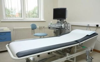
Ultrasound of the knee joints
In what cases is ultrasound of the knee joint necessary?
Echography is prescribed in case of questionable symptoms of the disease to confirm or refute the preliminary diagnosis. Indications for the procedure may be suspicion of the following diseases:
- damage to the lateral, medial or cruciate ligaments, meniscus;
- deforming osteoarthritis, arthritis of various etiologies;
- osteochondropathy:
- injuries and fractures of joint structures;
- new bone formations.
Based on the results of the ultrasound, a research protocol is drawn up, the transcript of which is analyzed by an orthopedic surgeon.
Early and competent diagnosis makes it possible to timely detect pathological processes occurring in the soft tissues, cartilage and bones of the knee joint and prescribe adequate treatment.
What can be seen on an ultrasound of the knee joint
When going for an examination, the patient usually has in hand a direction that indicates a preliminary diagnosis and clearly indicates which projections of the joint are necessary. Using ultrasound, it is possible to identify almost all joint deformities, namely the following pathological changes:
- Meniscus damage. Statistically, meniscal tears and bruises are the most common knee injuries.
- Expansion or narrowing of the joint space.
- Violation of the contours of the articulation.
- Ulceration of cartilage tissue and bone surface.
- Osteophyte formation
- The presence of loose bodies in the joint cavity.
- Thickening of the joint capsule or overgrowth with connective tissue.
Ultrasound of the knee ligaments
Using ultrasound, you can visually determine the composition and amount of synovial fluid, its homogeneity and the presence of foreign inclusions in it. In addition, using ultrasound, you can detect the presence of cysts in joint structures, diagnose sprains or ruptures of ligaments and tendons, and the appearance of pathological effusion.
After the ultrasound scan, the patient receives an examination report, which gives a complete picture of the condition of the knee joint. Based on these data, the specialist can confidently confirm or refute the preliminary diagnosis and prescribe treatment.
X-ray or ultrasound of the knee joint – which is better?
Often, patients who are undergoing diagnostic procedures using ultrasound have doubts about the advantages of this method. Many patients suffering from joint pathologies ask doctors to refer them to x-rays, believing that it is more informative.
Ultrasound has many advantages over x-rays
Let's compare these methods for diagnosing the knee joint. The capabilities of ultrasound examination in relation to the musculoskeletal system are in no way inferior to the radiographic method.
However, X-ray examination has a serious drawback: the method allows assessing the condition of only the bone elements of the joint. The x-ray does not reflect damage to the soft tissues of the knee. In addition, it is necessary to note the great information content of ultrasound in relation to small fractures and bone cracks, which often go unnoticed during radiography.
Thus, we can confidently say that an ultrasound examination of the knee joint is more accurate and informative than an x-ray.
Plus, ultrasound is much safer. Echography can be performed as often as required by the specifics of the disease. The procedure is safely prescribed to pregnant women and newborn babies.
Technique for performing ultrasound of the knee joints
A diagnostic examination using ultrasound does not require special training from a person. It is done on any day and at a time convenient for the patient. The only precaution is that the procedure should be delayed for those who have recently received intra-articular injections. It must take at least 5 days for the medicine to completely dissolve in the joint cavity.
During diagnostics, a special colorless and odorless gel is applied to the examination area and does not leave marks on clothing. It is necessary for better contact of the device’s sensor with the skin.
Method of performing ultrasound of the knee joint
Before the procedure, the patient is asked to lie down on the couch. A special cushion is placed under the knee to fix the leg in a bent position. Considering that the shape of the joint does not make it possible to assess the condition of the articulation elements in one projection, scanning is carried out from several sides.
Study of the knee joint in different projections
To obtain more reliable echogram results, the patient is asked to sequentially take different positions, changing the angle of flexion of the lower limb at the knee:
- View from the front. In this case, the patient lies on his back with his leg straightened and extended. The specialist assesses the condition of the ligaments and tendons, examines the patella and popliteal area.
- Medial scan. The patient does not change position, remaining in the same position. The examination allows you to clarify the presence of fluid and effusion in the internal cavities of the joint, examine the internal collateral ligaments, meniscus and medial capsule.
- Lateral scanning. The patient flexes the limb at an angle of 45°. This position allows examination of the lateral collateral ligaments, lateral meniscus, and capsule.
- Posterior knee examinations. The person lies on his stomach with his leg straightened. The doctor checks the condition of the calf muscles, internal ligaments and tendons.
The diagnostic procedure lasts no more than 15–20 minutes. Only a qualified specialist who has perfect knowledge of the anatomy of the knee and knows how to fill out a protocol can conduct an ultrasound examination.
Research protocol and its transcript
When conducting ultrasound diagnostics, the specialist pays attention to the smallest deviations of the parameters of the articular cavity from physiological norms, the condition of the cartilage and other fragments of the articulation. All these changes are recorded in the study protocol and compared with the norm.
For example, in chronic tendonitis, echography reveals pathological thickening of the ligaments. Traumatic injuries are characterized by the appearance of areas with heterogeneous echogenicity. When a meniscus ruptures, ultrasound shows a change in the contours of the cartilage; a “blank” stripe appears at the site of the damage.
Examination of a healthy knee demonstrates smooth and clear outlines of bones and cartilage, decreased echogenicity in the area of the joint capsules and inversions.
Normally, an ultrasound shows the following picture:
- absence of bone growths and soft tissue swelling;
- clear contour of the intraarticular membrane;
- clean cavity structures without pathological effusion.
If necessary, additional positions may be included in the protocol to create a more accurate clinical picture for non-standard joint disorders.
Cost of the procedure
Diagnostics using ultrasound is considered one of the most inexpensive and accessible procedures for the population. The research is also good because it does not require the purchase of additional consumables. It is enough to bring a towel or napkin with you to the procedure. Paid medical institutions do not even require this; they are equipped with everything necessary.
Patients often ask how much an ultrasound scan costs when directed by a doctor. In a regular district clinic, echography can be done for free. To do this, just get an appointment with a specialist and make an appointment at a convenient time. There are also paid services there. The current price list is usually located at the reception desk. The patient can undergo paid joint diagnostics on the day of treatment.
How much an ultrasound of the knee joint will cost depends on the equipment and qualifications of the specialists
On average, the price of an ultrasound procedure in different regions of the country ranges from 900 to 1,500 rubles.
It depends on the type of additional services provided, the class of equipment and the qualifications of specialists. Modern equipment allows you to see a color image of the knee joint in 3D format.
Conclusion
Ultrasound of the knee joints is a simple and informative procedure that today can be done both in a budget clinic and in a private medical institution.
Modern devices provide excellent image quality and allow for quick and accurate diagnosis of various joint diseases. This fact is very important.
Accurate and quick diagnosis ensures timely and adequate treatment, increasing the patient’s chances of a full recovery.
Source: https://artritu.net/uzi-kolennyx-sustavov
What does an ultrasound of the knee joint show?
Despite the fact that today there are such diagnostic methods as computer and magnetic resonance imaging, ultrasound examination still does not lose its relevance.
The demand for this method is due to the fact that it is as simple and safe as possible, but it also helps to identify the condition of soft and connective tissues, as well as cartilage.
If a doctor suspects the development of knee joint pathology in a child, an ultrasound examination will be performed first, due to its availability and relatively low cost.
What an ultrasound of the knee joint in an adult shows, and in what cases such an examination is carried out, every person should know.
What does an ultrasound of the knee show?
Since closer to 15-16 years of age the cartilaginous tissues localized in the knee joint are replaced by bone structures, there is practically no point in conducting such an examination in adult patients. This is because the ultrasound waves emitted by the applicator will reflect from the bone structures and return back, which is why the doctor will not be able to see anything.
Very often, traumatologists and rheumatologists are asked whether ultrasound of the knee joint is performed on adult patients or whether it is better to immediately sign up for another diagnostic method. If you receive serious injuries or the presence of pain of unknown etiology, most experts advise giving preference to X-rays or MRI.
Carrying out an ultrasound of the knee ligaments in an adult is practically meaningless, since the waves will be reflected back. The situation is completely different if it is necessary to examine the condition of soft and cartilaginous tissues in a child whose age does not exceed 14-15 years.
The knee joint ultrasound technique can be performed only if there is no other equipment in the medical institution or if the procedure is carried out by a qualified physician who can correctly “read” the information received. During the diagnosis, the physician will be able to visually assess the condition of the following tissues:
- cartilaginous structures (shape, thickness and degree of wear);
- ligaments;
- articulation contours;
- vessels and capillaries;
- joint space. To make an accurate diagnosis, a specialist must assess whether this value is deviated from the norm. The joint space can be narrowed, normal or widened;
- synovial fluid. Not only its quantity is assessed, but also its consistency and viscosity;
- appearance of bone structures;
- foreign bodies in the joint.
The undoubted advantage of this technique is that it takes no more than 15-20 minutes and is absolutely painless for the patient. During the procedure, the physician will bring an applicator to the affected joint, which will display an image on the monitor. Based on the data obtained, the doctor will be able to identify the overall clinical picture.
What diseases can be detected
Which is better, ultrasound or MRI of the knee joint, depends on what pathology is suspected in the patient. If a doctor suspects the development of bursitis, there is no point in performing magnetic tomography, since ultrasound can also help identify problems with the synovial fluid.
Also, this type of diagnosis will help to see tumors, cysts and blood accumulation in the knee joint. In a child, an ultrasound of the knee joint helps to determine ossification and the formation of tumors, indicating abnormal development.
After viewing the image and interpreting the ultrasound of the knee joint, the doctor will be able to determine what pathology the patient is faced with and make one of the following diagnoses:
- arthrosis;
- arthritis;
- tendinitis;
- bursitis;
- meniscal damage;
- tendon sprain and rupture;
- bone fracture;
- congenital lesions of the lower extremities (for example, dysplasia);
- the formation of neoplasms of benign or malignant origin.
Important! For arthrosis, ultrasound is prescribed quite rarely, due to the fact that this diagnostic method will not help identify all destructive changes, which is why the patient may be prescribed an inappropriate treatment regimen.
Indications
Since ultrasound examination of the knee joint is not informative in adult patients, it is most often prescribed to children. But in some cases, such an examination can also be prescribed for adult patients. Usually they resort to his help in the following cases:
- the need for urgent diagnostics (after injury);
- identification of professional suitability of athletes;
- constant pain that appeared after a recent injury;
- swelling of the joint caused by unknown reasons;
- constant discomfort in the knees when moving;
- suspicion of synovitis and fluid accumulation in the joint.
This examination is also regularly carried out for children who engage in active sports (gymnastics, figure skating, running). In this case, ultrasound helps to assess the condition of cartilaginous and connective structures and identify pathologies in the early stages.
Advantages and disadvantages
What is better to do, X-ray or ultrasound of the knee joint, depends on what preliminary diagnosis is made to the patient. Compared to other diagnostic methods, ultrasound has the following advantages:
- the procedure takes no more than minutes;
- the patient does not have to pre-prepare for diagnosis;
- such an examination has no contraindications, it can be carried out even during pregnancy;
- Ultrasound examination allows us to examine almost all elements of the joint;
- At the initial stage of pathology, ultrasound best determines the structure and thickness of cartilage tissue.
Another advantage of this type of diagnosis is that an ultrasound of the knee joint can be done almost anywhere, since such equipment is available in almost all modern clinics and municipal hospitals.
As for the weaknesses of ultrasound examination of the knee joint, these include the fact that the procedure does not allow a full examination of adult patients, which is why the choice often falls on other, more informative techniques.
Important! How much an ultrasound scan costs depends on where exactly the examination is performed. Most often they charge 550-700 rubles for such a service.
Which is the best option: ultrasound or MRI
As medical practice shows, many doctors first prescribe an ultrasound scan to the patient and only after that send him for an MRI or CT scan. The doctor should explain to the patient how an MRI differs from an ultrasound of the knee joint when issuing a referral.
Although both procedures are designed to assess the condition of soft, cartilaginous and connective tissues, they have significant differences:
- Price. Magnetic tomography costs several times more than ultrasound; the minimum cost of such a service is 4,000 rubles.
- An MRI will show a more detailed picture and help assess the condition of not only soft tissues, but also bones.
- MRI has a greater number of contraindications and limitations.
- Tomographs are not available in all clinics (most often private).
- Magnetic tomography is not always suitable for overweight patients.
- The images obtained during MRI are the most accurate and most informative.
Despite the fact that MRI provides more information, many patients prefer ultrasound (especially if it is necessary to assess the condition of the joint after a minor injury). This is due to the fact that ultrasound is a simpler and more accessible procedure that can be done in almost any medical institution.
Conclusion
The doctor will tell you where you can get an ultrasound of the knee joint after ordering the examination. If a doctor has prescribed this diagnostic method to a patient, you should not worry that it will be useless and ineffective.
As practice shows, ultrasound also helps to determine sprains and tears of ligaments, as well as to identify tissue deformation.
Orthopedist. Experience: 4 years. Education : Diploma in General Medicine (Medicine and Prevention), Izhevsk State Medical Academy (2015)
Advanced training courses: “Orthopedics”, Izhevsk State Medical Academy (2019)
Source: https://VashyNogi.com/bolezni/kosti/uzi-kolennogo-sustava.html
Ultrasound examination of the knee joint in adults and children
17.02.2019
Ultrasound - ultrasound examination - of the knee joints is prescribed if there is insufficient information or it is impossible to take x-rays of the knees.
Ultrasound is considered a safer technique. It helps to examine the soft tissues of the joint that x-rays do not show.
If necessary, ultrasound diagnostics are performed repeatedly to assess the effectiveness of treatment or disease progression.
How does ultrasound work?
A high-frequency ultrasound wave is emitted by the ultrasound machine's transducer, reflected off the body's tissues, and then returned.
A black and white image is formed on the monitor screen: black areas that absorb ultrasound, and white areas that repel. Liquid hypoechoic media absorb the wave, and hyperechoic bone tissue reflects it.
Ligaments, tendons, and cartilage have medium echogenicity and are visible in different shades of gray.
Ultrasound diagnostics of joints in children is especially informative, since their bones have not yet become completely compacted and are permeable to ultrasound waves.
In childhood, according to the results of ultrasound, a description of cartilage, ligaments, tendons, menisci, and the condition of the joint capsule is carried out.
In adults, ultrasound of joints does not always provide the necessary information, since bone structures overlap soft tissue formations.
The blood supply to the joint and the condition of the popliteal vascular bundle are assessed using Doppler ultrasound or color duplex scanning. These techniques show the speed and other characteristics of blood flow, as well as the structure of the vascular wall.
In what cases is ultrasound of the knee joint prescribed, are there any contraindications?
The knee joint has a complex structure. It is formed by three bones covered with cartilage: the tibia, femur, and patella. The latter is directly connected to the quadriceps tendon.
There are lateral, posterior, and intra-articular ligaments of the knee joint. In the area of the tendons there are synovial bursae filled with fluid. Between the articular surfaces of the bones there are two semilunar cartilages that perform a shock-absorbing function.
These are menisci. The top of the joint is covered with a capsule.
This complex structure is due to the fact that the knee has a supporting function and is subjected to heavy loads every day. Any joint structure can be damaged.
The indication for ultrasound diagnostic examination of the knees is the suspicion of their pathology:
- osteoarthritis, degeneration of articular cartilage;
- arthritis;
- injuries of the knee joint - fractures, ruptures, sprains of the ligament apparatus, tear of the meniscus of the knee joint;
- neoplasms;
- hemarthrosis;
- synovitis;
- bursitis;
- cysts.
The doctor prescribes an ultrasound if the following symptoms are present:
- swelling, redness of the knee;
- pain in the knees when walking;
- impaired mobility of the knee joint;
- volumetric formations in periarticular tissues;
- joint deformity;
- falling to one knee.
For children, an ultrasound is performed to determine the timing of joint development: the presence of ossification zones and their number are assessed.
Ultrasound of the popliteal fossa area is important not only for diagnosing joint pathology, but also for assessing the condition of the neurovascular bundle. Its integrity, blood flow speed, and vascular condition are determined.
There are no absolute contraindications to ultrasound of the knees; there are only some restrictions for its implementation:
- changes in the skin of the knee - rash, burns, wounds;
- state of motor, mental excitement;
- intra-articular block performed the day before;
- complete immobility of the joint - visualization of most structures is impossible.
How is the examination carried out, is preparation required?
No specific preparation is needed for an ultrasound of the joints. The only point that should be taken into account: the procedure is not carried out within 5 days after placing a drug blockade in the joint cavity. Therefore, if you had such a manipulation the day before, you need to wait 5-7 days before the ultrasound.
When going for an examination, take with you a referral, an insurance policy, an outpatient card with previous ultrasound results (if it was performed previously), dry or wet wipes, and a sheet.
Ultrasound is usually done in the supine position. The joint is examined from different sides, so the doctor asks the patient to turn on his stomach or back and bend his knees. The ultrasound machine sensor is lubricated with a neutral gel, which improves visualization, and different parts are sequentially examined with its help:
- On the anterior surface, the sensor is first placed above the upper edge of the patella and the condition of the femoral cartilage and tibial condyle is assessed. Then it is turned parallel to the thigh and the ligamentous apparatus, muscle tendons, and synovial joint capsule are examined.
- The sensor is installed at the rear in three projections. The medial projection makes it possible to assess the condition of the articular ends of the femur and tibia, part of the meniscus, blood vessels and nerves. In this case, the sensor is placed in an oblique-longitudinal position. It is then moved to the lateral edge to view the same structures from a different angle. When the sensor is positioned transversely in the popliteal fossa, the posterior condyles of the femur, cruciate ligaments, and the posterior surface of the menisci are clearly visible.
- Lateral projections make it possible to analyze changes in the inner and outer parts of the knee and evaluate the areas of attachment of the ligaments to the bones.
The menisci are better visible when the leg is bent. If necessary, the examination is complemented by examination of the surrounding muscles to exclude the spread of infection or blood into the intermuscular spaces.
The procedure takes 20 minutes. The sonologist records the diagnostic results in the protocol and makes a conclusion about the identified problems.
What abnormalities can an ultrasound detect and what is normal?
The joint ultrasound protocol will contain a description of the following parameters that the examination can show:
- structure of cartilage and bone tissue;
- thickness of cartilage on the surface of the condyles;
- pathological inclusions (calcifications, hemorrhages);
- joint capsule and synovial bursae;
- meniscus tissue;
- width and uniformity of the joint space;
- amount and nature of synovial fluid;
- presence of space-occupying formations – tumors, cysts;
- condition of ligaments, tendons, muscles;
- zones of ossification in the knee joint in a child;
- characteristics of the neurovascular bundle;
- blood flow in the knee area.
The following indicators correspond to the norm:
- the surface of the cartilage is smooth, homogeneous;
- bone elements without deformations, with a clear continuous contour;
- the cartilage structure is homogeneous, echogenicity is slightly above average;
- synovial fluid without pathological impurities, in normal quantities;
- the synovial membrane is not visible;
- the joint space is uniform, without narrowing;
- ligaments, muscles and tendons without signs of damage to integrity or pathological inclusions;
- there are no neoplasms or loose bodies in the articular cavity.
In some cases, ultrasound of the knee joints shows that there are pathological changes in the knees.
Osteoarthritis is the most common degenerative disease of the joints, in which thinning of the cartilage, proliferation of bone osteophytes, reduction in the width of the joint space, and uneven structure of the menisci are observed. Similar changes are characteristic of arthritis. But the contours of the articular structures will be blurred, and their echogenicity will be reduced.
Bursitis - inflammation of the joint capsule is manifested by swelling and the presence of inflammatory effusion in it. At the same time, a triangular dark area is visible on the monitor - the joint capsule filled with fluid.
Tendonitis is an inflammatory process in the ligaments that is manifested on ultrasound by a decrease in their echogenicity. When the synovium becomes inflamed, it thickens and becomes visible on ultrasound.
Cysts look like dark liquid formations with clear contours. Tumors are pathological inclusions of medium echogenicity.
An ultrasound allows you to see a fracture of the bones adjacent to the joint: femur, tibia, patella. However, compared to x-rays, it is less informative in such cases.
And ultrasound diagnoses ruptures or incomplete tendon injuries with high accuracy in the form of hypoechoic zones, just like a meniscus tear. When the meniscus is damaged in the joint cavity, parts of it are identified - loose bodies that cause discomfort and noise when moving.
These are the so-called joint mice. The areas of separation of the ligaments from the bone are also clearly visible.
The interpretation of the results of the ultrasound examination is carried out by the attending physician - an orthopedist, traumatologist, rheumatologist, therapist or surgeon. Based on them, he makes a diagnosis. If the data obtained is not enough, the doctor recommends further examination: x-ray, MSCT or MRI of the knee.
Ultrasound of the knee is considered the safest method of examination. Its only drawback is the low information content when assessing the condition of the bones. Ultrasound allows one to detect soft tissue pathology with high accuracy.
Therefore, the recommended research method directly depends on the intended diagnosis. In pediatric practice, ultrasound is considered the gold standard and is widely used even in infancy.
Ultrasound examination of the knee joint in adults and children Link to main publication
Source: https://diagme.ru/uzi/kostey-i-sustavov/kolena