Reticular neuritis is an inflammatory process in the optic nerve. Pathological transformations occur outside the eyeball.
How the optic nerve is formed
This element consists of a large number of processes that carry information to the brain. Data processing takes place in the occipital lobe.
The optic nerve consists of a disc or head located in the eyeball. It extends through the orbit and into the skull. Then the fibers intertwine and connect to the brain. Several membranes make it possible to quickly conduct nerve impulses, protect against damage and supply useful microelements.
The substance under such a coating interacts with the brain, if inflammation occurs, damage is observed in the optic nerve. Neuritis can be classified based on the location of the source of infection:
- Normal - develops in the internal department.
- Retrobulbar. An inflamed area of nerve outside the eye.
- Papillitis. Differs in combined characteristics.
Let us list the forms of neuritis:
- Peripheral. The problem is localized in the subdural and subarachnoid nerve tissue.
- Axial. The inflammation is located in the central uneven bundle. This type of disease is detected most often.
- Transversal. The most complex form of neuritis, causing severe dysfunction of the visual apparatus. Inflammation develops in the direction from the axial bundle to other parts of the nervous tissue.
What is the difference between retrobulbar neuritis
This is inflammation of the nerve fibers. The lesion is located outside the eye in the skull. A distinctive feature of this disease is a slow decline in the quality of vision.
Often this form of pathology is detected in patients 25-35 years old. Retrobulbar neuritis is thought to be a symptom of multiple sclerosis. Typically, pathology develops against the background of nearby inflammatory processes. The negative external impact of the disease causes demineralization and irritation of the nerves.
Without good therapy, the fibers atrophy and the quality of vision deteriorates. In regular neuritis, the discs in the optic nerve are not involved in inflammatory conditions. Destructive processes are localized outside the eye. Inflammation of nervous tissue is characterized by proliferation of cellular structures and slight infiltration, in which painful cells enter normal tissue.
Inflammation begins from the soft membrane of the brain and spreads to the nerve fibers. When the problem is located in the nerve trunk and the process of tissue destruction occurs in a nonspecific manner, swelling appears and healthy tissues are affected.
Atrophic processes occur again after therapy; work in the damaged area can resume, and the quality of vision improves.
Etiology
The causes of retrobulbar neuritis may vary. Often the development of this pathology is provoked by multiple sclerosis. Inflammatory processes are among the symptoms indicating the occurrence of this disease.
Therefore, it is necessary to conduct a correct diagnosis to determine what exactly is causing vision problems. There are situations when the real reason cannot be found out. We list the main provoking factors:
- Brain injury.
- Diseases of the nervous system.
- Inflammation of the membranes of the brain.
- Pregnancy.
- Chronic disorders cause neuritis.
- Intoxication.
- Infectious viral disease.
Symptoms
The severity of symptoms in such a disease determines the degree of intensity of inflammation.
The disorder can develop in several ways:
- Spicy. Signs appear within a few days. Patients' vision deteriorates sharply and pain occurs in the eyes.
- Chronic. Vision declines slowly, the symptoms are not as pronounced as in the acute form of the pathology.
Signs of acute form:
- The quality of vision sharply decreases.
- Some colors are difficult to recognize.
- When a person moves the eye, pain occurs.
In difficult situations, the symptom appears even with complete rest.
- Signs of the chronic form:
- A blind spot appears in the center of the visual field.
- The patient experiences symptoms as if he sees some objects in the twilight, but the lighting is normal.
- Hyperthermia.
- Various visual illusions may occur, represented as dots and figures.
- Weakness.
- The person feels sick and vomits.
- All of the above symptoms can worsen after intense physical activity or in stressful situations.
Initially, the disease affects one eye and gradually spreads to the second.
How is diagnostics carried out?
When the first signs indicating the appearance of retrobulbar neuritis occur, you need to go for an examination to a doctor to identify pathological processes. Determining the cause of the development of pathology.
The diagnostic procedure involves:
- Visual inspection. The specialist determines the quality of vision and the intensity of color perception.
- Ophthalmoscopy. Allows you to diagnose the optic disc.
- Tests to determine the reaction of the eye to external stimuli. This examination technique makes it possible to determine the slowdown in signal conduction.
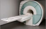
Magnetic resonance scanning is considered one of the most detailed ways to evaluate a patient's condition and identify retrobulbar neuritis. The principle of this technique is that a specialist injects a contrast liquid into the nerve, then photographs are taken. The optic fiber is clearly visible in the images.
Therapy
The best results are obtained with medication treatment. The drugs help reduce inflammation and prevent infection. Experts prescribe the following medications:
- Antibiotics.
- Medicines to stimulate blood circulation.
- Sulfonamides.
- Corticosteroids.
- Antihistamines.
The release form of medications may vary. Doctors prescribe medications individually for each patient. If everything is done correctly, the quality of vision improves within 3 weeks, unless there are complications such as complete loss of visual function and the occurrence of multiple sclerosis.
Consequences
The complications may be due to damage to the optic nerve. Many people cannot recover from inflammation. Visual acuity may deteriorate, but in some patients the problem resolves within a few months.
Side effects of medications
The main difficulties are caused by the occurrence of damage. In most patients, after inflammation, various kinds of irreversible processes occur that do not have characteristic symptoms. Visual acuity may decrease, but most patients return to normal or acceptable perception of surrounding objects.
Steroid medications used to treat neuritis worsen the immune system, making the body more susceptible to infections. Long-term use of steroids can also lead to bone deformation.
To prevent such a disorder, an examination by an ophthalmologist is required, which involves studying the condition of the fundus, determining the quality of vision, perimetry, and an indicator of pressure inside the eye.
Problems in the area of the optic nerves may show test results provoked by potentials.
This is a neurological technique; an ophthalmologist does not perform such examinations. If there is any sudden deterioration in the quality of vision, you should contact a specialist to identify the cause and take action.
Where does retrobulbar neuritis come from?
Often some pathologies arise as a result of others. Retrobulbar neuritis may appear as a sign of other health problems.
Let's list these diseases:
- Multiple sclerosis.
- Meningitis.
- Encephalomyelitis.
- Meningoencephalitis.
Vision deteriorates due to numerous pathological processes. Alcohol and tobacco can affect the quality of nerve tissue functioning. Often the cause cannot be determined. Neuritis has a negative effect on the eyes and nerve endings.
Consideration of the occurrence of pathology as symptoms of multiple sclerosis requires special attention. This condition occurs in people often. Approximately 50% of patients diagnosed with retrobulbar neuritis have problems with cerebral vessels.
The location of cholesterol plaques may determine the relationship between these two pathological processes.
In multiple sclerosis, cholesterol accumulates in front of the orbit in the intracranial part of the nerve fiber. The myelin sheaths are destroyed.
Despite complete destruction, the axial cylinders are not deformed. This is how ascending and descending trophic signs can manifest themselves.
The most common symptoms of retrobulbar neuritis are:
- Simultaneous improvement in the quality of vision.
- Changing the color of the disc.
Healing may occur on its own. Other signs coincide with the classic form of retrobulbar neuritis.
How to recognize the disease
To determine effective treatment, it is necessary to initially determine the causes and distinguish pathology from other processes. Classic options are easily identified. But sometimes a situation arises when retrobulbar neuritis develops without worsening visual function, sometimes edema is added to the main symptoms. Such situations can easily be confused with a stagnant disk.
With pseudoneuritis, vision does not deteriorate. With a stagnant disc, swelling occurs. Minimal bleeding indicates the presence of neuritis. While the causes of the development of pathology are determined and diagnostic procedures are performed in a medical institution, restorative treatment methods are used to reduce inflammation and eliminate infectious processes.
The following procedures are carried out:
- Desensitization.
- Metabolism stimulation.
- Dehydration.
- Strengthening the immune system.
- Improvements in the functioning of the central nervous system.
For approximately a week, patients take extended-spectrum antibiotics parenterally. Dexamethasone and various corticosteroids are prescribed
Antibiotics that affect the optic nerves should not be taken, since such treatments worsen the disease.
In such situations, specialists prescribe regular injections with a consistent reduction in dosage.
A glycerin solution helps get rid of edema and prevents its subsequent occurrence. Adrenaline tamponade in the nasal cavity has a positive effect on health. Mustard plasters and leeches for blood cleansing also help.
After this, additional diagnostics are carried out, and doctors can change the nature of the therapy. It will be aimed at eliminating the underlying pathology.
Vision problems caused by diseases of internal organs are solved through direct action on the main problem.
For alcohol poisoning, the disease is treated in a similar way. But you should not use antibiotics. Doctors carry out intoxication, wash the stomach and take other measures to eliminate toxic trace elements.
In such a situation, approximately 4 weeks are allotted for therapy. The nature of the destruction of the optic nerve does not affect the chosen method of treatment. For various types of intoxication, the patient should not drink alcohol or use tobacco products.
Additionally, vitamin complexes and medications containing glucose are prescribed.
Proper therapy and a positive attitude of the patient allows you to get good results without delay. Visual function is quickly restored. The degree of healing is determined by how advanced the condition was. In the chronic form of the disease, the prognosis is not so comforting; qualified doctors can correct everything.
Conclusion
When the optic nerve is damaged outside the eye in the area of the skull, retrobulbar neuritis develops; the main feature of this pathology is the deterioration in the quality of vision until it is completely lost.
The disease is one of the manifestations of multiple sclerosis and manifests itself at a young age. With this disease, the functioning of the optic nerves is disrupted. Fibers protected by the myelin sheath pass through the sclera, where meningeal formations appear, creating a nerve trunk.
The visual tract approaches the subcortical region, in which primary information processing is carried out and a visual reflex is created. After this, the impulses are transmitted to the corresponding area of the brain.
Treatment of retrobulbar neuritis involves the use of antibiotics and other drugs. It all depends on what factor affects the development of inflammatory processes in the brain.
Source: https://nevrology.net/sindromy-i-zabolevaniya/perifericheskoj-nervnoj-sistemy/nevrit/retrobulbarnyj.html
Danger and prognosis for retrobulbar optic neuritis
Optic neuritis is an inflammation of this element with suppression of visual function. There is specifically optic neuritis, when inflammation affects the nerve disc, and retrobulbar neuritis - pathological processes spread outside the eyeball.
Structure of the optic nerve
The human optic nerve contains more than a million nerve cell projections that transmit information from the retina to the brain and provide vision. The transformation of information occurs in the occipital lobe of the brain. This is where response signals are generated.
The optic nerve originates from the disc (head) in the eyeball and runs through the tissues of the orbit. Once in the cranial cavity, the optic nerves from different eyes connect and then run as part of another structure.
The nerve is covered with several membranes, which allows for high-speed conduction of impulses, as well as protection and nutrition of the element. The membranes and intrathecal space have a connection with the brain; accordingly, when the latter is inflamed, the optic nerve can be affected.
Depending on the damaged part of the optic nerve, ordinary neuritis (inflammation of the intraocular part), retrobulbar (damage to parts that lie outside the eye) and papillitis (mixed pathology) are distinguished.
Forms of neuritis:
- Peripheral. Exudate accumulates in the subdural and subarachnoid (between the hard and soft membrane) spaces of the optic nerve.
- Axial. Inflammation is localized in the axial bundle (central nerves). This form of neuritis is most often diagnosed.
- Transversal. The most severe form of neuritis, which provokes severe dysfunction of the visual apparatus. Inflammation spreads from the axial fascicle to the rest of the nerve.
Features of retrobulbar neuritis
Retrobulbar neuritis is an inflammatory process that affects the nerve fibers of the optic nerve outside the eyeball. A characteristic feature of the pathology is a gradual depression of visual function.
Most often, this form of neuritis is diagnosed in patients 25-35 years old. Retrobulbar neuritis is believed to be an early sign of multiple sclerosis.
Typically, retrobulbar neuritis develops against the background of existing inflammation of any localization. Unfavorable external and internal factors provoke demineralization and inflammation of the nerve fibers that line the optic nerve. Without treatment, inflammation gradually atrophies the fibers, which causes a decrease in visual acuity.
It is noteworthy that specifically in retrobulbar neuritis, the optic disc is not involved in the inflammatory process; destruction occurs outside the eyeball.
Nerve inflammation is manifested by cell proliferation (division) and pinpoint infiltration (penetration of abnormal cells into healthy tissue).
Inflammation begins from the pia mater, moving to the nerve fibers.
When the process is localized in the nerve trunk, the inflammation is nonspecific. Swelling and infiltration develop, many vessels and connective tissue are formed. Atrophy of nerve fibers occurs secondary. After treatment, the functionality of the affected area can be restored, and accordingly, visual acuity will improve.
Causes of inflammation of the optic nerve
As a rule, the progression of retrobulbar neuritis is associated with the development of multiple sclerosis: this pathology primarily provokes inflammation of the nerves. However, there are other causes of neuritis, so diagnosis plays a critical role in the process of choosing therapy.
It happens that it is not possible to reliably establish the cause of neuritis. In this case, they speak of an idiopathic (independent) form of neuritis.
Causes of retrobulbar neuritis:
- head injuries;
- brain pathologies;
- inflammation of the membranes of the eye and orbit;
- infectious and non-infectious lesions of the lining of the brain (encephalitis, meningitis);
- pathological pregnancy;
- chronic diseases (diabetes mellitus, gout, nephritis);
- viral and infectious pathologies (tuberculosis, brucellosis, typhus, malaria, smallpox, syphilis, influenza, tonsillitis, otitis media, sinusitis, tonsillitis, erysipelas);
- multiple sclerosis.
Intoxication of the body aggravates the course of neuritis. This applies to drugs, alcohol and some medications.
Symptoms of acute and chronic retrobulbar neuritis
At the initial stage of retrobulbar neuritis, the fundus of the eye may not be deformed, but most often there is hyperemia and distortion of the boundaries of the optic nerve head. Retrobulbar neuritis typically affects only one eye, but the disease can spread to the second. Simultaneous inflammation of both eyes is rarely diagnosed.
With this type of nerve inflammation, a central scotoma (blind area) occurs. Initially, the spot is large, but gradually decreases with adequate treatment. Sometimes the central scotoma turns into a paracentral annular scotoma.
The severity of neuritis symptoms is determined by the extent of inflammation. Typically, acute and chronic forms of neuritis are distinguished.
In acute pathology, a few days are enough for symptoms to develop. A person experiences visual discomfort and a sharp decrease in visual acuity occurs. In acute neuritis, the pain intensifies from pressing the eyeball into the orbit.
The acute form of neuritis also causes impaired color vision and acute pain when moving the eyes. In severe cases, pain persists even at rest. Visual acuity usually returns after treatment, but in rare cases the eye remains almost completely blind.
Chronic neuritis is characterized by gradual depression of visual function. The clinical picture is not expressed. As the disease progresses, a blind spot may appear in the field of vision, and patients complain of a feeling of blurred vision.
In addition, chronic neuritis is accompanied by nausea and vomiting, visual illusions, and general weakness. Symptoms will intensify as the disease progresses and after physical activity. People with neuritis should also avoid worries and stress.
Retrobulbar neuritis caused by diabetes mellitus is usually chronic. The disease is more common in men and affects both eyes. Vision is slowly depressed, and absolute or relative central scotomas appear.
Clinical picture of toxic neuritis
The clinical picture of retrobulbar neuritis, which is caused by intoxication, has some of its own characteristics. The most common cause is poisoning with methyl alcohol and mixtures containing it.
In mild poisoning, only nausea and vomiting are observed, in severe cases - unconsciousness. After a few days, vision begins to deteriorate (up to complete blindness).
On examination, the pupil is dilated and the reaction to light is weakened. The fundus is not deformed, although symptoms of ischemic neuritis may appear.
Without treatment within a month, visual acuity may be restored, but with subsequent deterioration.
Alcohol and tobacco intoxication develops with chronic alcoholism and abuse of strong varieties of tobacco. Neuritis affecting the papillomacular bundle is diagnosed mainly in men over 30 years of age. The course of inflammation is similar to retrobulbar neuritis: mild hyperemia of the disc, the presence of a central scotoma. It is possible to improve visual acuity by giving up bad habits.
Diagnosis of retrobulbar neuritis
Any manifestations of retrobulbar neuritis are a good reason to consult an experienced doctor and undergo a full examination. It is very important to make an accurate diagnosis and study in more detail the specifics of the development of the disease in a particular patient. Before choosing a treatment method, you need to find out the cause of the neuritis.
Typically the examination includes the following procedures:
- ophthalmological examination;
- visometry (determining visual acuity);
- light perception test;
- ophthalmoscopy (examination of the fundus of the eye);
- additional tests that help examine the reaction of the visual system to visual stimuli and determine the conduction speed of nerve impulses.
Magnetic resonance scanning is considered the most informative method for diagnosing optic neuritis. The procedure consists of obtaining images of the optic nerve with the preliminary introduction of a special contrast agent into it. MRI is a safe procedure and allows for detailed examination of the optic fiber.
Differential diagnosis of neuritis
Diagnosis of typical cases of retrobulbar neuritis is quite simple; it is more difficult to identify mild forms of inflammation that do not affect visual abilities, and swelling of the optic nerve. The most difficult part of the diagnosis is to rule out edema and congestive disc complications. Both of these conditions, like neuritis, are accompanied by a decrease in visual acuity.
Differential diagnosis consists of excluding pseudoneuritis and optic disc congestion. In the case of pseudoneuritis, vision does not deteriorate, there is no progression of the condition. The initial stage of a stagnant disc is also characterized by preservation of vision and partial marginal swelling of the nerve disc. As the disease progresses, intracranial pressure increases.
Differentiation of retrobulbar neuritis from ordinary neuritis occurs by assessing the condition of the disc and checking visual acuity. Sharp blindness, the presence of a central scotoma and minor changes in the disc indicate a retrobulbar type of inflammation.
First aid for retrobulbar neuritis
Before determining the cause of retrobulbar neuritis, antibacterial and anti-inflammatory therapy should be carried out. Urgent desensitization, dehydration, correction of immunity and measures to improve metabolism in the tissues of the central nervous system are prescribed.
Broad-spectrum antibiotics are prescribed by injection for a week. Do not use ototoxic drugs (Streptomycin, Gentamicin, Neomycin), which have a negative effect on the optic nerve. Corticosteroids are considered safe: Dexamethasone 1 ml every day (10-15 procedures), Prednisolone 0.005 g (4-6 times a day) for five days.
Therapy for retrobulbar neuritis:
- Diacab (Acetazolamide). 0.25 g of the drug 2-3 times a day. The course of treatment is three days. In parallel, Panangin is prescribed two tablets three times a day.
- Glycerol. 1-1.5 g/kg.
- Magnesium sulfate 25%. Intramuscularly 10 ml.
- Glucose 40% intravenously and Solcoseryl (Actovegin) intramuscularly.
- Hexamethylenetetramine solution 40%.
- Adrenaline solution 0.1%. Tampons in the middle nasal passage for 20 minutes.
- Piracetam (Nootropil). Up to 12 g/day.
- Dibazol. 10 mg twice a day for 2-3 months.
Similar therapy is indicated for bilateral toxic neuritis, which developed against the background of methyl alcohol poisoning. Only antibiotics are excluded.
How to treat retrobulbar neuritis
Therapy for inflammation of the optic nerve has a favorable prognosis only if it is started on time. If the disease is ignored, severe complications can develop, including multiple sclerosis and even complete loss of vision.
Retrobulbar neuritis requires hospitalization of the patient. Inpatient treatment helps to avoid complications. If multiple sclerosis is suspected, the patient must be admitted to a hospital. Only constant monitoring of the patient’s condition and timely adjustments to the treatment plan guarantee complete recovery.
The course of treatment for neuritis should be determined by the doctor depending on the cause of the development of the pathology. In cases where the cause has not been established, complex anti-inflammatory therapy is recommended. It is very important to stop the spread of the process and promote the restoration of the affected fibers.
Drugs for the treatment of retrobulbar neuritis:
- anti-inflammatory;
- antihistamines;
- antiviral;
- antibacterial;
- hormonal (for local use);
- vitamin supplements (especially group B).
It is recommended to combine drug therapy and physiotherapy. To eliminate inflammation of the optic nerve, magnetic therapy and laser stimulation are used.
Noticeable improvement occurs only after a month of regular treatment. In severe cases, regression of inflammation can only be achieved through surgery.
The procedure involves opening the nerve sheath, which helps relieve pressure on its fibers. However, neuritis requiring surgery is extremely rare.
As a rule, conservative therapy is effective.
After determining the etiology of neuritis, drugs are added to treat the cause. This could be specific therapy for tuberculosis, drugs for herpes, surgical relief of sinusitis.
Consequences of retrobulbar neuritis
Most often, pathological changes caused by inflammation of the optic nerve can be reversed. In most patients, visual function is restored almost completely within a year.
However, in cases where inflammation appears against the background of neuromyelitis or multiple sclerosis, the risk of relapse of neuritis increases. It is noteworthy that 70% of women who have had retrobulbar optic neuritis subsequently develop multiple sclerosis. In the absence of these or similar pathologies, the chances of re-inflammation are minimal.
Prevention of inflammation of the optic nerve
To avoid toxic neuritis, you need to give up bad habits, or reduce the amount of alcohol consumed and replace strong cigarettes with light ones. You also need to protect yourself from toxic liquids.
Since inflammation of the optic nerve develops for various reasons, prevention must be comprehensive. It is important to complete treatment of acute forms of pathologies and control chronic ones.
Retrobulbar neuritis may resolve on its own, but there is an increased risk of nerve atrophy and complete blindness in one or both eyes. If any symptoms occur, you should immediately undergo examination and consult with an experienced specialist.
Source: https://BeregiZrenie.ru/rogovitsa-setchatka/retrobulbarnyj-nevrit/
Retrobulbar optic neuritis: symptoms and treatment
With retrobulbar neuritis, the optic nerve is affected. It is represented by the axons of the retinal ganglion. Bunches of fibers, separated from each other by myelin, pass through the sclera of the eyes, where they are surrounded by meningeal formations and form the nerve trunk.
The visual tract approaches the subcortical centers, where primary information processing is carried out and visual reflexes are formed. Next they go to the visual zone of the cerebral cortex.
- At the base of the brain, a partial decussation occurs, so the visual analyzers are connected to the opposite halves of the visual areas.
- The functions of the optic nerve include the ability to transmit impulses associated with the perception of objects, colors, and light and shade.
- If completely damaged, functions are not restored.
Retrobulbar neuritis
Signs of decreased visual function are associated with inflammation of the optic nerve, which can occur in any part of it. If the process occurs inside the eye, they talk about papillitis, outside it - about retrobulbar neuritis. There are three forms of the disease:
- Axial. Long processes - axons - are affected. It is characterized by the appearance of blind spots (in a crowd) in the field of view. This form is the most common.
- Peripheral. Inflammation occurs in areas of the myelin sheath of the optic nerve. It is progressive in nature and spreads deeper into the trunk over time. Exudate accumulates under the membrane, which leads to pain during eyeball movement. Central vision is intact, but the fields are narrowed.
- Transversal. The periphery and the axon bundle are affected first, then the remaining areas, including the disc. Leads to complete loss of vision.
In retrobulbar optic neuritis, the disc becomes affected as the disease progresses.
Causes
The etiology of optic neuritis is not fully understood. Probably one of the main factors provoking its development is a genetic predisposition that contributes to the erroneous perception of myelin proteins as proteins of pathological microorganisms. As a result, T-lymphocytes accumulate in the affected area and immune defense is activated.
The risk of getting sick increases at a young age up to 40 years (the average age of those who first noticed symptoms is 25-28 years). Women are more often affected by pathologies.
The range of causes that lead to optic neuritis is wide. It includes infectious and non-infectious factors.
Viral infections lead to primary inflammation: herpes, cytomegalovirus, infectious mononucleosis. It is often caused by sinusitis, tonsillitis, otitis media, tonsillitis, and less commonly by pyelonephritis. Separately, inflammation of the orbit of the eyes is distinguished, in particular, uveitis and chorioretinitis.
Damage to the brain leads to secondary retrobulbar neuritis. Such causes include neurosyphilis, HIV infection, inflammation of the brain and its membranes, and Lyme disease.
Signs of pathology develop against the background of blood diseases, diabetes, injuries, poisoning, vaccinations, autoimmune diseases, and taking certain medications.
Neoplasms exert a compressive effect on the vessels, the intersection of nerve fibers.
In two thirds of patients with multiple sclerosis, secondary optic neuritis is diagnosed. The reasons for this process lie in demyelination. However, at the initial stage, atrophy of the myelin sheath is not always obvious. Its signs are not detected even on MRI. This may be due to the low resolution of the device used and fatty tissue.
Signs of myelin destruction are diagnosed in the brain. They are noted in all patients up to 5 years from the onset of the disease. In half of the patients, the images also show symptoms of demyelination of both visual pathways, more than 37% - on one side. In the remaining 12% of patients, the visual pathways are intact.
Symptoms
The disease occurs in acute and chronic forms. In the first case, signs of neuritis are noted in the first two to three days. Painful sensations appear sharply, intensifying with eye movement.
Vision decreases. The perception of some colors deteriorates. In the first stages, the disease usually affects one eye, and as the inflammatory process develops, it spreads to the second.
Sometimes swelling and hyperemia appear.
The chronic form is characterized by a gradual decrease in the ability to see. Over time, atrophy of the optic nerve develops, leading to the formation of a blind spot (scopoma) in the center of the eyes.
It is found in approximately 60% of cases. The patient notes that everything around is in twilight. Sometimes they talk about the appearance of non-existent points and figures. There is redness in the eyes.
The patient may feel nauseous and sometimes complain of pain in the head.
The symptoms of neuritis intensify after severe mental stress and heavy physical exertion.
Common symptoms include gradual deterioration of vision, increased sensitivity of the retina, pain in or around the eyes, and photophobia. Signs appear with varying strength. Thus, vision may decrease slightly, in other cases lead to possible blindness, while the person will perceive only light. The latter is observed mainly in the axial and transversal form.
Pain is most severe with peripheral neuritis. With this form of visual impairment, the so-called lateral vision disappears, but visual acuity may remain intact.
Interestingly, few people indicate a violation of color perception. More often this sign is discovered when research is carried out.
Symptoms largely depend on where in the area from the eyes to the visual centers the optic nerve is damaged. It is associated not only with deterioration of vision, but also with its bifurcation. Noteworthy is the presence of signs of nystagmus (involuntary movements) in many patients. Sometimes symptoms of optic neuritis are accompanied by loss of control over the position of the head in space.
Diagnostics
The appearance of the first symptoms of visual impairment and pain in the eye area is the basis for contacting an ophthalmologist. During the examination, he will ask what worries the patient, clarify the symptoms of inflammation of the optic nerve, and prescribe treatment.
If there are signs of optic neuritis, blood pressure and temperature change, and a consultation with a neurologist is prescribed.
The following studies are recommended:
- Perimetry. Dynamic allows you to clarify whether there is impairment of peripheral vision, the presence of scotomas, and their type. With static, the ability to perceive changing brightness is examined.
- Magnetic resonance imaging of the brain. Typically, this type of examination allows you to see signs of demyelination of the brain. Neurology recommends diagnostics to rule out multiple sclerosis.
- Electrophysiological study. The goal is to evaluate the functioning of the optic nerve, retina, and areas of the brain that are responsible for recognizing visual images.
- Optical coherence tomography. Allows you to measure the diameter of the optic nerve.
- Fundoscopic examination. The main purpose is to see the fundus.
- Blood analysis. An additional method that allows you to see nonspecific signs of inflammation.
- Study of cerebrospinal fluid. Allows you to detect neuroinfections.
For neuritis that has become chronic, ophthalmoscopy is performed with special drops. Before the examination, medicine is instilled into the eye. The specialist examines the fundus through the pupil. This allows you to see the disc, detect swelling and redness.
Fluorescein angiography is also performed. A special drug is injected into the vein to illuminate the vessels.
Transverse myelitis: what is this disease and how to treat it
The last two methods are effective only if the inflammatory process has gone far enough to affect the disc. At the initial stages of the disease they are not informative.
To assess vision, its acuity, changes, and the presence of masses, additional methods are used: metamorphopsia, hemianopsia, captimetry, control method of examination. No equipment is needed to implement them.
Treatment
Treatment tactics depend on the cause that caused the disease. If it cannot be found out, complex therapy is carried out. The patient is indicated for hospitalization. In the hospital, the doctor will be able to monitor the patient’s condition and adjust the treatment of inflammation of the nerve tissue.
For a viral infection, antiviral agents (Amiksin) are prescribed; a bacterial infection is treated with broad-spectrum antibiotics. In parallel, the use of antihistamines is indicated. The consequences of poisoning are relieved with detoxification drugs. Hemodez is used.
To reduce inflammation and swelling, glucocorticosteroids (Prednisolone, Dexamethasone) are used. In case of significant impairment of visual function, the drugs are administered with a syringe behind the eyeball.
Actovegin and Trental help improve nutrition and normalize the supply of oxygen to the brain and optic nerve tissues. Vitamins PP, B1, B6 normalize metabolism.
To improve the transmission of nerve impulses, cholinesterase inhibitors, for example Neuromidin, are prescribed.
If optic neuritis accompanies multiple sclerosis, interferons (Extavia, Betaferon), Glatiramer acetate, and immunosuppressants are used.
During the recovery period, the use of traditional medicine is recommended. A few aloe leaves are cut and placed in the refrigerator. After three days, peel them and chop them finely.
Add 5 g of medicinal eyebright, the color of cornflower, as well as 200 g of honey. Pour the resulting mixture with 700 ml of dry red wine, stir, place in a water bath for 40 minutes, and strain.
Drink one tablespoon before meals.
Complications
With acute retrobulbar neuritis and properly selected therapy, vision can be restored in some cases. The disease, which occurs against the background of multiple sclerosis, often becomes chronic, accompanied by exacerbations and nerve atrophy.
In case of optic nerve neuritis, the prognosis is unfavorable.
Prevention
The main way to detect the development of pathology in a timely manner is to undergo regular examinations with an ophthalmologist. The methods this doctor uses are painless. They will allow you to quickly assess visual acuity, its features, and measure the thickness of the optic nerve.
Eye injuries should be avoided whenever possible. All infections and diseases must be treated in a timely manner, preventing their transformation into pathological forms and avoiding complications. Smoking and alcohol cessation is indicated.
With retrobulbar optic neuritis, vision deterioration, a decrease in its acuity, and color perception are observed. Diagnosis of the disease is carried out by an ophthalmologist based on the observed symptoms and using special research methods. Treatment tactics and consequences depend on the cause of the pathology.
To prepare the article, the following sources were used: Nugumanova A. M., Khamitova G. Kh. Features of the course of retrobulbar neuritis in multiple sclerosis (clinical case) // Journal of Practical Medicine - 2013. Ignatova Yu. N., Smagina I. V., Gridina A. O., Sidorenko V. A., Popovtseva A. V.
, Smirnova O. V., Elchaninova S. A., Fedyanin A. S. Retrobulbar neuritis in patients with multiple sclerosis // Journal Bulletin of Siberian Medicine - 2009. Kukhtik S. Yu., Popova M. Yu., Tantsurova K. S. Retrobulbar optic neuritis // Journal Bulletin of the Council of Young Scientists and Specialists of the Chelyabinsk Region - 2016.
Source: https://neuromed.online/retrobulbarnyy-nevrit/
Retrobulbar neuritis
Retrobulbar neuritis is an inflammatory lesion of the area of the optic nerve located between the orbit and the optic chiasm. Accompanied by a decrease in visual acuity, the appearance of loss, limitations of the visual fields, pain when moving the eyeball. Diagnosis includes a comprehensive neurological and ophthalmological examination. Treatment is carried out with antibacterial/antiviral pharmaceuticals, glucocorticosteroids, antihistamines, diuretics, neuroprotective agents, and, if necessary, detoxification and physiotherapy.
Retrobulbar neuritis rarely has a primary true inflammatory nature; it is usually a secondary pathology associated with various brain lesions (primarily multiple sclerosis). In this regard, the disease is the subject of study in neurology.
Optic neuritis in the intraorbital area is called intrabulbar and is considered within the framework of ophthalmology. In both options, diagnosis and therapeutic measures require the joint participation of neurologists and ophthalmologists. Retrobulbar neuritis occurs at different ages, mainly at 20-40 years.
Statistics show that the incidence among women is 2 times higher than among men.
Retrobulbar neuritis
The etiology of the disease is varied. Retrobulbar neuritis of true inflammatory origin occurs during infectious, chronic inflammatory processes in the body against the background of immunodeficiency states. The etiofactors are:
- Chronic viral infections . The leading role belongs to cytomegalovirus, the causative agent of infectious mononucleosis and herpes infection. Viruses that persist in the body cause distortion of immune reactivity. As a result of the production of antibodies to one's own nerve tissues, autoimmune inflammation of the extraorbital part of the optic nerve occurs.
- Chronic infectious foci. More often, the source of infection becomes nearby foci (sinusitis, otitis, chronic tonsillitis), less often - more distant ones (pyelonephritis, cystitis). Hematogenous introduction of infectious agents into the optic nerve tissue leads to the development of inflammation.
- Inflammatory processes of the orbit : uveitis, iridocyclitis, chorioretinitis. Hematogenous, perineural spread of the process causes damage to the retrobulbar portion of the optic nerve. Total optic neuritis is more common.
Secondary retrobulbar neuritis develops due to damage to cerebral tissue. The main causative pathologies are the following diseases:
- Multiple sclerosis . The process of demyelination of nerve fibers extends to the optic nerves and is accompanied by an inflammatory reaction. Retrobulbar neuritis is the first symptom in 65-80% of cases of multiple sclerosis.
- Brain tumors . Volumetric formations of the optic chiasm (chiasmal gliomas), neoplasms of other localization, compressing the chiasmal area, disrupt the blood supply and metabolism of the optic nerve tissue. The result is the development of symptoms of neuritis.
- Neuroinfections (neuroAIDS, neurosyphilis, tuberculous meningitis, Lyme disease) can occur with damage to the retrobulbar part of the optic nerve. Inflammatory changes are caused by direct damage by infectious agents.
In some cases, retrobulbar neuritis occurs due to toxic damage to the optic nerve. Exogenous intoxications are caused by exposure to pesticides, methyl alcohol, iodine preparations, and some medications. Endogenous toxicoses develop against the background of dysmetabolic diseases, renal failure, and pregnancy.
The optic nerve consists of millions of fibers that originate in the retina and end in the subcortical optic ganglia.
Various etiofactors lead to ischemia, demyelination, and inflammatory changes in the optic fibers in the area from the orbit to the chiasm (optic chiasm).
These processes cause disruption of the function of transmitting information from the image-perceiving receptors of the retina, which is clinically manifested by deterioration of visual acuity, color perception disorder, and loss of optical fields.
Further progression of the process is accompanied by irreversible damage to an increasing number of optical fibers and filling of the resulting areas of destruction with connective tissue growths. Such atrophic changes lead to irreversible loss of vision.
The clinical picture varies significantly with damage to the central (axial) and peripheral parts of the nerve trunk. Depending on the location of the pathological process, retrobulbar neuritis is classified into three main forms:
- Axial . Characterized by predominant damage to the axial bundle. It manifests itself as a sharp decline in central vision and the appearance of central scotomas. The axial form is most common.
- Peripheral . Inflammation is localized in the peripheral fibers and is accompanied by the formation of intrathecal exudate, which provokes pain. Central vision is preserved, with typical narrowing of the visual fields.
- Transversal. The inflammatory process that begins in the peripheral or axial part spreads throughout the entire diameter of the nerve trunk. Visual function is impaired up to amaurosis (complete blindness).
The following classification is based on the variability of the course of the disease, according to which two options are distinguished:
- Spicy . Sudden, rapidly progressing loss of vision. An acute variant of the course is characteristic of primary neuritis. Occurs mainly at young ages.
- Chronic . Symptoms increase gradually against the background of cerebral damage. Subsequently, periods of remission and exacerbation are observed. The transition from acute to chronic is possible.
The leading manifestations of the disease are variable visual disorders, which are unilateral or bilateral in nature. In 60% of cases there is a significant deterioration in visual acuity.
In the acute form, vision decreases in a period from several hours to two days; in the chronic form, the process takes 1-2 weeks. Patients describe visual dysfunction as a “veil before the eyes”, “blurred image”.
Some patients complain of sudden “flashes of light” appearing in their eyes. The gradual onset of the disease occurs with deterioration of visual adaptation at dusk (hemeralopia).
Changes in the visual fields are characterized by the appearance of dark spots (spots) before the eyes and a narrowing of the visible area. They can be combined with a decrease in visual acuity.
In most cases, retrobulbar neuritis occurs with impaired color perception (dyschromatopsia).
Due to a pronounced decrease in visual function, dyschromatopsia is not felt by patients and is diagnosed only during an ophthalmological examination.
According to various data, 53-88% of patients complain of pain behind the eyeball, which intensifies when moving the eyes, especially when looking upward.
The severity of the pain syndrome varies from moderate discomfort to intense pain, and cephalgia may occur. Pain syndrome is most typical for the peripheral form.
Secondary retrobulbar neuritis is combined with signs of brain damage; in the case of multiple sclerosis, it can remain the only clinical manifestation for a long time.
In the absence of timely therapy and limited ability to relieve the causative cerebral pathology, retrobulbar neuritis becomes chronic.
Mass death of nerve fibers is observed, they are replaced by connective tissue that is unable to conduct nerve impulses.
Descending atrophy of the optic nerve develops, leading to a persistent decrease in vision, disabling the patient. Total atrophy of the optic nerve leads to complete amaurosis.
The diagnostic task is to establish a diagnosis of “retrobulbar neuritis”, determine its etiology, and necessarily exclude multiple sclerosis. The diagnostic algorithm includes a thorough medical history (initial symptoms, rate of progression, previous diseases), a complete ophthalmological and neurological examination.
Ophthalmological examinations:
- Visometry. Detects a decrease in visual acuity to 0.2-0.1 diopters, sometimes to light perception. The resulting disturbances cannot be corrected by lenses.
- Determination of color perception . A decrease in brightness and saturation of color perception leads to patient errors when viewing color tables. In the absence of changes in the optic disk (OND) in the fundus, color vision disorder is an important indicator
- Perimetry. Central, paracentral, and peripheral scotomas are detected in the visual fields. Bitemporal narrowing of the visual fields indicates a transition of the process to chiasmus.
- Ophthalmoscopy. Determines swelling, hyperemia of the optic disc, petechial hemorrhages, and with the development of atrophy - disc blanching. In 65% of cases, acute retrobulbar neuritis in multiple sclerosis is not accompanied by changes in the fundus.
Neurological studies:
- Assessment of neurological status . A neurologist's examination of the motor, sensitive, reflex, cognitive, and mental spheres allows one to identify/exclude general cerebral and focal neurological symptoms indicating the presence of cerebral pathology.
- Visual VPs . The progression of the process is characterized by an increasing increase in latency time (by 31-35% of normal), a decrease in the amplitude of evoked visual potentials on the affected side.
- MRI of the brain. Allows you to visualize the primary pathology of the brain (inflammatory foci, tumor, areas of demyelination) or verify its absence.
- Ultrasound of the optic nerve . It has been used relatively recently and is an auxiliary diagnostic method. Ultrasound measurement of the diameter of a section of the nerve trunk located at a distance of 3 mm behind the orbit reveals its increase by 30-60%.
- Cerebrospinal fluid examination. Necessary if a neuroinfection is suspected. Along with the study of the chemical and cellular composition of the cerebrospinal fluid, PCR studies and ELISA are carried out aimed at identifying the pathogen.
It is necessary to differentiate retrobulbar neuritis from ischemic optic neuropathy, intrabulbar lesions, and neuromyelitis optica.
Ischemic neuropathy develops against the background of vascular disorders due to atherosclerosis, hypertension, and diabetes. Differential diagnosis may require retinal angiography and ultrasound examination of the ophthalmic arteries.
Intrabulbar neuritis is characterized by changes in the optic disc from the first days of the disease. Opticomyelitis occurs with symptoms of spinal lesions - myelitis.
Therapy is based on the etiopathogenetic principle and corresponds to the etiology of the lesion. Retrobulbar neuritis of secondary origin requires elimination of the root cause: removal of the tumor, immunocorrective treatment of multiple sclerosis, therapy for neuroinfection. The main components of complex therapy are:
- Etiotropic treatment. In the case of a bacterial infection, broad-spectrum antibiotics are used; in case of a viral infection, antiviral drugs and interferon inducers are used.
- Anti-inflammatory therapy. It is carried out by retrobulbar administration of glucocorticosteroid drugs. In severe cases, systemic corticosteroid therapy is indicated.
- Anti-edematous therapy. Aimed at reducing swelling of the affected nerve trunk. Diuretics (acetazolamide, furosemide), antihistamines (chloropyramine, desloratadine) are prescribed.
- Neuroprotective treatment . In order to maintain and quickly restore nerve function, B vitamins, nicotinamide, vitamin C, and microcirculation-improving agents (pentoxifylline) are used.
- Detoxification. Indicated for toxic damage, neuroinfection, severe inflammatory processes. It is carried out by intravenous drip administration of dextran and saline solutions.
- Physiotherapy. In the absence of contraindications, magnetic therapy and laser therapy are used. At the first signs of atrophy, electrical stimulation is advisable.
The primary acute variant of the disease has a predominantly favorable course with almost complete restoration of visual function. Chronic forms are characterized by a persistent relapsing course, accompanied by loss of vision.
The progression of the underlying pathology leads to persistent disabling disorders. On average, the number of optic nerve atrophies is 22-25%.
Prevention is aimed at increasing the body's resistance to infectious agents, timely elimination of inflammatory foci (sinusitis, otitis, cystitis), adequate treatment of diseases of the eyeball.
Source: https://www.KrasotaiMedicina.ru/diseases/zabolevanija_neurology/retrobulbar-neuritis