- Kidney tomography is a modern, high-tech examination method that reveals both early pathological changes and developed complications of the disease process.
- The high speed and accuracy of the data obtained during scanning allows the use of CT for routine diagnostics and in emergency situations.
- The significant information content of the technique is sufficient for making a diagnosis and prognosis of the disease.
Description of the technique
Computed tomography refers to x-ray diagnostic examination methods. To obtain information about the state of organs and systems, ionizing radiation and modern computer data processing technology are used.
- Tomographs consist of a frame with a built-in X-ray emitter, receiving devices, a couch, and a computer for controlling and interpreting images.
- To carry out diagnostics, the patient is placed inside the annular hole of the frame to the level of the area being examined.
- Scanning takes place step by step, layer by layer, or continuously at a given speed, along a fan-shaped trajectory.
When launched, the emitter-sensor system located in the frame rotates around the patient. The emitter emits a beam of X-rays that travels through the body in different directions.
At the exit from the body, sensors detect signals, then record uneven absorption of rays by tissues and local attenuation of the signal.
The array of received data is transferred to a central computer station, where it undergoes processing and reconstruction.
During one study, the doctor receives 500-2000 layer-by-layer images. To visually represent the anatomical features of the organ structure and the localization of the pathological process, the pictures are converted into a three-dimensional three-dimensional model.
Pros and cons of CT
The introduction of CT into clinical practice has increased the level of disease detection. With its advent, the number of invasive procedures has decreased, and the need for additional expensive studies to clarify the diagnosis has disappeared.
Positive aspects of the technique:
- high sensitivity;
- clear, high-quality image of the studied structures;
- the absence of a “summation effect”, that is, on CT scans, images of organs do not overlap each other;
- construction of virtual 3D models;
- high speed of data acquisition allows for research in emergency situations;
- standardization of the process;
- absence of negative subjective feelings.
The negative aspects of CT include: the harmful effect of X-rays on cells, the appearance of artifacts from built-in metal structures, and low information content in the study of soft tissue structures.
CT with contrast agents
X-ray contrast agents are used to differentiate organs, pathologically changed and normal tissues.
The contrast indicates the exact localization of the pathogenic process, the nature of the changes, and increased blood flow in the tumors.
CT scan of renal vessels can reveal stenosis, complete blockage of blood flow. The drugs of choice include nonionic iodine substances, in which the incidence of unwanted side reactions is reduced to a minimum.
When studying with a contrast agent after intravenous administration, the doctor monitors the progress of the drug on the monitor screen.
When performing MSCT, the following phases of contrast are distinguished:
- Arterial phase - at this stage, the condition of the renal arteries is assessed, vascular formations and developmental anomalies are diagnosed.
- Nephrographic phase - reveals tumor-like formations of the kidneys, areas of purulent melting, foci of destruction of renal tissue.
- Urographic phase - diagnoses the pathology of the pyelocaliceal system.
- Delayed urographic phase - used to identify cystic formations in the perinephric tissue.
CT without contrast enhancement
Tomography without contrast, or native CT, is performed to diagnose urolithiasis. A native scan reveals stones in the renal pelvis system. The method allows you to determine the location of stones, shape, size and quantity.
By assessing the densitometric characteristics of identified stones, the chemical nature of the stones is recognized.
When the stone size is more than 0.5 cm, the accuracy of determining the composition based on density indicators reaches 100%.
Kidney and adrenal gland examination
In modern times, CT occupies a leading place in the diagnosis of kidney and adrenal gland lesions. Scanning reveals diseases of a purulent-inflammatory, parasitic, or tumor nature.
- Contrast enhancement on CT shows impaired organ perfusion, identifies the location and causative factor of the blood flow disorder.
- The results of the study are used to determine treatment tactics, patient management, monitor the dynamics of the process, and plan the scope of surgical interventions.
- CT scanning of the kidneys and adrenal glands is used for the primary diagnosis of diseases leading to dysfunction of organs and systems.
- When the information content of other examination methods is low, CT is used to determine the location and clarify the nature of previously identified pathological changes.
- Tomography is indicated for patients with the following diseases:
- malignant and benign kidney tumors;
- tumors of the adrenal cortex and medulla: incidentaloma, pheochromocytoma;
- metastatic damage to the kidneys and adrenal glands;
- inflammatory, parasitic cystic formations;
- hemorrhages, hematomas;
- stenosis, occlusion of renal vessels due to thromboembolism, systemic atherosclerosis;
- renal arteriovenous fistulas;
- infectious processes of a nonspecific nature: abscess, infiltrates;
- granulomatous lesions in sarcoidosis, tuberculosis;
- suspected dilatation of the urinary tract;
- diagnosis of adrenal hyperplasia;
- injuries - bruise, crushing of an organ, rupture with damage to blood vessels;
- attacks of acute pain in the lumbar region;
- the appearance of blood in the urine.
Normal Normal Bilateral hydronephrosis Nephrosclerosis and calculus of the left kidney Parapelvic cysts of the left kidney
Under what conditions is CT not performed?
The radiation that a patient receives during research can have short-term or long-term negative consequences.
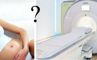
The most susceptible and sensitive to the effects of X-rays are women during pregnancy and children under 18 years of age. Tomography is not performed for this category of people.
- In some cases, CT is prescribed for health reasons with the use of personal protective equipment against ionizing radiation.
- Patients weighing 180 kg or more will also be denied the procedure for technical reasons, since most machines have a maximum load capacity of 150 kg.
- Tomography using intravenous contrast is not performed on patients:
- with reduced functional capacity of the kidneys, as determined by calculating the level of creatinine in the blood and calculating the glomerular filtration rate (creatinine more than 130 mmol/l);
- with a history of allergic reactions to iodine preparations;
- with severe persistent bronchial asthma;
- with thyrotoxicosis.
How can patients prepare for the study?
During a routine examination, 2-3 days before the procedure, the doctor gives recommendations on proper preparation, compliance with which will ensure the receipt of high-quality images and accurate results.
The patient is required to:
- stick to a diet, excluding foods that cause excess gas;
- take carminatives for severe flatulence (activated carbon, simethicone);
- Plan your last meal no later than 6-8 hours before the procedure;
- Appear for diagnostics on an empty stomach.
- When examining with contrast, you must first ensure that there is no impairment of renal function.
- To do this, biochemical tests are carried out 3-7 days to determine the level of creatinine in the blood serum.
- The day before tomography, avoid taking medications that have a nephrotoxic effect: non-steroidal anti-inflammatory drugs, ACE inhibitors, diuretics, hypoglycemic drugs (biguanides).
- To reduce the level of side effects and speed up the removal of the contrast agent, adequate hydration is carried out 24 and 2 hours before the study.
- To do this, the patient must drink 3-4 liters of clean water without gas on the eve of the scan and 2 liters before the procedure.
How does the scan work?
For comfort during diagnosis and to prevent the appearance of artifacts, the patient changes into loose clothing without metal elements.
A peripheral catheter is inserted into the cubital vein to administer a contrast agent. The iodine preparation is administered manually before the study, or an automatic injector is attached to the catheter for dosed administration of contrast during scanning.
The standard position on the tomograph table is the supine position, with the upper limbs raised up. To prevent involuntary movements, the patient is secured to the couch with belts.
When starting, the table slides into the frame of the device and stops at the level of the lumbar region.
Depending on the scanning technology, the table with the patient moves continuously or in short “steps”.
The radiologist gives commands to hold your breath through the intercom. Upon completion of the data collection cycle, the couch moves to its original position.
The laboratory assistant helps the patient get up, assesses the condition and escorts him to a special room for observation for 30 minutes.
They do a CT scan for 15-20 minutes. Contrast enhancement takes an additional time of up to 40 minutes. The patient receives the study protocol, conclusion and tomograms with identified changes within two hours.
CT scans are prescribed to children according to strict indications in complex clinical cases. The main application is the diagnosis of traumatic injuries, tumors, and identification of stones.
Newborns are examined without the use of drugs or premedication. The scan is performed immediately after feeding. The presence of a parent wearing radiation protection equipment is allowed.
In older children, CT scans are performed under sedation to exclude motor activity and anxiety.
The anesthesiology team is present during the examination. The office is equipped with the necessary equipment for carrying out resuscitation measures.
Alternative diagnostic methods
MRI is preferable for detecting malignant and benign neoplasms and assessing regional lymph nodes.
The use of gadolinium drugs increases the sensitivity of the method to 91%, specificity to 94% in recognizing the nature of tumors.
The method identifies adrenal hyperplasia in patients with Cushing's syndrome. It is highly accurate in detecting perirenal hematomas.
- Ultrasound visualizes the position of the kidneys, changes in size, thickness of the parenchyma, and the condition of the pyelocaliceal system.
- It has high accuracy in diagnosing renal failure, identifying stones, obstruction, tumors with lesion sizes of 1 cm in the adrenal glands and 2 cm in the kidneys.
The method will determine the shape, size, contours of tumors, fluid formations and calcifications. To make a histological diagnosis, a biopsy is taken from the lesion under ultrasound guidance.
Video
The high sensitivity and specificity of CT has reduced the number of diagnostic errors to a minimum. Early detection of pathological processes allowed doctors to prescribe adequate treatment and avoid unnecessary surgical interventions. Thanks to this, recovery rates, survival rates and quality of life for patients have improved.
Source: https://osnimke.ru/tazovye-organy/kt-pochek.html
Why is a kidney CT scan with contrast performed?
Kidney CT with contrast is a diagnostic method used in urology to identify various pathologies and is highly accurate. CT cannot be classified as “ordinary” studies, such as urine analysis or ultrasound, since it is more expensive and is prescribed as necessary, because it is not completely harmless.
In this article we will talk about the intricacies of the CT procedure, for which kidney diseases it is prescribed, what advantages this diagnostic method has, and also consider the existing contraindications and learn about the rules for preparing for the study.
CT even detects metastases
About computed tomography and its features
Contrast tomography of the kidneys is a diagnostic method that examines organs with X-rays and creates a computer image of them. The introduction of contrast improves image quality and is necessary when examining the kidneys due to the peculiarities of their structure.
The method is very informative and easy to carry out: during the procedure, the patient is placed on a mobile table, which is placed inside the scanner. The device makes movements that create a detailed image of the kidneys.
For contrast, iodine-containing substances are used, which tend to quickly spread throughout the body and, in fact, increase the contrast of the image.
The introduction of contrast can be:
- intravenous - through a catheter, at a rate of 2-7 mls;
- oral – used in the diagnosis of the pelvis and gastrointestinal tract;
- rectal – for detailed visualization of the large intestine;
- inhalation - not common, used to study the brain and lungs.
Examination of the kidneys with a contrast agent has many advantages, for example:
- Highly informative – allows you to identify diseases in the early stages.
- Painlessness – during the procedure there is no intervention in the body, there is no pain.
- Low probability of false results thanks to high-tech equipment.
- Does not cause adverse reactions.
There are also disadvantages to kidney CT with contrast:
- Relative harmfulness of X-rays. However, in minimal doses there is no harm to the body, so the frequency of the procedure should be determined by the doctor, based on the patient’s health condition.
- Contraindications to the use of contrast media.
- It is not advisable to perform a CT scan of the kidneys and bladder with contrast in children under 14-16 years of age, since the children's body is most susceptible to X-ray radiation.
- High demands are placed on the specialists who carry out the procedure and interpret the results, as well as on the engineers working with the device.
Compared to the proven effectiveness of this diagnostic method and its high accuracy, the described disadvantages are minor, but still, before conducting this study, it is necessary to clearly discuss the possible risks with the doctor.
Computed tomography of the kidneys with contrast is used:
- for diagnosing tumors;
- for organ injuries;
- in cases of kidney dysfunction due to infectious diseases;
- for cysts;
- to detect hydronephrosis;
- for kidney stones;
- to detect congenital and acquired anomalies;
- as part of monitoring the condition of the organ after treatment or kidney infarction.
Many patients are frightened by the procedure for administering a contrast agent, and they ask the question: what will a kidney CT scan without contrast show? Without the substance, the image quality will be much worse; there are cases in which the substance is not used, the main reason for this is intolerance to the components of the drug.
The contrast contains iodine
The tomograph can be closed or open.
Using tomography, many kidney pathologies are detected.
The contrast is excreted by the kidneys.
How to properly prepare for the procedure
A specialist should tell you how to prepare for a kidney CT with contrast a day before the procedure. In fact, there is no special preparation. 3-6 hours before the administration of a contrast agent, you should refrain from eating; drinking clean water is allowed. Before getting into the device, you should remove all metal jewelry.
Medical staff should check with the patient whether there is an allergy to iodine and seafood. You need to know this in advance, since the contrast contains iodine, and when administered, it can provoke an allergic reaction.
This concludes the preparation for a kidney CT scan with contrast. The substance is activated after about 20-30 seconds, the patient may feel the urge to urinate, but they are false and pass quickly.
The diagnostic instructions indicate that the patient should not move in the machine; he is fixed and the table is pushed into the tomograph. The doctor is in the next room behind glass, and the patient can use a special device inside the tomograph to communicate with him.
Before the study begins, the doctor talks about the rules for being in the device.
Who is contraindicated for kidney CT?
Like many other diagnostic procedures, not all patients can have a kidney CT scan with contrast. First of all, the administration of contrast is prohibited for people who have problems associated with removing the substance from the body, as well as those with individual intolerance to the components of the drug.
In addition, the procedure is contraindicated for:
- Pregnancy - X-ray radiation can have a negative effect on the fetus, so before CT scanning it is necessary to clearly ensure that there is no early pregnancy. If a woman expecting a child urgently needs an examination, an MRI without contrast agent is acceptable.
- Obesity - in this case, the use of a conventional tomograph is impossible, since it is designed for patients weighing up to 120 kg. For obese patients, an open type tomograph is required.
- Claustrophobia and other severe mental disorders - these diseases are a direct contraindication to CT scanning of the kidneys and urinary tract with contrast. Such disorders do not allow the patient to remain motionless for a long time or stay in close spaces, which is necessary for research.
- Renal failure in diabetes mellitus - with a high concentration of sugar, blood vessels are affected; in this case, a CT scan of the kidneys with a contrast agent cannot be performed, since the kidneys will not be able to remove the drug from the body.
Important! If you suspect any contraindications, you must inform your doctor before the study. The specialist will assess the possible risks and benefits and make the final decision.
The method has some contraindications
Development of a diagnostic method
Nowadays, in addition to standard computed tomography, there is also multislice computed tomography. This method appeared in 1992 and allows the use of several elements to capture X-ray waves that travel in a spiral through the areas of interest.
Compared to a conventional kidney CT with contrast, MSCT has greater image detail and allows you to make a larger number of sections.
Using this study you can diagnose:
- abscesses;
- occlusion of renal vessels;
- hemorrhages;
- polycystic disease;
- kidney cancer;
- stones;
- bruises and kidney injuries;
- establish the stage of the tumor;
- identify the cause of renal colic.
The price of this diagnostic method is about 3-4 thousand rubles, and has contraindications similar to conventional computed tomography. 2-3 hours before the procedure you should not eat food or carbonated drinks.
MSCT is a more advanced version of CT
From the photos and videos in this article, we learned about the features of computed tomography of the kidneys, considered recommendations for prescribing this diagnostic method, and how to prepare for it.
Frequently asked questions to the doctor
Only pros
Hello. Tell us, what advantages does MSCT of the kidneys have?
Good evening. Unlike simple CT, there is less radiation exposure, the examination is more accurate, and at the same time less time is spent on the process of obtaining images. Thus, MSCT has greater productivity, but at the same time a higher cost.
Source: https://bolyatpochki.ru/diagnostika/drugie-metodi/kt-pochek-s-kontrastirovaniem-270.html
Kidney CT scan with contrast
Computed tomography of the kidneys with contrast is a layer-by-layer x-ray examination of organs, which is of high diagnostic value.
Thanks to the ability to obtain three-dimensional images of tissue sections in increments of 3 to 10 mm, a specialist can accurately diagnose a wide range of pathologies.
Using a kidney CT with contrast, urolithiasis, tumor processes, cysts and abscesses are determined.
The best offers from clinics for CT scans of the kidneys with contrast
CT scan of the kidneys on the map of Moscow:
Administration of contrast agent (bolus method)
Price: 6300 rub.
Recording a CT scan to disk
For free
- Issue of film with photograph
- Price: 500 rub.
- Price: 400 rub.
- Recording research on flash
- Price: 1000 rub.
- Price: 650 rub.
Main indications:
- trauma, blow, bruise of the renal area;
- anatomical anomalies in the development of structures of the urinary system;
- suspicion of urolithiasis;
- symptoms of an infectious process;
- malignant and benign tumors of renal tissue.
Pain in the lower back for no apparent reason is also considered an indication for diagnosis.
Computed tomography of the kidneys with contrast is more informative regarding neoplasms.
Why is it better to have a kidney CT scan with contrast done here?
Available
There is a flexible discount system
Qualitatively
The equipment meets international standards
Comfortable
We work around the clock within walking distance from the metro
Fast
The result is ready in 30 minutes in electronic form
- pregnancy at any stage;
- age less than 14 years;
- severe obesity (weight more than 150 kg);
- diabetes mellitus in the stage of decompensation;
- allergy to iodine-containing compounds.
The patient should refuse food 2-3 hours before the test.
Progress of the procedure
In the specialist’s office, having previously removed all jewelry and metal objects, the person lies down on the moving table of the apparatus. Next, the table moves into the tomograph ring. The device takes several pictures and then turns off.
A contrast agent containing iodine is injected into the patient's body using an automatic injector, and the procedure is repeated again. This two-stage diagnosis increases the diagnostic value of the procedure.
A kidney CT scan with contrast takes 20 to 30 minutes. The person does not feel pain, inconvenience or discomfort. The only requirement for the patient is to remain completely still inside the tomograph.
The high accuracy and information content of computed tomography in our medical center is based on the use of the modern “Brilliance CT 64” device manufactured by Philips.
Diagnostic cost
The price of an examination at the Open Clinic includes the entire range of diagnostic procedures, including interpretation of the results.
Our partners
Source: https://OpenClinics.ru/kt/kt-pochek-s-kontrastirovaniem/
How is a kidney CT scan done with contrast?
Beam scanning is a universal method, as it allows you to study any internal organ of the body without instrumental penetration by visualizing the processes occurring inside the body. Abdominal organs such as the kidneys and adjacent systems and structures are clearly visualized. To enhance image clarity, a scanning mode such as kidney CT with contrast is used.
The operation of scanning machines is based on the production of ionizing radiation, which, penetrating into biological tissues, is partially absorbed by them. The denser the fiber structure, the more X-rays it absorbs.
Hollow and liquid substances, on the contrary, transmit rays almost unhindered. Based on the analysis of the difference in such absorption, the equipment processes the signals and transmits them in the form of images to the computer screen.
Features of CT scan of the kidneys with contrast
Radiation screening is carried out step by step on a special tomograph. The machine produces multiple scanning sections in a longitudinal position at any given depth.
This allows you not only to obtain many static images of the studied area, but also to combine photos into a multidimensional projection to view the area from all sides, rotating it at any angle and precisely zooming in on anomalous zones.
The study allows you to quickly determine the source of the disease, since in most cases the foci of the anomaly are visible immediately, at the first diagnosis. If the pathology is microscopic in size or the cause of the inflammation is not clear, specialists prescribe a CT scan of the kidneys with contrast.
This procedure differs little from the standard screening protocol. The main feature is the use of a special coloring amplifier - contrast - in the process. The solution is a “dye” that has a ferromagnetic base and shows up well in photographs, making them more contrasty and clear.
Images are displayed on the monitor in black and white, denser formations appear light, and hollow and liquid structures appear dark. Some pathologies, such as cancerous lesions, may be similar in density to adjacent tissues and are more difficult to distinguish in photographs. Staining is used to identify them.
There is no point in being afraid of this mode of procedure. In the absence of individual contraindications, dye preparations are absolutely harmless and do not leave any side effects.
When studying this area, an intravenous solution is administered. Distributed throughout the bloodstream, the substance accumulates in soft tissues, tinting them, especially vascular channels and capillaries.
CT with staining helps to clearly see even microscopic pathologies in detail.
The entire process of scanning and intravenous administration of the amplifier is supervised by experienced physicians. At the slightest deviation from the norm, the procedure is stopped; all actions of doctors are aimed at avoiding complications.
To determine whether the patient has a negative reaction to the drug, drug testing is performed in advance.
If the immune system has a positive response (expressed in an allergic reaction - itching, redness at the injection site), enhanced screening is not prescribed; alternative diagnostic methods are proposed.
When is a kidney CT scan with contrast prescribed?
Most often, screening is indicated in controversial situations when other methods have not given an effective result. Scanning is a more informative method if the cause of the disease has not been discovered, if the results of an ultrasound examination or urography turned out to be controversial, and also if there are suspicions about the course of oncological processes in this area.
Contrast is also used only when absolutely necessary. The treating doctor will definitely explain and justify the choice of this diagnostic regimen. Kidney CT scan with contrast is performed for the following indications:
- displacement, change in location of organs;
- cystic formations on tissues;
- infection of unknown etiology;
- congenital or acquired pathologies of the configuration of vascular canals;
- disturbance in the processing of protein compounds;
- acute or chronic diseases of the body’s natural “filters”;
- post-traumatic pathologies;
- monitoring the effectiveness of treatment;
- control of the rate of formation of thrombotic barriers in blood vessels;
- study of adrenal formations;
- disturbance in organ development;
- preoperative period, assessment of the patient’s operability;
- stagnation of fluid, leading to stretching of the walls of organs;
- visual control during tissue sampling (biopsy);
- determination of the nature and nature of neoplasms.
CT does not involve penetration into biological tissues, therefore it is absolutely painless and does not require anesthesia in the standard mode. Suspicions of improper operation of filtrates arise when deviations from the norm in laboratory tests are detected; such methods cannot detect the root cause of the pathology. Computer scanning is designed to cope with this task.
Are there any prohibitions on performing a CT scan of the kidneys with contrast?
- The period of pregnancy at any stage of the course. Regardless of a woman’s confidence that she is not pregnant, she will definitely have to take an analysis or a rapid pregnancy test. This is necessary in order to eliminate any possibility of harm to the intrauterine fetus. The study is carried out under the influence of X-rays, which aggressively affect any actively growing tissue, which can negatively affect the health of the unborn baby. Enhancing staining is also contraindicated during pregnancy, since the drug will penetrate through the mother’s blood into the child’s organs, which can lead to intoxication of his body. Dyes are also not used during breastfeeding, as they easily penetrate into any liquid substance, including milk.
- Children's age up to 12-14 years. During this period, the young patient actively develops. A child should not be exposed to harmful radiation.
- Urinary dysfunction. With reduced urinary function, the dye can remain in the body for a long time, which will negatively affect the functioning of organs and can lead to poisoning.
- Allergy to medications. CT scan of the kidneys with contrast is contraindicated in case of allergic rejection to the components of the drug. During the procedure, mild nausea, an acidic taste in the mouth, hot flashes as the solution spreads throughout the body, and a slight burning sensation at the injection site are considered normal. You should not be afraid of such manifestations; they will pass quickly. But if the patient feels difficulty breathing, gagging, or swelling, the procedure must be stopped immediately.
- Severe anxiety, mental or neurological disorders, fear, excessive pain, shock. Such factors will not allow the patient to remain motionless for a long time, which is required to obtain the correct result. In these situations, a person’s motor activity cannot be controlled. The solution may be to use sedatives or complete anesthesia.
- Excess body weight by more than 120-200 kg. It is difficult for people of a large weight category to undergo a CT scan, since tomographs are not designed for such weight. An obese person will not fit inside the installation. In this case, it is recommended to resort to the use of special equipment of an open configuration, which has no weight restrictions.
Patients are often afraid to undergo radiation examination, worrying that they may receive a high dose of radiation. There is no point in worrying about this, since any ionizing effect is recorded in the patient’s medical record, and doctors carefully monitor the annual level received by the person. The X-radiation in the device is so low that even with repeated access to diagnostic scanning, it is very difficult to exceed the annual radiation exposure rate.
Are preparatory measures necessary for a CT scan of the kidneys with contrast?
A kidney CT scan with contrast is prescribed only by a doctor based on the patient’s medical history. The subtleties and features of the procedure, important recommendations and training instructions will be provided by both the doctor and the radiologist on duty at the diagnostic center.
To eliminate the risks of critical complications, blood tests for hormonal levels are mandatory. This is necessary in order to determine the possibility of using contrast.
As general recommendations for preparing for the procedure, it is necessary to prepare a package of documents, including a medical record, a referral from the treating specialist, the results of other examinations and previous images if CT was performed more than once.
It is necessary to take care of comfortable clothing during the screening and remove metal objects from the body. Once the session is over, the person can return to their normal lifestyle.
A rehabilitation period is not required, since the procedure does not affect the patient’s well-being.
How is a kidney CT with contrast performed?
Screening is carried out according to the standard protocol for this diagnostic mode. At the preparatory stage, the client is registered and questioned on issues related to the disease. Next, detailed instructions are given.
A person is invited into a room with equipment, placed on the moving part of the tomograph, and sensors are attached to the area being studied. If necessary, the limbs are secured with straps built into the unit.
This is necessary to maintain stillness throughout the session.
In a kidney CT scan with contrast, a healthcare professional inserts a catheter connected to an auto-injector into a patient's vein. Through which saline solution is injected into the patient’s blood.
Next, the conveyor with the human body is located inside the installation, contrast is added to the saline solution, which spreads throughout the body within half a minute. The installation begins a step-by-step scan.
The dye enters the blood throughout the entire session, which lasts about half an hour.
When the examination is completed, the patient is helped up, the sensors are removed, the catheter is removed, and he is asked to go to the waiting room and wait for the CT results. This may also take 30-50 minutes, please be patient as correct interpretation of the results is worth the time spent.
What are the pros and cons of a kidney CT scan with contrast?
- No pain during the procedure.
- There is no injury to the skin or internal organs.
- obtaining a quick conclusion on which the diagnosis will be based.
- A visual picture of the processes occurring inside the body, clear photographs.
- Determination of pathological zones with high resolution of the device.
- The study is possible even with metal implants implanted into the body.
- Relatively cheap (more financially advantageous than MRI).
The disadvantages of beam scanning include:
- Increased dose of radiation.
- Proportion of probability of developing allergic rejection to a medicinal dye.
- Impossibility of studying pathologies in children and pregnant women.
- Lower quality of photographic images than similar MRI.
What are the consequences after a CT scan of the kidneys with contrast?
Consequences may be associated with insufficient preparation for the study. If doctors do not detect an allergic reaction to contrast in time, this can lead to a severe rejection of the immune system to the components of the drug, up to Quincke's edema and a severe drop in blood pressure.
Such cases are extremely rare, as doctors carefully monitor the progress of the procedure. At the slightest suspicion of deviations from the norm, prompt measures are taken to prevent complications.
Kidney CT scan with contrast during pregnancy
Source: https://mrt-v-msk.ru/kt-pochek-s-kontrastom/
Computed tomography of the kidneys
Computed tomography of the kidneys allows you to obtain their diagnostic images, including in 3D format. This study is used for early detection of pathological processes in the kidneys and adrenal glands.
Kidney CT is usually prescribed for:
- suspected congenital anomalies;
- suspicion of the presence of calculi (stones) in the urinary tract;
- assessing the density of kidney stones before the crushing procedure;
- injuries to the kidney area;
- diagnosis of neoplasms and metastases;
- monitoring the condition of the kidneys after surgery;
- determining the site for taking a biopsy;
- inflammatory and other kidney diseases.
At the European Medical Center, computed tomography of the kidneys is performed using modern equipment that fully complies with European quality and safety standards. The Phillips iCT 256 multislice CT scanner combines superior image quality with maximum patient comfort. Our MSCT device is equipped with the IDose software package, which can significantly reduce radiation exposure during examinations.
One of the advantages of performing a kidney CT scan at the European Medical Center in Moscow is the round-the-clock operation of the radiology department.
The department is headed by Professor Evgeniy Isaakovich Libson (Israel). He is an honorary fellow of King's College (UK), a member of the European and North American radiology societies, and before joining EMC, he headed the radiology department of the Hadassah Medical Center, one of the largest in Israel.
EMC uses modern research protocols that comply with the recommendations of the American and European radiological communities ESR and RSNA.
The impeccably polite and attentive administrative staff of EMC does everything to make you feel comfortable and safe throughout your stay in the clinic.
Computed tomography of the kidneys with contrast
In some cases, for better visualization of vascular structures and the urinary tract (pyelocalyceal complex, ureters), a CT scan of the kidneys with intravenous contrast is necessary.
In this case, an iodine-containing contrast agent is used, which allows one to more clearly examine the small structures of organs, evaluate blood supply, urination, as well as other features of the functioning of the kidney.
Only with contrast does it become possible to study lymph nodes, which is very important in the diagnosis and staging of kidney cancer.
The question of the need to administer contrast is decided by the radiologist, based on the purpose of the study.
Contraindications
Talk to your doctor about having a kidney CT scan with contrast if you are pregnant or if you:
- renal failure;
- diabetes;
- thyroid diseases;
- allergy to iodine and iodine-containing drugs.
In some cases, before the study you need to undergo tests and carry out medication preparation, but sometimes you have to refuse computed tomography and select a different method of examination.
Progress of the study
Before the examination, you will be asked to remove any metal items that fall into the scanning area. The x-ray technician will tell you about how the procedure will take place and answer your questions. You will need to lie down on the CT scanner table.
If necessary, to make it more convenient for you to fix a stationary position, the x-ray technician will offer you special pillows and bolsters. Next, the x-ray technician will control the examination from the control room. You will hear it on speakerphone.
The table will slide into the arch of the tomograph, after which the x-ray technician will take pictures of the required area. The study is carried out with a short breath hold.
If necessary, after the first series of images, you will be given a contrast agent intravenously and the study will continue.
The entire study will take no more than 15 minutes.
Kidney computed tomography results
Images from the tomograph are transmitted to professional workstations of radiologists. Most studies in our center are independently analyzed by two specialists, after which you are given a final conclusion. If necessary, research data can be submitted for discussion within councils of doctors (in this case, urologists and radiologists).
The turnaround time for results depends on the complexity of the clinical situation. Your doctor will tell you about the preliminary results immediately after the study.
- You can receive the results in any form convenient for you.
- Factors influencing the result of the study
- The quality of images obtained with CT scans of the kidneys may be affected by patient movement during the examination, the presence of surgical clips or catheters, and barium suspension in the gastrointestinal tract after radiography.
Computed tomography of the kidneys for children
Both adults and children, including newborns, can undergo computed tomography of the kidneys at EMC. To obtain clear images, patient immobility during the examination is of great importance.
For active children, being completely at rest often becomes an impossible task, so at EMC it is possible to perform such studies under medicated sleep in the presence of an experienced anesthesiologist.
In addition, one of the parents will be able to be with the child during the study. You only need to wear an apron that protects against X-ray radiation.
Source: https://www.emcmos.ru/articles/kompyuternaya-tomografiya-pochek
Kidney CT scan with contrast
Kidney CT is a modern method for diagnosing a paired organ located in the retroperitoneum on both sides of the spine using X-rays.
Computed tomography is performed without contrast and with contrast, depending on the purpose pursued. Examination with contrast is highly informative in visualizing neoplasms and vascular pathologies.
What does a kidney CT scan with contrast show?
Kidney CT scan with bolus contrast can diagnose the following pathologies:
- Neoplasms;
- Diseases of the excretory system;
- Tumor. Location, stage and size;
- Thrombus;
- Cyst;
- Inflammatory process;
- Pathologies in blood vessels
- Changes in the size of lymph nodes.
The contrast agent accumulates in the tissues, which makes the images clear and separates pathological cells from healthy ones.
When examining the kidneys, the images show the condition of the adrenal glands, some organs of the retroperitoneal space, the vertebrae of the lumbar spine and soft tissues.
Preparation
A referral for examination is issued by a neurologist, gynecologist, urologist, neurologist and therapist in order to refute or confirm a previously made diagnosis.
Special preparation for the study is required. It is important to stop eating and drinking 6-8 hours before the test.
As for diagnostics without contrast enhancement, it is necessary in advance to select clothing that does not contain metal elements. Otherwise, the medical staff of the diagnostic center can provide a disposable medical gown upon request.
Process
The process of performing a CT scan of the kidneys with contrast is no different from a CT scan of the kidneys without the injection of contrast.
Contrast is administered intravenously in 2 ways:
- Before the start of the examination or at the time of its implementation;
- During the entire examination using a catheter.
After contrast administration, the patient may experience a metallic taste in the mouth, nausea, and pain in the area where the contrast agent was administered.
The study takes about 45-55 minutes.
Indications
It is necessary to undergo examination for the following indications:
- Frequent increase in blood pressure (blood pressure);
- Inflammatory processes;
- Damage to internal organs;
- Clarification of the location, size and stage of the tumor;
- Pathologies in blood vessels.
Contraindications
There are the following contraindications when administering contrast enhancement:
- Allergy to iodine. The contrast contains iodine-containing substances, which can cause an allergic reaction in the patient (swelling of the face, itching, redness, etc.);
- Some kidney diseases. Contrast agent is slowly eliminated from the body of a patient with kidney disease, which can cause poisoning;
- Diabetes.
Mothers of infants are not recommended to feed the child for 2 days after diagnosis with contrast, as it can enter the child’s body and cause unpleasant consequences. Within 2 days, the contrast agent will be eliminated from the body naturally.
Source: https://KTdiagnostik.ru/kt-pochek/contrast/